
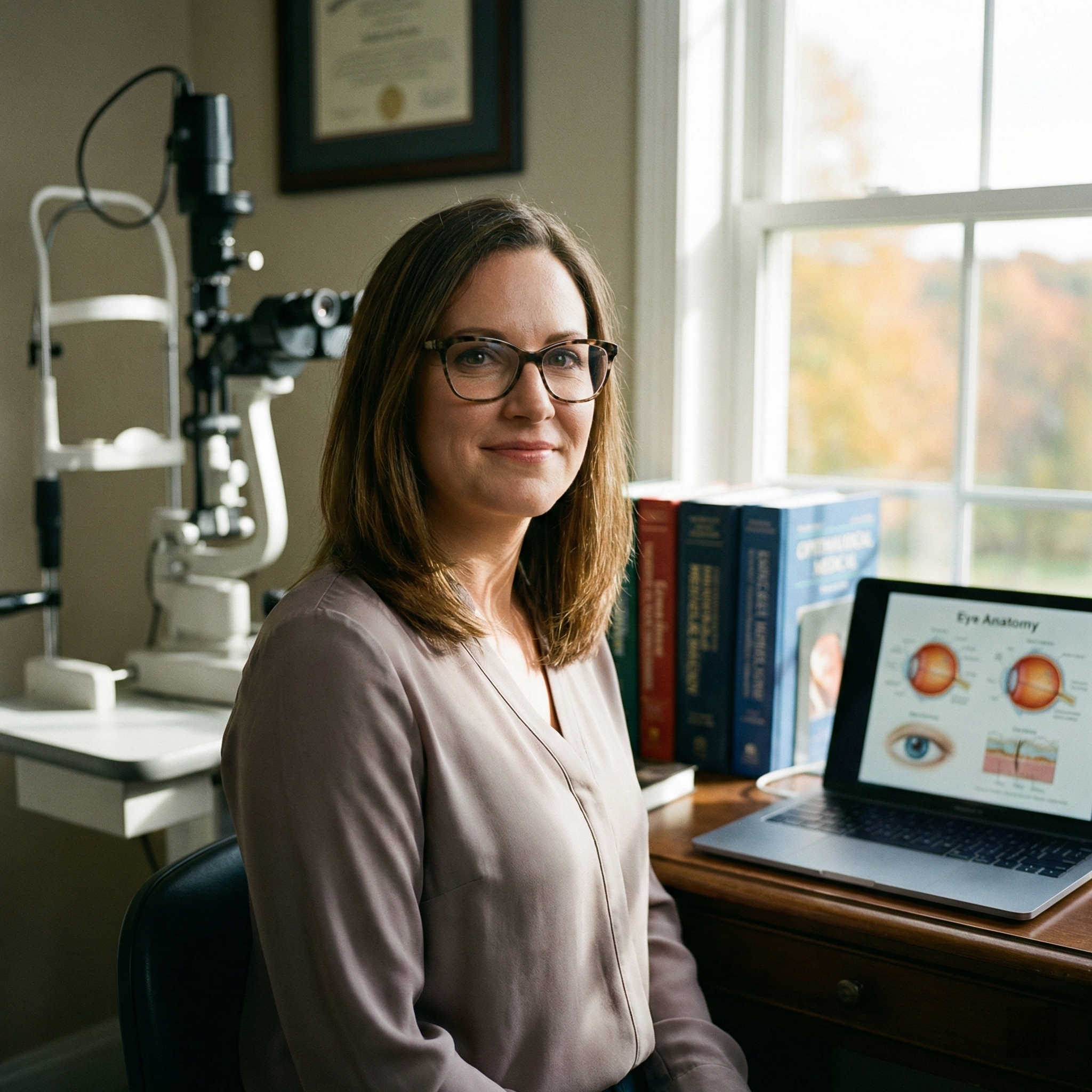
At Liv Hospital, we stress the importance of ophthalmoscopy as a key tool. This easy, non-invasive method lets doctors see the eye’s inner parts. This includes the retina, optic disc, and blood vessels.
Ophthalmoscopy is key for spotting eye problems and diseases that affect the eyes. Knowing how to use an ophthalmoscope well helps doctors give correct diagnoses. They can then create good treatment plans.
Key Takeaways
- Ophthalmoscopy is a vital diagnostic procedure for visualizing the inner eye structures.
- It helps in diagnosing eye conditions and detecting systemic diseases.
- Proper use of an ophthalmoscope is essential for accurate diagnoses.
- Liv Hospital guides healthcare professionals through comprehensive fundus examination techniques.
- Ophthalmoscopy is a simple, non-invasive procedure.
Understanding Ophthalmoscopy and Equipment

Knowing about ophthalmoscopy and its tools is key for healthcare workers. It helps them check the retina and other eye parts. Ophthalmoscopy lets them see inside the eye, like the retina, macula, and optic disc.
What is Fundus Examination
A fundus exam, or fundoscopy, looks at the eye’s inside. It checks the retina, macula, and optic disc. This is important for spotting and treating eye problems like diabetic retinopathy and glaucoma. We use an ophthalmoscope, a tool like a flashlight, to see these parts.
Types of Ophthalmoscopes and Their Applications
There are many ophthalmoscopy types, like direct, indirect, and slit-lamp ophthalmoscopy. Each has its own use and benefits.
| Type of Ophthalmoscopy | Magnification | Field of View | Application |
| Direct Ophthalmoscopy | 15 times | Narrow | Detailed examination of the fundus, useful for the optic disc and macula. |
| Indirect Ophthalmoscopy | 2-5 times | Wide | Looks at the retina’s edges, good for finding retinal detachments. |
| Slit-Lamp Ophthalmoscopy | Variable | Variable | Uses a slit lamp for detailed views, often with extra lenses. |
For more on fundoscopic exams, check out Stanford Medicine’s guide.
Key Components and Settings of an Ophthalmoscope
Knowing an ophthalmoscope’s parts and settings is vital. The aperture and filter settings must match the patient’s needs. The aperture size controls light and area seen. Filters help reduce glare or improve contrast.
Examination of the Fundus of the Eye: Step-by-Step Technique

To check the fundus of the eye, doctors need to follow a detailed method. This ensures they get a complete and correct look. It involves several steps, from getting the room and patient ready to using the ophthalmoscope right.
Preparing the Environment and Patient
Getting the room and patient ready is key for a good fundus check. The test needs a semi-dark room and takes 5 to 10 minutes. The patient sits in a dark room, and the doctor uses the ophthalmoscope to shine light through the pupil.
The room should be lit well for the patient but dark enough for the ophthalmoscope. The patient sits comfortably, with their eyes at the doctor’s level. This makes the test less uncomfortable and helps the patient stay steady.
Proper Ophthalmoscope Handling and Approach
Using the ophthalmoscope correctly is important for a clear fundus view. We hold the ophthalmoscope with the hand for the eye being checked (right hand for the right eye). It should be close to the eye but not touching, for a clear view.
The patient looks straight ahead and slightly down. We approach from 15 degrees to the side, shining the light into the pupil. The patient looks past us, focusing on something far away.
Systematic Visualization Technique
Using a systematic way to look at the fundus is essential. We first find the optic disc, then check the retinal vessels, and lastly the peripheral retina. The optic disc is recognized by its color, shape, and blood vessels.
| Structure | Normal Findings | Abnormal Findings |
| Optic Disc | Sharp margins, pink color | Blurred margins, pallor |
| Retinal Vessels | Normal caliber, no tortuosity | Narrowing, tortuosity |
| Macula | Normal pigmentation | Abnormal pigmentation, lesions |
By following these steps and using the ophthalmoscope right, we can see the fundus clearly. This helps us make accurate diagnoses and manage eye problems well.
Conclusion
An ophthalmoscope is key for eye exams. It lets doctors see inside the eye. Knowing what is fundus examination helps them spot and treat eye problems.
Ophthalmoscopy is very good at finding eye issues. Studies show it’s right 90 to 95 percent of the time. It can spot glaucoma, diabetic retinopathy, and age-related macular degeneration early.
To get the best results, doctors must use the ophthalmoscope right. They need to look at the retina, macula, and optic disc carefully. This way, they can find and fix problems, like those seen in glaucoma.
With practice, ophthalmoscopy becomes a vital tool for doctors. It helps them give better care to patients. Learning how to use it well can really help patients.
FAQ
What is the purpose of a fundus examination using an ophthalmoscope?
A fundus examination helps diagnose eye conditions and detect diseases that affect the eyes. It gives a detailed look at the eye’s inner structures.
What are the different types of ophthalmoscopes used for fundus examination?
Direct ophthalmoscopy magnifies about 15 times. Indirect ophthalmoscopy offers a wider view.
How do I properly handle and approach the patient with an ophthalmoscope?
Be comfortable and make sure the patient is relaxed. Hold the ophthalmoscope right and approach from the correct angle for a clear view.
What are the key components and settings of an ophthalmoscope that I should be aware of?
Know the aperture and filter options for a clear fundus view. Learn the different settings to improve your exam.
How do I interpret the findings from a fundus examination?
Understanding normal and abnormal anatomy is key. Look for signs of conditions like cataracts or glaucoma. Consider the patient’s history and symptoms.
What is the significance of a normal fundoscopic exam?
A normal exam means the eye’s inner structures look healthy. But, it doesn’t rule out other eye or systemic diseases.
Can a fundus examination detect systemic diseases such as hypertension?
Yes, it can show signs of diseases like hypertension. These can cause changes in the retina’s blood vessels.
What is the difference between a fundoscope and an ophthalmoscope?
Fundoscope and ophthalmoscope are often used the same way. They both examine the eye’s fundus.
How is fundoscopy used in the diagnosis of glaucoma?
Fundoscopy checks the optic nerve head for damage. This can indicate glaucoma.
References
National Center for Biotechnology Information. Evidence-Based Medical Insight. Retrieved from https://www.ncbi.nlm.nih.gov/books/NBK221/


