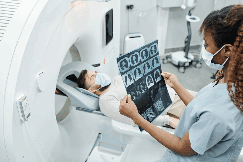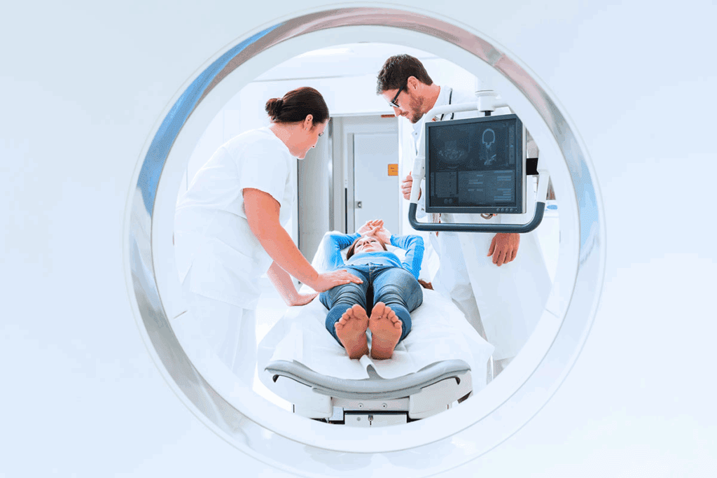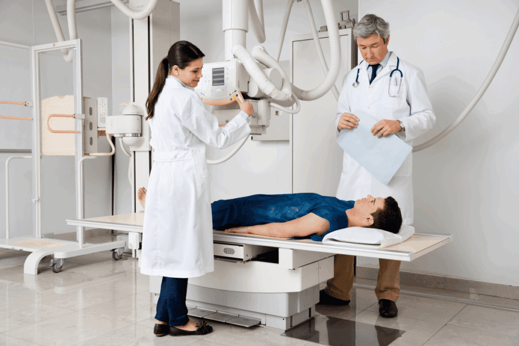
At Liv Hospital, we know how vital diagnostic images are in today’s healthcare. Radiology plays a key role in diagnosis. It captures images of the body, showing what’s happening inside. Need imaging examples? We explain 5 powerful and common medical imaging methods, their uses, and how they work (e.g., X-ray, MRI).
We employ five main medical imaging methods for diagnosis and treatment. These methods are essential in healthcare, allowing for assessments without surgery. Our team is dedicated to providing top-notch care by using the latest medical standards.
Key Takeaways
- Five primary medical imaging techniques are used in diagnosis and treatment.
- Diagnostic images support assessments without invasive procedures.
- Liv Hospital adopts the latest academic protocols for international medical outcomes.
- Medical imaging is critical in modern healthcare systems.
- Our team is committed to delivering high-quality care.
The Evolution of Medical Imaging Technology

Medical imaging technology has come a long way, changing how we find and treat diseases. Wilhelm Conrad Röntgen discovered X-rays in 1895. This discovery started a journey that has made it easier to see inside the human body.
Historical Development of Diagnostic Imaging
Diagnostic imaging has grown slowly but surely, with big steps forward. Some major milestones include:
- The introduction of X-rays for seeing bones and finding fractures.
- The 1970s brought Computed Tomography (CT) scans for detailed cross-sections.
- In the 1980s, Magnetic Resonance Imaging (MRI) gave us clear views of soft tissues.
These breakthroughs have greatly improved our ability to diagnose diseases early and treat them effectively.
Impact on Modern Healthcare
Diagnostic imaging has had a huge impact on healthcare today. Early detection and diagnosis are now more precise, thanks to new imaging tech. This has led to:
- Better patient outcomes from timely treatments.
- More tailored treatment plans for better care.
- Lower healthcare costs by avoiding unnecessary surgeries.
As we keep pushing the boundaries of medical imaging, we’ll see even more advanced tools. These will make healthcare even better.
Understanding Medical Imaging Examples in Diagnostics

Medical imaging has changed how we find and treat diseases. It gives doctors detailed views of the body’s inside. This helps them diagnose and keep track of many health issues.
Role in Disease Detection and Diagnosis
Medical imaging is key in finding and diagnosing diseases. Tools like X-rays, CT scans, MRI, ultrasound, and PET scans give vital info. For example, a study in the Journal of Clinical Oncology shows imaging’s role in cancer diagnosis.
“Imaging modalities have become essential tools in the diagnosis and staging of cancer.”
Journal of Clinical Oncology
These tools help doctors:
- See tumors and how big they are
- Spot vascular diseases and track them
- Find and understand musculoskeletal problems and injuries
Importance in Treatment Planning
Medical imaging is also key in planning treatments. It gives detailed views of the body’s inside. This helps doctors plan better treatments.
In cancer treatment, imaging is very helpful. It helps in:
| Imaging Modality | Role in Cancer Treatment |
| CT Scans | Help in planning radiation therapy by providing detailed images of tumors and surrounding tissues. |
| MRI | Assists in assessing the extent of tumor spread and monitoring response to treatment. |
| PET Scans | Provides information on metabolic activity within tumors, aiding in assessing treatment efficacy. |
As medical imaging tech gets better, its role in diagnostics and treatment planning will grow. This will lead to better patient care and outcomes.
X-Ray Imaging: The Foundation of Medical Diagnostics
X-ray imaging is key in medical diagnostics. It gives us quick and clear views of what’s inside our bodies. This technology is vital for understanding many health issues, making it a must-have in today’s healthcare.
How X-Ray Technology Works
X-ray technology uses special radiation to show us what’s inside our bodies. When an X-ray beam hits a body part, different tissues absorb it differently. For example, bones absorb more than soft tissues, creating a contrast that lets us see bones.
Getting an X-ray image is simple. The patient stands between the X-ray machine and a detector. The machine sends out a controlled dose of radiation, which goes through the body and is caught by the detector. This creates an image that doctors can use to diagnose many conditions.
Clinical Applications: Bone Fractures and Chest Evaluation
X-rays are used a lot for diagnosing bone fractures and checking the chest. For fractures, X-rays show us the bones clearly, helping doctors decide how to treat them. For chest checks, X-rays help spot lung problems like pneumonia by showing lung tissue issues.
X-ray technology is also used in many other areas. It’s chosen because it’s easy, fast, and works well. Here’s a list of some common uses of X-ray imaging:
| Clinical Application | Description |
| Bone Fractures | Diagnosing and assessing the severity of fractures |
| Chest Evaluation | Diagnosing lung conditions such as pneumonia |
| Dental Imaging | Examining teeth and jawbone structures |
Limitations and Safety Considerations
Even though X-ray imaging is very useful, it has some downsides and safety issues. One big concern is the radiation it uses, which can be harmful. We must think carefully about when to use X-rays, keeping in mind the risks, like for pregnant women and kids.
“The use of X-rays in medical diagnostics has revolutionized the way we understand and treat various health conditions, but it’s vital to use this technology wisely to reduce radiation exposure.”
To lower these risks, we follow strict rules to use the least amount of radiation needed. Also, new diagnostic imaging equipment is making X-rays safer and more effective.
In summary, X-ray imaging is a key part of medical diagnostics. It helps us see inside the body quickly and effectively. By knowing its uses, limits, and safety, we can keep using it to help patients get better.
CT Scans: Advanced Cross-Sectional Imaging
Computed Tomography (CT) scans have changed how we diagnose diseases. They give detailed images of the body’s inside. This technology is key in healthcare, helping doctors find and treat many health issues.
Technical Principles of Computed Tomography
CT scans use X-rays from different angles to show the body’s inside. A rotating X-ray tube and detectors capture this data. Then, computers turn it into detailed images.
CT scans have gotten better over time. They now scan faster, show more detail, and use less radiation. Modern scanners can scan big parts of the body quickly, which is great for emergencies.
Applications in Trauma Assessment
In trauma cases, CT scans are very important. They quickly and accurately find injuries like bleeding or organ damage. This helps doctors make fast, important decisions for patients.
CT scans are great for checking complex injuries. They can look at the head, neck, chest, belly, and pelvis all at once. This is very helpful in cases of car accidents or falls.
Role in Cancer Detection and Staging
CT scans are also key in fighting cancer. They help find tumors, see how big they are, and if they’ve spread. This info is vital for planning treatment.
Using contrast agents in CT scans makes some tissues show up better. This helps doctors see more clearly and find problems easier. It’s very useful for spotting different tissues and finding issues.
CT scans are a big part of modern medicine. They help doctors diagnose and treat many conditions. As technology keeps improving, CT scans will keep getting better at helping patients.
MRI: Detailed Soft Tissue Visualization
Magnetic Resonance Imaging (MRI) has changed how we diagnose diseases. It shows detailed images of soft tissues. This helps us understand and treat many health issues.
Magnetic Resonance Technology Explained
MRI machines use strong magnets and radio waves to see inside the body. They align hydrogen atoms and then disturb them. This disturbance creates signals that build detailed images.
The science behind MRI is complex. But, it’s easy to use. MRI is great for seeing soft tissues, which are hard to spot with other methods like X-rays.
Neurological Applications and Research Advances
MRI is key in neurology, helping us see the brain and spinal cord. It helps find problems like multiple sclerosis, stroke, and spinal injuries. MRI images help doctors understand damage and choose the right treatment.
- Diagnosing and monitoring neurological disorders
- Assessing the extent of injuries and conditions affecting the brain and spinal cord
- Guiding neurosurgical procedures
Research keeps improving MRI for the brain. It includes functional MRI (fMRI) and diffusion tensor imaging (DTI) for studying brain activity and white matter.
Musculoskeletal and Organ System Imaging
MRI is also used for muscles, tendons, and ligaments. It’s great for spotting sports injuries and other muscle problems.
It also looks at organs like the heart. MRI can see heart function and find vascular diseases. It’s safe because it doesn’t use harmful radiation.
- Assessing musculoskeletal injuries and conditions
- Imaging organ systems for disease diagnosis
- Monitoring the progression of diseases and the effectiveness of treatments
Using MRI helps us diagnose better and treat more effectively. It’s a big help in many medical areas.
Ultrasound: Real-Time Imaging Without Radiation
We use ultrasound imaging to safely see inside the body in real-time. It’s a key tool in modern medicine. This technology is non-invasive and doesn’t use radiation.
The Science Behind Sonography
Ultrasound imaging, or sonography, uses sound waves to see inside the body. A device called a transducer sends out high-frequency sound waves. These waves bounce off body parts and return to the transducer, creating images.
The science of sonography relies on acoustic impedance. Different tissues reflect sound waves in unique ways. This allows for detailed images. It’s not just for looking; it also gives real-time feedback. Doctors can see how organs move and blood flows.
Prenatal and Pregnancy Monitoring
Ultrasound is vital for prenatal and pregnancy monitoring. It lets us safely see the fetus, track its growth, and spot any issues. This is key for:
- Confirming pregnancy and figuring out how far along you are
- Watching the fetus grow and develop
- Finding out if there are twins or more
- Spotting any problems with the fetus
Ultrasound is a must-have in obstetrics. It gives important information for managing pregnancy and planning for birth.
Diagnostic Applications for Internal Organs
Ultrasound is also used for diagnosing issues with internal organs. It’s great for checking:
- The liver and gallbladder for problems like gallstones
- The kidneys for stones, cysts, or tumors
- The thyroid gland for nodules or issues
- The reproductive organs for cysts, tumors, or other problems
Ultrasound is versatile and safe. It’s often the first choice for many medical tests. This reduces the need for more invasive or radiation-based tests.
PET Scans: Metabolic Activity Visualization
Positron Emission Tomography, or PET scans, is a top-notch tool for checking how our bodies work at a tiny level. It uses a bit of radioactive stuff to show us what’s going on inside. This helps doctors find and treat diseases better.
Detecting Disease through Metabolic Activity
PET scans spot how active our body’s tissues and organs are. They’re great for finding where things aren’t working right, like in diseases. They use a special radioactive tracer that shows up in busy areas.
Key Benefits of PET Scans:
- They catch diseases early, like cancer, before we even feel sick
- They check if treatments are working
- They see if a disease has spread
Oncological Applications and Cancer Staging
In cancer care, PET scans are super helpful. They find the main tumor and see if it’s spread. This info helps doctors plan the best treatment and guess how well we’ll do.
| Application | Description |
| Cancer Diagnosis | Find the main tumor and where it’s spread |
| Cancer Staging | See how far the disease has spread |
| Treatment Monitoring | Check if treatment is working |
Neurological and Cardiac Assessment
PET scans also help in brain and heart health. They spot problems like Alzheimer’s, Parkinson’s, and epilepsy in the brain. In the heart, they check if it’s working right and find heart disease.
“PET scans have become an indispensable tool in modern medicine, giving us insights into the body’s metabolic processes that were previously unimaginable.” – A Nuclear Medicine Specialist
In short, PET scans are a key tool in medicine. They give us important info on how our bodies work. This helps doctors in many areas, like cancer, brain, and heart health.
Comparing Imaging Examples: Selecting the Optimal Diagnostic Method
Choosing the right diagnostic imaging method is complex. We must consider many clinical factors. This ensures accurate diagnosis and effective treatment planning.
Clinical Factors Influencing Imaging Selection
Several factors affect the choice of diagnostic imaging. These include the patient’s medical history and the suspected condition. For example, X-ray imaging is often used first for bone fractures. It provides quick and clear images of bones.
MRI is best for soft tissue imaging. It’s great for diagnosing brain, spinal cord, and muscle issues. The choice between CT scans and ultrasound depends on the need for detailed images without radiation.
- The type of diagnostic imaging needed
- The patient’s condition and medical history
- The availability of imaging modalities
- The need for contrast agents
- Cost and insurance considerations
Complementary Use of Multiple Imaging Technologies
Using multiple imaging technologies together improves accuracy. For instance, PET scans with CT scans (PET/CT) offer metabolic and anatomical details. This helps in detecting and staging cancer better.
Combining ultrasound with MRI provides real-time guidance during procedures. It also benefits from MRI’s detailed soft tissue imaging. This approach helps healthcare providers make better treatment decisions.
Understanding the strengths and limitations of different imaging methods improves patient care. The use of various imaging modalities in clinical practice shows how technology and expertise enhance healthcare.
Advanced Medical Imaging at Leading Healthcare Facilities
We are dedicated to top-notch healthcare, starting with the latest diagnostic imaging tools. At Liv Hospital, we know that advanced imaging is key for quality care.
State-of-the-Art Equipment and Protocols
We spend a lot on the newest imaging tech. This means our patients get precise diagnoses and effective treatments. Our gear includes high-resolution MRI machines and advanced CT scanners.
These tools help us see even the toughest conditions clearly. Our protocols make sure every image is top-notch. We use methods that cut down on radiation, keeping patients safe and helping us make accurate diagnoses.
Quality Assurance and International Standards
At Liv Hospital, we aim for the highest in diagnostic imaging quality. We follow international standards for quality assurance. This means our equipment is always up to date and ready to go.
Our radiologists and technicians get constant training. They learn about the latest in medical imaging. This keeps our care at the highest level.
We use the latest tech and follow strict quality rules. This way, we meet the world’s top standards in diagnostic imaging.
Conclusion: The Essential Role of Diagnostic Imaging in Modern Medicine
Diagnostic medical imaging has changed healthcare a lot. It helps find diseases early and treat them well. The main imaging methods—X-ray, CT, MRI, ultrasound, and PET scans—have changed how we care for patients. They help us make accurate diagnoses and plan treatments that fit each patient.
At Liv Hospital, we know how key imaging and diagnostics are for top-notch healthcare. We use the latest imaging tech to make sure our patients get the right diagnosis and treatment. Knowing about diagnostic imaging is important for doctors and patients. It helps make better decisions and ensures the best care.
We keep working to improve in diagnostic medical imaging. We’re committed to giving our international patients the best support and care. Our goal is to make sure every patient gets the best possible treatment.
FAQ
References
What are the primary medical imaging techniques used in diagnosis and treatment?
The main techniques are X-rays, CT scans, MRI, ultrasound, and PET scans. They help doctors see inside the body. This is key for planning treatments.
How has medical imaging technology evolved?
It has changed a lot. Wilhelm Conrad Röntgen found X-rays in 1895. Then, CT scans and MRIs were developed. Each new tech has helped doctors find and treat diseases sooner.
What is the role of medical imaging in disease detection and diagnosis?
Medical imaging is very important. It helps doctors see inside the body. This lets them plan better treatments.
How does X-ray imaging work, and what are its clinical applications?
X-rays create images of the inside of the body. Doctors use them to check for bone breaks and chest problems.
What are the benefits of using CT scans in medical diagnosis?
CT scans give detailed images of the body. They are key for checking injuries, finding cancer, and planning treatments. They are a big step forward in medical imaging.
What is the principle behind MRI technology, and what are its applications?
MRI uses magnetic fields to show soft tissues. It’s used for brain, muscle, and organ imaging. It’s a powerful tool for doctors.
How does ultrasound imaging work, and what are its diagnostic applications?
Ultrasound uses sound waves for real-time images. It’s used for checking on babies during pregnancy and for looking at organs inside the body.
What is the role of PET scans in disease detection and diagnosis?
PET scans show how active the body’s cells are. They help find diseases and are used for cancer and heart checks.
How do healthcare professionals select the appropriate imaging modality for diagnosis?
Doctors choose based on the disease and what each imaging method offers. They often use more than one to get the best view.
What measures are taken to ensure high-quality diagnostic images?
Liv Hospital uses top-notch equipment and strict protocols. We follow international standards to get the best images.
What is diagnostic medical imaging, and why is it important?
It’s using imaging tech to diagnose and treat diseases. It’s key in modern medicine. It helps doctors plan better treatments.
What are the different types of diagnostic imaging?
There are X-rays, CT scans, MRI, ultrasound, and PET scans. Each has its own uses and benefits.
Reference
Hussain, S. (2022). Modern diagnostic imaging technique applications and advances. PMC, https://www.ncbi.nlm.nih.gov/pmc/articles/PMC9192206/


