
Did you know over a million vascular procedures are done every year in the U.S.? Angioplasty and stenting are among the top ones. These treatments are changing medicine, making recovery faster and results better.
Image-guided treatments have made a big leap forward. This has led to more accurate and successful care. Interventional radiology is now a key part of healthcare, giving new options instead of old surgeries.
Angioplasty and stenting are leading the way in treating vascular diseases. Knowing about these treatments helps patients choose the best care for themselves.
Key Takeaways
- Angioplasty and stenting are among the most common vascular procedures performed today.
- Interventional radiology offers minimally invasive alternatives to traditional surgery.
- Image-guided treatments improve patient outcomes and reduce recovery times.
- Vascular procedures are increasingly prevalent, with over a million performed annually in the U.S.
- Understanding these procedures can help patients make informed decisions about their healthcare.
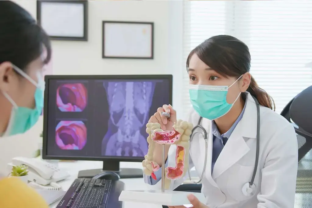
The Field of Interventional Radiology

Interventional radiology started in diagnostic radiology and has grown a lot. It now includes many treatments. These treatments are new and help patients a lot.
Definition and Scope of Interventional Radiology
Interventional radiology uses imaging and catheters to treat diseases. It’s a big field that helps patients in many ways. It’s a new way to treat diseases instead of surgery.
Doctors use tools like fluoroscopy and MRI to guide treatments. This makes treatments safer and more effective. So, interventional radiology is very important in healthcare today.
The Evolution of Minimally Invasive Image-Guided Treatments
Interventional radiology has gotten better over time. It now offers treatments that are less invasive and more effective. The field keeps getting better with new research and technology.
It’s helping with complex conditions like Cpc-PH. Doctors use many ways to find and treat these conditions. This makes treatments more precise and effective.
|
Procedure Type |
Description |
Benefits |
|---|---|---|
|
Vascular Interventions |
Treatments for vascular diseases, including angioplasty and stenting. |
Minimally invasive, reduced recovery time. |
|
Non-Vascular Interventions |
Procedures for conditions not related to blood vessels, such as biopsies and drainages. |
Less risk of complications, precise targeting. |
|
Image-Guided Treatments |
Use of imaging technologies to guide interventions. |
Enhanced precision, improved outcomes. |
Interventional radiology will keep getting better. We’ll see even better care for patients. New technologies and methods will be key in this progress.
Imaging Technologies in Interventional Radiology

Interventional radiology uses advanced imaging to guide treatments. These technologies help doctors see what they’re doing in real time. This makes treatments more accurate and safe.
Fluoroscopy and Digital Subtraction Angiography
Fluoroscopy shows X-ray images of the body in motion. It helps doctors see where instruments are inside the body. Digital Subtraction Angiography (DSA) makes blood vessels stand out by removing background images.
Together, fluoroscopy and DSA make vascular treatments more precise. They help place devices like stents correctly, lowering the chance of problems.
CT, Ultrasound, and MRI Guidance
Other tools like Computed Tomography (CT), ultrasound, and Magnetic Resonance Imaging (MRI) also guide treatments. CT is great for finding the right spot for biopsies and drainages.
- Ultrasound is good for dealing with fluid or superficial issues.
- MRI is top-notch for soft tissue and precise tumor treatments.
Fusion Imaging Techniques
Fusion imaging mixes data from different scans for a clearer view. It makes treatments more accurate by showing more details.
This method has improved patient results by cutting down on risks and making treatments work better. It’s key for complex tasks like vascular interventions and tumor treatments.
Angiography: The Foundation of Vascular Interventions
Angiography is key in vascular interventions, used for both diagnosis and treatment. It’s a vital tool in interventional radiology, helping us see the vascular system clearly.
Diagnostic Angiography Techniques
Diagnostic angiography uses contrast agents and imaging to see blood vessels and find vascular problems. We use different methods, like selective and superselective angiography, to get detailed images of blood vessel anatomy.
These methods help us spot issues like stenosis, aneurysms, and malformations. This is important for planning treatments.
Therapeutic Applications of Angiography
Angiography is also vital for treating vascular diseases. It helps us guide catheters and devices to the right place for treatment.
Therapeutic uses include angioplasty, stenting, and embolization. These treatments are done under angiography to ensure they’re done right and safely.
Patient Preparation and Recovery
Getting patients ready for angiography is key. We make sure they’re well-hydrated and review their medical history to lower risks.
After the procedure, we watch for any problems and manage side effects. We also give patients care instructions to help them recover well.
Angioplasty and Stenting: The Most Common Interventional Radiology Procedure
Angioplasty and stenting are key in interventional radiology. They treat vascular diseases in a less invasive way. This improves patient results and cuts down on recovery time.
Peripheral Arterial Disease Interventions
Peripheral arterial disease (PAD) narrows or blocks arteries in the legs. Angioplasty and stenting fix this, bringing back blood flow and easing symptoms. We use top-notch imaging to place the stent just right.
These treatments are less invasive and safer than surgery. They help patients with PAD live better lives.
Coronary and Carotid Stenting
Coronary and carotid artery diseases can cause heart attacks and strokes. Angioplasty and stenting are vital vascular procedures for these issues. They keep blood flowing and prevent serious heart and brain problems.
In coronary stenting, drug-eluting stents are used to prevent blockages. Carotid stenting uses special devices to protect against stroke. We pick the best stenting plan for each patient.
Types of Stents and Their Selection Criteria
Choosing the right stent is key for success. There are many stents, like bare-metal, drug-eluting, and bioabsorbable ones. The right one depends on the blockage, patient health, and risk of blockage coming back.
- Bare-metal stents offer support but may cause more blockages.
- Drug-eluting stents release medicine to stop blockages from coming back.
- Bioabsorbable stents dissolve over time, aiming to avoid long-term issues.
Knowing each stent’s strengths and weaknesses helps us customize treatment. This ensures the best results for our patients in interventional radiology procedures.
Embolization Therapies in Clinical Practice
Embolization therapies are key in modern interventional radiology. They offer targeted treatments with quick recovery times. We use these treatments to manage many conditions, improving patient outcomes and quality of life.
Uterine Fibroid Embolization: Procedure and Outcomes
Uterine fibroid embolization (UFE) has changed how we treat uterine fibroids. It involves injecting agents into the arteries that feed the fibroids. This reduces their size and relieves symptoms.
The procedure is done under local anesthesia and sedation. We use a small catheter to reach the uterine arteries. Then, we deliver the embolic material, cutting off the fibroids’ blood supply.
Research shows UFE greatly improves life for women with fibroids. It reduces heavy bleeding and pelvic pain.
Gastrointestinal Bleeding Management
Embolization therapies are also vital for managing gastrointestinal bleeding. When other methods fail, we use catheter-based treatments to stop the bleeding.
We find the bleeding source through angiography and then block the vessel. This method is very effective and can save lives in severe cases.
Key benefits of embolization for gastrointestinal bleeding include:
- Minimally invasive approach, reducing recovery time
- High success rate in controlling bleeding
- Ability to target the bleeding site directly
Trauma-Related Embolization Procedures
In trauma cases, embolization therapies help control bleeding and stabilize patients. We use catheter-based methods to find and block bleeding vessels. This reduces the need for surgery.
These procedures require great skill and teamwork. Interventional radiologists, trauma surgeons, and other healthcare professionals work together closely.
Embolization therapies offer a non-invasive solution. They help lower morbidity and mortality in trauma patients, leading to better outcomes.
Common Vascular Access Procedures
Vascular access procedures are key in interventional radiology. They help doctors give treatments and therapies. These steps are vital for patient care, making sure access to the blood system is safe and effective.
Central Venous Catheter Placement Techniques
Central venous catheters are used for giving medicines, fluids, and blood products. To place these catheters, doctors use imaging to guide them. Ultrasound guidance is now the best way to do this, making it safer.
The process starts by finding the internal jugular or subclavian vein under ultrasound. A catheter is then put in over a guidewire. Its position is checked with fluoroscopy or chest X-rays.
Port Placement for Long-Term Therapy
Ports are devices for long-term access to the blood system. They are used for chemotherapy, antibiotics, or nutrition. The process involves making a pocket under the skin and tunneling a catheter to a vein.
We use imaging to place the port and catheter accurately. The procedure is done under local anesthesia and sedation. Ports are a good option for patients needing long-term access.
Dialysis Access Creation and Maintenance
Dialysis access is essential for patients on hemodialysis. Interventional radiologists help by creating and keeping dialysis access open. This includes making arteriovenous fistulas (AVFs) and using angioplasty.
Making an AVF involves surgically joining an artery and vein in the forearm. Radiologists use angioplasty to keep the AVF open. This ensures good blood flow for dialysis.
|
Procedure |
Description |
Indications |
|---|---|---|
|
Central Venous Catheter Placement |
Placement of a catheter into a large vein for medication or fluid administration |
Short-term vascular access, medication administration, fluid resuscitation |
|
Port Placement |
Implantation of a subcutaneous port for long-term vascular access |
Long-term chemotherapy, antibiotic therapy, total parenteral nutrition |
|
Dialysis Access Creation |
Creation of an arteriovenous fistula or graft for hemodialysis |
Hemodialysis in patients with end-stage renal disease |
The Society of Interventional Radiology says vascular access procedures are very important. They require skill and precision. We aim to provide the best care in these procedures, ensuring good results for our patients.
Biliary and Hepatic Interventional Radiology Procedures
Biliary and hepatic interventional radiology procedures have changed how we diagnose and treat liver and bile duct issues. These methods are key in managing complex liver and bile duct problems.
We use advanced imaging to guide our treatments. This ensures we are precise and safe. We offer a variety of procedures to meet each patient’s needs. This approach improves results and shortens recovery times.
Percutaneous Transhepatic Cholangiography (PTC)
PTC is a vital diagnostic tool for seeing the bile ducts. It involves inserting a needle into the liver to inject contrast material. This gives us clear images of the biliary system.
PTC is used to find out why bile ducts are blocked, to diagnose jaundice, and to plan treatments. The info from PTC helps us choose the best treatment.
Biliary Drainage and Stenting Methods
Biliary drainage and stenting help with blocked bile ducts. We use stents or drainage catheters to keep bile flowing. This relieves symptoms and improves liver function.
Whether to use internal or external drainage depends on the patient’s situation. Internal stenting is often better because it allows for natural bile flow and lowers the risk of complications.
Management of Biliary Complications
Handling biliary complications is a big part of interventional radiology. Issues like bile leaks, strictures, and stent blockages need quick and effective solutions. This prevents more problems.
We use methods like balloon dilation and more stenting to fix these problems. Keeping a close eye on patients and following up is key. This ensures our treatments work and manages long-term issues.
Genitourinary System Interventions
The genitourinary system is a key area where interventional radiology (IR) plays a big role. IR techniques help diagnose and treat many conditions. These methods offer new, less invasive ways to manage diseases compared to old surgery methods.
Nephrostomy Tube Placement and Management
Nephrostomy tube placement is a common IR procedure. It helps by creating a direct path for urine to drain from the kidney to outside the body. This is very helpful for patients with blockages in the urinary tract.
We do this procedure under imaging guidance, like ultrasound or fluoroscopy. This ensures the tube is placed correctly and reduces risks. After the procedure, we watch the tube for any signs of problems or infection.
Key Steps in Nephrostomy Tube Placement:
- Pre-procedure imaging to plan the access route
- Local anesthesia and sedation for patient comfort
- Real-time imaging guidance for precise tube placement
- Post-procedure monitoring for possible complications
Ureteral Stenting Techniques
Ureteral stenting is another important IR intervention. It keeps the ureter open, ensuring urine flows freely from the kidney to the bladder. Stents are great for dealing with blockages caused by stones, strictures, or external pressure.
The procedure involves placing a stent in the ureter under fluoroscopy. The stent keeps the ureter open, relieving blockages and protecting kidney function.
|
Procedure |
Indications |
Benefits |
|---|---|---|
|
Nephrostomy Tube Placement |
Urinary tract obstruction, kidney stones |
Relieves obstruction, preserves renal function |
|
Ureteral Stenting |
Ureteral obstruction, ureteral strictures |
Maintains ureteral patency, ensures urine flow |
Interventions for Renal Transplant Complications
Patients with kidney transplants often need IR interventions. These are for issues like vascular stenosis, fluid collections, or urinary leaks. These interventions are key to keeping the transplanted kidney working well.
We use IR techniques like angioplasty, stenting, and drainage to tackle these problems. For example, angioplasty and stenting can fix vascular stenosis. Drainage procedures help with fluid collections or abscesses.
In conclusion, interventions in the genitourinary system are essential for treating many urological conditions. IR techniques offer effective, less invasive solutions. These improve patient outcomes and quality of life.
Interventional Oncology: Image-Guided Cancer Treatments
Interventional oncology is a key part of cancer treatment. It uses new methods that are less invasive than old surgeries. Experts in interventional radiology and advanced imaging work together to treat cancer.
Image-guided treatments are changing how we fight cancer. These methods are safer and let patients recover faster. They also help doctors target tumors more precisely, protecting healthy tissue.
Tumor Ablation Technologies
Tumor ablation kills cancer cells with heat, cold, or chemicals. Radiofrequency ablation (RFA) and microwave ablation (MWA) are popular for treating tumors. These treatments are guided by images, ensuring the device is placed correctly.
RFA uses electrical currents to heat up and kill tumor cells. MWA uses microwave energy for the same effect. Both are good for treating small to medium tumors and can be used with other treatments.
Transarterial Chemoembolization (TACE)
TACE delivers chemotherapy directly to tumors through their blood supply. It involves injecting chemotherapy drugs mixed with an embolizing agent into the tumor’s artery. This blocks blood flow, trapping the drugs inside the tumor.
TACE is great for liver cancer and has shown good results. It’s done under fluoroscopy, allowing for precise targeting of the tumor’s blood supply.
Radioembolization with Y-90 Microspheres
Radioembolization uses radioactive microspheres to kill tumor cells. These microspheres, filled with Yttrium-90 (Y-90), emit beta radiation. It’s also known as selective internal radiation therapy (SIRT).
Y-90 microspheres deliver a high dose of radiation to the tumor while protecting healthy tissue. This method is effective for liver tumors and is used in various cases of liver cancer.
In conclusion, interventional oncology brings new treatments to cancer patients. It uses image-guided technologies for targeted therapies. These advancements improve patient outcomes and quality of life. As the field grows, we expect more progress in cancer treatment through interventional oncology.
Venous Interventional Procedures
Venous interventional procedures are key in today’s vascular care. They help diagnose and treat venous conditions, improving patient results. These methods are less invasive, cutting down on surgery and recovery times.
IVC Filter Placement and Retrieval Indications
IVC filter placement stops pulmonary embolism in high-risk patients. Filters in the inferior vena cava catch blood clots before they hit the lungs. It’s used when anticoagulation is not safe, or when pulmonary embolism keeps happening despite treatment.
Removing IVC filters is considered when the risk of pulmonary embolism goes down. The decision to remove depends on the patient’s risk and any complications.
|
Indication |
Description |
|---|---|
|
Contraindication to Anticoagulation |
Patients who cannot be anticoagulated due to risk of bleeding. |
|
Recurrent Pulmonary Embolism |
Patients who experience PE despite being on anticoagulation therapy. |
|
Severe Trauma |
Trauma patients at high risk of developing deep vein thrombosis. |
Catheter-Directed Thrombolysis for DVT
Catheter-directed thrombolysis (CDT) treats deep vein thrombosis (DVT) by injecting thrombolytic agents into the clot. It reduces clot size and improves blood flow.
A study in the Journal of Vascular Interventional Radiology found CDT is a great option for acute iliofemoral DVT. It has a high success rate and improves patient outcomes.
“Catheter-directed thrombolysis has revolutionized the treatment of DVT, providing a minimally invasive solution with significant benefits for patient recovery.”Journal of Vascular Interventional Radiology
Endovenous Treatments for Varicose Veins
Endovenous treatments like endovenous laser therapy (EVLT) and radiofrequency ablation (RFA) are top choices for varicose veins. They use heat to close off the vein, reducing symptoms and improving looks.
Choosing between EVLT and RFA depends on the vein size and patient preference. Both are effective, with high patient satisfaction.
Frequency Analysis of Interventional Radiology Procedures in the United States
Medical technology keeps getting better, leading to more interventional radiology procedures in the U.S. This rise shows a move towards less invasive treatments. These treatments help patients recover faster and have fewer side effects than old surgeries.
Statistical Data on Procedure Prevalence
Recent studies have given us insights into how common certain interventional radiology procedures are. In the U.S., some procedures are now more common than before.
|
Procedure Type |
Number Performed Annually |
Growth Rate |
|---|---|---|
|
Angioplasty and Stenting |
Over 500,000 |
5% annual increase |
|
Embolization Therapies |
Around 200,000 |
8% annual increase |
|
Venous Interventions |
Approximately 150,000 |
10% annual increase |
The numbers show a big jump in interventional radiology procedures. Venous interventions are growing the fastest.
Demographic Patterns in Procedure Utilization
Looking at who gets these procedures, we see big differences. Older adults are more likely to get them. This is because vascular diseases are more common in this age group.
Trends in Procedure Growth and Adoption
Several things are driving the growth in interventional radiology procedures. New technology, more diseases, and a preference for less invasive treatments are key. This is why more doctors and patients are choosing these procedures.
- Increased availability of advanced imaging technologies
- Growing awareness among healthcare providers and patients
- Expanding applications of interventional radiology techniques
These trends are likely to keep going. This means interventional radiology will play an even bigger role in medicine.
Drainage Procedures in Interventional Radiology Practice
Drainage procedures are key in interventional radiology. They help manage fluid collections and abscesses. These methods have changed how we treat complex medical issues, making treatments less invasive.
Abscess Drainage Techniques
Abscess drainage is a common procedure. It uses imaging to place a catheter in the abscess. This helps remove the abscess contents, reducing complications and aiding in healing.
The techniques for abscess drainage include:
- Ultrasound-guided drainage for superficial abscesses
- CT-guided drainage for deeper or more complex abscesses
- Fluoroscopy-guided drainage for certain cases where real-time imaging is beneficial
Thoracic Interventions: Pleural Effusion Management
Thoracic interventions, like managing pleural effusions, are vital. Pleural effusion drainage involves placing a catheter or chest tube to remove fluid from the pleural space.
The benefits of these thoracic interventions include:
- Lower risk of complications compared to surgery
- Improved patient comfort and shorter recovery times
- More accurate catheter placement with real-time imaging
Percutaneous Abdominal Fluid Collections Drainage
Percutaneous drainage of abdominal fluid collections is essential. It’s used for conditions like ascites, bilomas, and infected fluid collections. The method uses imaging to place a catheter in the fluid collection.
Important aspects of percutaneous drainage include:
- Choosing the best drainage route and catheter size
- Using imaging to ensure accurate catheter placement
- Monitoring drainage output and fluid characteristics to assess treatment response
These drainage procedures offer effective, minimally invasive treatments. They improve patient outcomes and quality of life.
Vertebral Augmentation for Spinal Fractures
Spinal fractures management has improved a lot with vertebral augmentation techniques. These are key in interventional radiology. We use advanced imaging to make sure these procedures are precise and safe.
Vertebroplasty: Technique and Outcomes
Vertebroplasty is when bone cement is injected into a fractured vertebra. This makes it stable and less painful. It’s done under local anesthesia and sedation, so recovery is quick. Vertebroplasty greatly improves life quality for those with painful vertebral compression fractures.
Kyphoplasty: Procedure and Patient Selection
Kyphoplasty involves a balloon in the fractured vertebra to restore height before cement is injected. This method stabilizes the fracture and corrects the deformity. Choosing the right patient is key, considering age, health, and symptoms.
Post-Procedure Care and Complications
Patients are watched for hours after the procedure and then go home. They should avoid heavy lifting and bending for weeks. While safe, risks include cement leakage, infection, and new fractures. Choosing the right patient and careful technique reduce these risks.
|
Procedure |
Key Benefits |
Potential Complications |
|---|---|---|
|
Vertebroplasty |
Pain relief, stabilization of fracture |
Cement leakage, infection |
|
Kyphoplasty |
Restoration of vertebral height, deformity correction |
New fractures, cement extravasation |
We keep improving our methods and care to get the best results for vertebral augmentation patients.
Nutritional Support: Gastrostomy and Feeding Tube Placement
For those who can’t eat by mouth, interventional radiology offers a solution. Gastrostomy and feeding tube placement are key. They help people get the nutrients they need for a long time.
Percutaneous Gastrostomy Techniques
Percutaneous gastrostomy puts a feeding tube into the stomach through the skin. It’s done under imaging like fluoroscopy or ultrasound. This makes sure the tube is in the right spot.
We use local anesthesia and sedation to make the process as painless as possible. This method has fewer risks than surgery and lets patients start eating again quickly.
Jejunostomy Tube Placement
Jejunostomy tube placement is another way to get nutrients. The tube goes into the jejunum, a part of the small intestine. It’s good for those with stomach problems or who are at risk of choking.
Like gastrostomy, it’s done with imaging to place the tube correctly. This avoids problems and helps nutrients get to the body right.
Long-Term Management of Enteral Access
After tubes are put in, managing them long-term is key. This means keeping the tubes clean, watching for infections, and teaching patients how to care for them.
We also stress the need for regular check-ups. This is to make sure the tubes are working and to adjust care as needed.
Training and Specialization in Interventional Radiology
Interventional radiology needs a lot of skill, which comes from hard training and focus. Doctors in this field must learn a lot to master the complex methods used.
Educational Pathways for Interventional Radiologists
Interventional radiologists start with a solid base in diagnostic radiology. They finish medical school and then do years of residency in radiology. After that, they get specialized training in interventional radiology through fellowships. These steps help them learn the skills needed for many procedures.
Key parts of their training include:
- Practical experience with imaging and interventional methods
- Learning how to choose patients and prepare for procedures
- Understanding different IR techniques and when to use them
- Learning to handle problems and care for patients after procedures
Subspecialty Areas Within Interventional Radiology
Interventional radiology has many subspecialties, each needing its own skills and knowledge. Some areas include:
- Vascular interventions, like angioplasty and stenting
- Oncological interventions, like tumor ablation and chemoembolization
- Non-vascular interventions, such as drainage and biopsies
- Neurointerventions, for the brain and spinal cord
Doctors can pick one or more areas to focus on, based on their interests and practice needs.
Maintaining Competency and Certification
To keep up with new techniques and advancements, interventional radiologists need ongoing education. They can do this through:
- Going to conferences and workshops on interventional radiology
- Doing continuing medical education (CME) programs
- Reading peer-reviewed journals and online resources
- Getting certified by organizations like the American Board of Radiology
Benefits and Risks of Common Interventional Radiology Procedures
Interventional radiology has changed medicine by giving new ways to treat patients. These methods use imaging like CT and ultrasound. They help patients heal faster and with fewer problems.
Advantages Over Conventional Surgery
Interventional radiology is less invasive than surgery. This means less damage and pain for patients. For example, angioplasty and stenting are now common for blood vessel diseases.
Patients often face fewer complications and less pain after these procedures. This is because imaging helps doctors be more precise and accurate.
Potential Complications and Their Management
Though safe, interventional radiology has risks like bleeding and infection. But, these can be lowered by choosing the right patients and using careful techniques.
It’s key to check patients well before the procedure. This helps spot risks like bleeding problems or kidney issues.
- Careful patient selection to minimize risks
- Meticulous procedural technique
- Appropriate post-procedural care
Patient Selection and Contraindications
Choosing the right patient is vital in interventional radiology. Some conditions make certain procedures too risky. For instance, severe bleeding problems are a big no-no.
We look at many things when deciding if a patient is right for these procedures. This includes their health, any other health issues, and how the procedure might help.
By weighing the good and bad, we can make sure patients get the best care. This care is based on the latest in interventional radiology.
Conclusion: The Future of Interventional Radiology
The field of interventional radiology is changing fast. New technology and techniques are making patient care better. We’re moving towards more minimally invasive procedures.
These changes mean patients can recover faster and have better results. Radiology interventions are getting more precise and effective. The future looks bright for interventional radiology.
We’re excited about new trends and technologies. Expect more innovation in imaging, embolization therapies, and vascular interventions. This will keep improving patient care.
We’re dedicated to providing top-notch healthcare for international patients. By using the latest in interventional radiology, we’re creating a better future for patients everywhere.
FAQ
What is interventional radiology?
Interventional radiology is a medical field that uses small, guided procedures to treat diseases. We use advanced imaging like CT and ultrasound to guide these treatments.
What is the most common procedure in interventional radiology?
Angioplasty and stenting is very common. It treats blocked blood vessels. This helps restore blood flow.
What are the benefits of interventional radiology procedures?
These procedures have many benefits. They reduce recovery time and pain. They also improve outcomes compared to surgery.
What imaging technologies are used in interventional radiology?
We use many imaging tools like CT and MRI. These help us see inside the body. They make our treatments safer and more accurate.
What is embolization therapy?
Embolization therapy blocks blood flow to certain areas. It treats conditions like uterine fibroids and bleeding. It’s a minimally invasive method.
What is the role of interventional radiology in cancer treatment?
It plays a big role in cancer treatment. We use image-guided therapies like tumor ablation. We work with oncologists to help cancer patients.
What are the risks and complications associated with interventional radiology procedures?
While safe, there are risks like bleeding and infection. We take steps to minimize these risks. This ensures the best outcomes for patients.
How do I prepare for an interventional radiology procedure?
Preparation varies by procedure and patient. We give detailed instructions. This includes what to eat and take, and other important steps.
What is the recovery process like after an interventional radiology procedure?
Recovery time and process differ. We provide personalized care. This helps ensure a smooth recovery and the best results.
Are interventional radiology procedures available worldwide?
Yes, they are available globally. We help international patients with advanced medical treatments. We offer full care and support.
References
Centers for Disease Control and Prevention. Evidence-Based Medical Insight. Retrieved from https://www.cdc.gov/nchs/fastats/vascular-procedures.html


