At Liv Hospital, we understand how crucial accurate diagnoses are for kidney and urinary tract health. A kidney CT scan plays a vital role by providing detailed images that help doctors identify various conditions.
Using advanced imaging technology, we can detect kidney stones, tumors, cysts, and other abnormalities with precision. When contrast agents are used, they enhance the visibility of blood vessels and soft tissues, making it easier to identify even small issues.
Understanding the key findings in kidney CT scan results allows our specialists to create personalized treatment plans for each patient. At Liv Hospital, we combine cutting-edge technology with expert care to ensure the best outcomes for kidney health.
Key Takeaways
- CT scans help diagnose kidney stones, tumors, and cysts.
- Contrast agents enhance the visualization of blood vessels and soft tissues.
- Accurate diagnoses enable personalized care and effective treatment plans.
- Liv Hospital uses advanced CT scan technology for precise diagnoses.
- Understanding CT scan results is key to managing kidney and urinary tract conditions.
Understanding Kidney CT Scan Technology and Applications
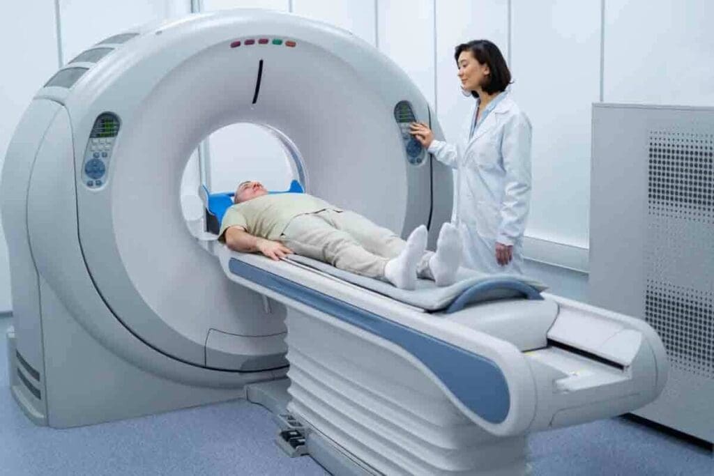
Kidney CT scans are key in medical diagnosis. They use X-ray technology to show the kidneys and urinary tract clearly. This helps doctors make accurate diagnoses and treatment plans.
How Kidney CT Scans Work
Kidney CT scans use X-rays to make detailed images of the kidneys. They can spot problems like stones, cysts, or tumors. A CT scan with contrast of the kidneys uses a contrast agent to see blood vessels and tissues better.
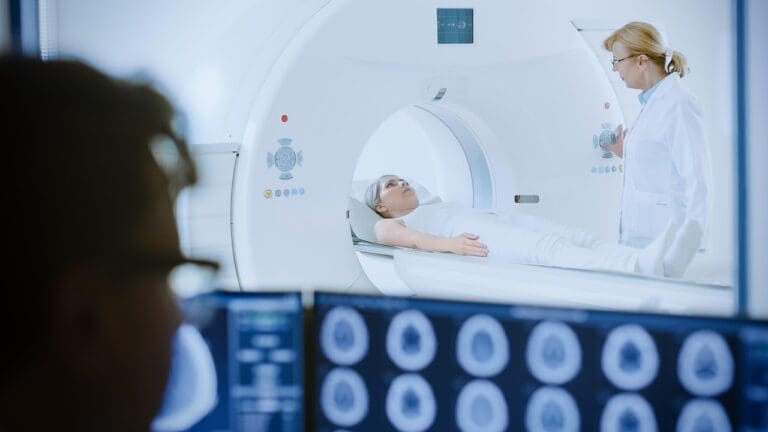
The scan involves lying on a table that slides into a CT scanner. The scanner is a large, doughnut-shaped machine. It rotates around the body, taking X-ray measurements from different angles. A computer then turns these measurements into images.
Types of Kidney CT Examinations
Kidney CT scans come in two types: with and without contrast. A CT renal with contrast shows blood vessels and kidney structures more clearly. It’s great for spotting blood flow issues and certain tumors or cysts.
A kidney CT scan without contrast is used to find kidney stones or other abnormalities. It doesn’t need contrast to see these issues.
Diagnostic Accuracy and Limitations
Kidney CT scans, with contrast, are very accurate for many kidney and urinary tract problems. But, they have some limits. The scan’s quality, the radiologist’s skill, and the patient’s health can affect the results.
Also, contrast agents might not be safe for everyone. Patients with kidney issues or allergies should talk to their doctors before a CT scan.
Analyzing Kidney CT Scan Results: What Radiologists Look For
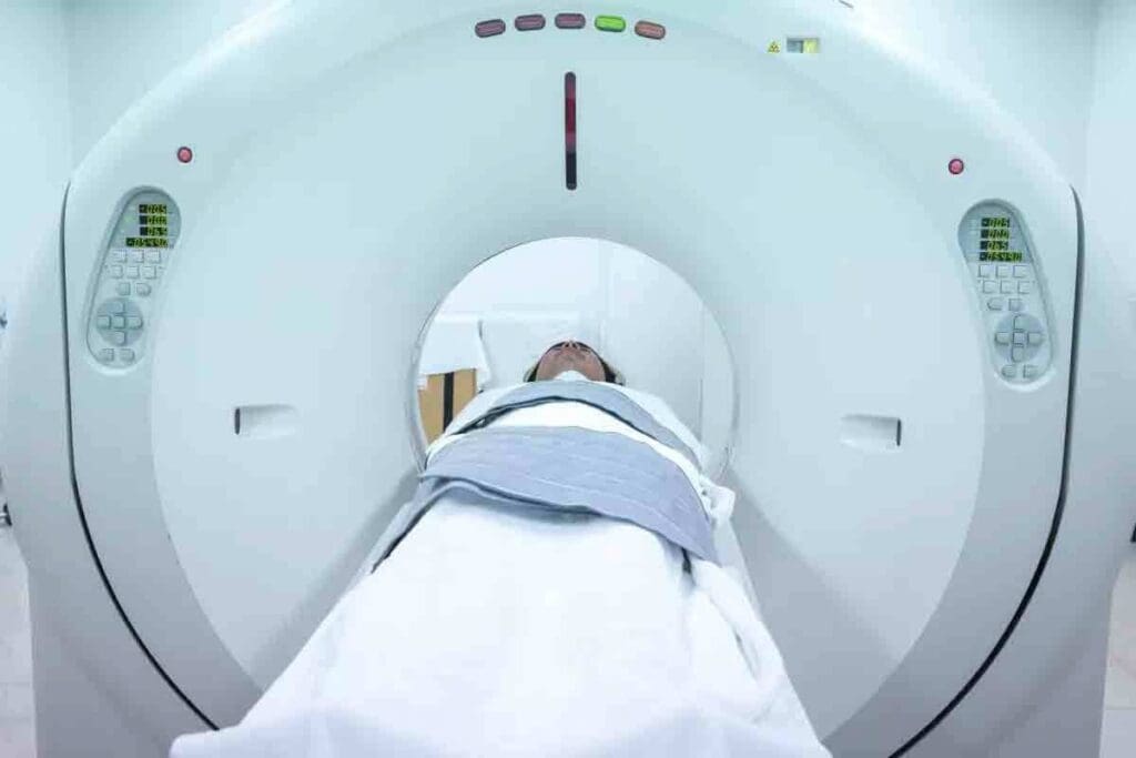
Radiologists are key in looking at the kidney CT scan results. They check for any problems and help decide on treatments. They look at the images to see how the kidneys and urinary tract look.
Normal Kidney Anatomy on CT
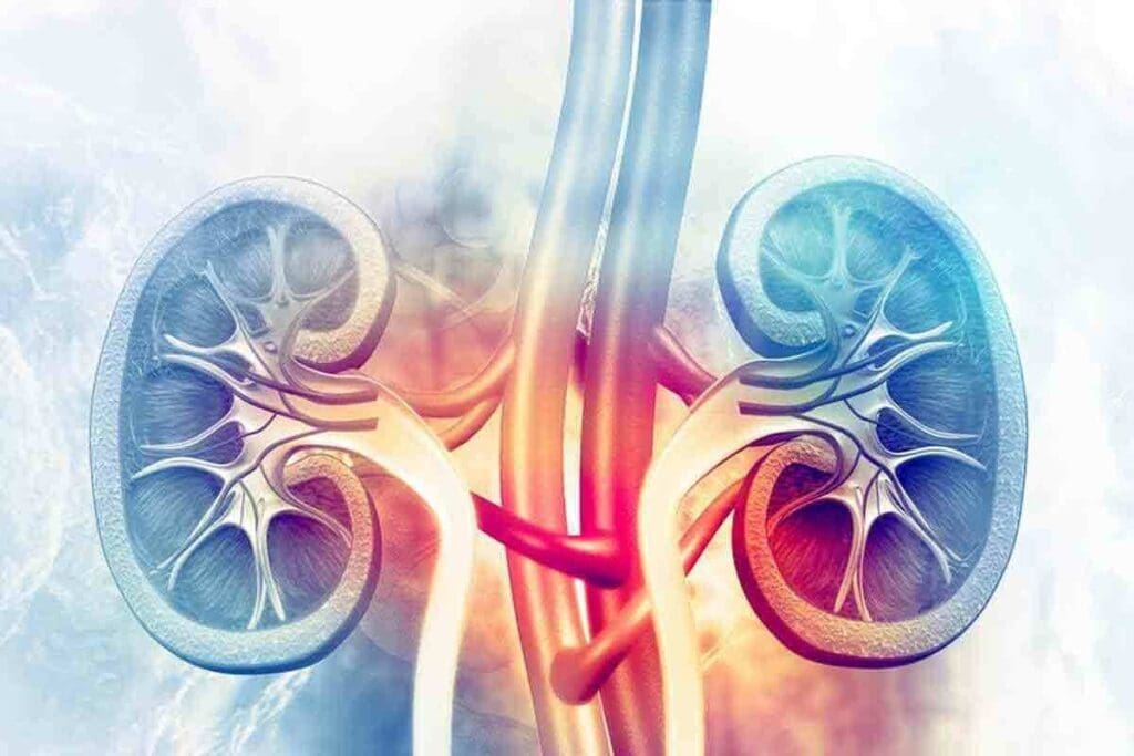
A normal kidney CT scan shows the kidneys, renal pelvis, and ureters. The kidneys should look the same on both sides. They should have a smooth shape and be the right size.
The kidney parts, called the cortex and medulla, should be clear. They should also show a clear difference between them.
Common Variations in Normal Anatomy
CT scans can show different normal kidney shapes and sizes. Some people might have bigger or smaller kidneys. They might also have extra blood vessels.
| Variation | Description |
| Kidney Size | Normal variation in kidney size can be seen, with some individuals having slightly larger or smaller kidneys. |
| Renal Shape | The shape of the kidneys can vary, with some individuals having lobulated or irregular kidneys. |
| Accessory Vessels | Accessory renal arteries or veins can be present, which can be a normal variation. |
Role in Treatment Planning
CT scan findings are very important for planning treatments. They help doctors figure out the best way to help patients with kidney and urinary tract problems. Radiologists can spot problems and tell how serious they are.
This helps doctors decide if surgery, a biopsy, or other treatments are needed. For example, if a kidney stone is found, the scan shows how big and where it is. This helps doctors choose the right treatment.
Kidney Stones: Detection and Characterization on CT
CT scans are great at finding kidney stones, even the tiny ones. They give doctors a lot of information about these stones. This is very important for treating kidney stone disease, which affects many people.
CT scans show the size, type, and where the stones. This info helps doctors decide the best treatment. We’ll look at how CT scans do this and why it’s good for patients.
Size and Composition Analysis
CT scans are good at measuring stone size and what they’re made of. This info is key for choosing treatment. For example, big stones might need help passing.
CT scans can also tell what kind of stone it is. Different stones show up differently on CT scans. For example, uric acid stones are different from calcium oxalate stones. A study on PMC shows that this helps doctors decide how to treat.
- Size Analysis: Helps determine the likelihood of spontaneous passage.
- Composition Analysis: Guides treatment decisions, such as the use of urinary alkalization for uric acid stones.
CT Stonogram Technique
The CT stonogram technique makes detailed images of kidney stones. It’s great for looking at stone characteristics and planning treatment.
With the CT scanogram, doctors can see the stone’s size, shape, and where it is. This is important for treatments like ESWL. It helps doctors choose the best treatment and predict how well it will work.
Predictors of Stone Passage
CT scans can tell if a stone will pass on its own. They look at the stone’s size, where it is, and the symptoms. Smaller stones closer to the ureterovesical junction are more likely to pass.
Treatment Implications Based on CT Findings
CT scan info is very important for treatment plans. Big stones or those blocking the flow might need surgery or ESWL. But small stones might just need to drink more water and take pain meds.
By knowing what the stones are like, doctors can make treatment plans that fit each patient. This helps improve treatment results and lowers the chance of problems.
Contrast vs. Non-Contrast Kidney CT Scans: When Each is Used
It’s important to know the difference between contrast and non-contrast CT scans for kidney checks. The right choice depends on what doctors need to see. This helps them make the best decisions for patients.
Benefits of Kidney CT With Contrast
Contrast CT scans show blood vessels and soft tissues better. They’re great for finding and understanding tumors and blood vessel problems. The contrast makes these areas stand out, helping doctors diagnose and plan treatments.
Enhanced visualization of blood vessels is a big plus of contrast CT scans. This is key for spotting issues like narrowed renal arteries or blood vessel malformations.
| Condition | Contrast-Enhanced CT | Non-Contrast CT |
| Renal Tumors | Highly visible due to contrast enhancement | Limited visibility |
| Kidney Stones | Visible but not as clear as non-contrast | Highly visible |
| Vascular Abnormalities | Highly visible due to contrast enhancement | Limited visibility |
Indications for Kidney CT Without Contrast
Non-contrast CT scans are best for finding kidney stones. They show up well without contrast. This method is also safer for people with kidney issues or those at risk of kidney problems from contrast.
“Non-contrast CT scans are the gold standard for diagnosing kidney stones due to their high sensitivity and specificity.” –
Radiological Society of North America
Contrast Safety Considerations
Contrast agents are usually safe, but there are risks. These are bigger for people with kidney disease or iodine allergies. We check these risks carefully to protect our patients.
Enhanced Visualization of Vascular Structures
Contrast makes blood vessels show up better on CT scans. This helps find problems like narrowed arteries or aneurysms. Knowing this is key to planning treatments and surgeries.
Knowing when to use contrast or non-contrast CT scans helps doctors get better results. The right choice depends on the patient’s needs and what doctors need to see for the best care.
Cystic Abnormalities on Kidney CT Scans
Kidney CT scans often show cystic abnormalities. These can be simple cysts or complex lesions needing more checks. Cystic abnormalities are fluid-filled sacs in the kidneys, seen more often with CT scans.
Simple Renal Cysts
Simple renal cysts are common on kidney CT scans. They are usually harmless, fluid-filled, and have thin walls. Simple cysts are often not a problem and don’t need treatment, but big or complex ones can cause confusion.
Complex Cystic Lesions
Complex cystic lesions are different. They might show signs of being more serious. Features like thick walls, septations, or solid parts inside the cyst are concerning. It’s important to check these lesions closely to decide what to do next.
Bosniak Classification System
The Bosniak system helps sort cystic kidney lesions by their CT scan looks. It helps figure out if a cyst might be cancerous. The system goes from Category I (safe cysts) to Category IV (likely cancer).
Follow-up Recommendations
What to do next depends on the Bosniak score. Lesions rated I and II usually don’t need to be checked again. But lesions rated IIF need regular scans to watch for changes. Lesions rated III and IV might need surgery or more tests because they’re more likely to be cancerous.
Knowing about cystic abnormalities on kidney CT scans and using the Bosniak system is key. It helps doctors decide the best course of action. This way, they can avoid unnecessary tests while keeping patients safe.
Solid Masses and Tumors Identified Through Kidney CT Scan Results
Kidney CT scans can spot different types of kidney masses and tumors. This is key for checking and managing kidney health.
Characteristics of Benign Renal Masses
Benign kidney masses, like angiomyolipomas, are often seen on CT scans. These masses have fat that shows up well on CT images. We look at certain signs to tell if these are harmless or not.
Having fat in a kidney mass usually means it’s not cancer. But not all harmless masses have fat. So, we need to check them more closely.
Malignant Renal Tumors
Malignant kidney tumors, mainly renal cell carcinoma (RCC), are a big worry. RCC is the most common kidney cancer in adults. CT scans are key in finding and figuring out how far it has spread.
We check the tumor’s size, where it is, and how it looks on CT scans to spot RCC. Knowing how far it has spread is key to photoretreatment.
Staging of Renal Cell Carcinoma
Staging RCC means looking at the tumor’s size, if it’s in the ph nodes, and if it has spread. We use the TNM system to classify how far it has spread.
- Tumor size and extent (T stage)
- Lymph node involvement (N stage)
- Distant metastasis (M stage)
Differentiating Between Benign and Malignant Lesions
Telling benign from malignant kidney lesions is very important. We look at CT scan features like how the lesion reacts to contrast, if it has calcium, and if it has fat. Contrast CT scans help see how the blood vessels are in the lesion.
Sometimes, we need more tests or a biopsy to be sure. Our aim is to give accurate diagnoses to help choose the right treatment.
Structural and Anatomical Abnormalities Detected on CT
CT scans have changed how we find kidney problems. They help spot structural and anatomical issues. These can affect how well the kidneys work and our overall health.
Congenital Kidney Anomalies
Kidney problems that start at birth are called congenital anomalies. CT scans can find many of these, like:
- Horseshoe kidney
- Renal ectopia
- Renal agenesis
These issues might also affect other parts of the urinary system. So, getting a full scan is key.
Acquired Structural Changes
Kidneys can also change due to disease or injury. CT scans spot these changes, such as:
- Cysts and cystic diseases
- Scarring from infections or a lack of blood flow
- Changes after surgery
Knowing about these changes helps doctors plan the right treatment.
Hydronephrosis and Obstruction
Hydronephrosis is when the kidney’s pelvis and calyces get too big because urine can’t flow. CT scans are great at finding this and figuring out why it’s happening.
Things like stones, tumors, and narrow spots can block urine. Finding the problem early is important to avoid kidney damage.
Renal Trauma Assessment
Kidney injuries can happen from accidents or violence. CT scans are the best way to see how bad the injury is.
| Grade | Description |
| I | Contusion or subcapsular hematoma |
| II | Laceration |
| III | Laceration >1 cm deep without urine leak |
Knowing the injury’s grade helps doctors decide the best treatment. This can range from just watching it to needing surgery.
Vascular Findings on Contrast-Enhanced Kidney CT
Vascular findings on contrast-enhanced kidney CT scans are key to diagnosing kidney issues. Contrast agents make blood vessels more visible. This helps spot problems that might not show up on regular scans.
Renal Artery Stenosis
Renal artery stenosis occurs when the renal arteries narrow. This can cause high blood pressure and harm the kidneys. CT scans with contrast can spot this narrowing.
Key Features:
- Narrowing of the renal artery lumen
- Post-stenotic dilation
- Collateral circulation
Aneurysms and Vascular Malformations
CT scans can find aneurysms and vascular malformations in the kidneys. These can cause pain or bleeding and might need treatment.
Diagnostic Features:
- Aneurysms appear as focal dilations of the renal artery or its branches
- Vascular malformations show abnormal connections between arteries and veins
Renal Infarction
Renal infarction happens when blood flow to the kidney stops, causing tissue death. CT scans with contrast can show where the kidney isn’t getting enough blood.
CT Findings:
- Wedge-shaped areas of decreased enhancement
- Global or segmental renal infarction
Arteriovenous Fistulas
Arteriovenous fistulas are abnormal connections between arteries and veins in the kidney. They can be present at birth or develop later. They might cause bleeding or high blood pressure.
| Condition | CT Findings | Clinical Implications |
| Renal Artery Stenosis | Narrowing of the renal artery, post-stenotic dilation | Hypertension, kidney damage |
| Aneurysms | Focal dilation of the renal artery or its branches | Pain, hematuria, possible rupture |
| Renal Infarction | Decreased or absent enhancement | Acute kidney injury, pain |
| Arteriovenous Fistulas | Early venous filling, abnormal A-V connection | Hematuria, hypertension |
Contrast-enhanced kidney CT scans are key for checking the kidneys’ blood vessels. They help diagnose and manage many vascular problems.
Can a CT Scan Detect Kidney Disease? Capabilities and Limitations
CT scans give valuable insights into kidney health. But knowing their limits is key to accurate diagnosis. They are a powerful tool, but can only detect certain types of kidney disease.
Structural vs. Functional Kidney Disease
CT scans are great at finding structural kidney diseases like cysts, tumors, and stones. These changes in the kidney’s anatomy are visible on a CT scan. For example, they can show the size, location, and type of kidney stones, which helps in choosing the right treatment.
But T scans are not as good at finding functional kidney diseases. These diseases affect how the kidney works, not its structure. Conditions like chronic kidney disease (CKD) or acute kidney injury (AKI) might not be diagnosed with a CT scan alone.
When Other Imaging Modalities Are Preferred
While CT scans are versatile, other imaging methods might be better in some cases. For instance:
- Ultrasound is often used for patients with kidney disease who need frequent monitoring. It doesn’t use radiation.
- MRI can show detailed images of the kidney’s structure and function without ionizing radiation. It’s useful for certain diagnoses.
- Functional imaging tests like renal scintigraphy can assess kidney function. They are great for evaluating conditions like renal artery stenosis.
| Imaging Modality | Advantages | Disadvantages |
| CT Scan | High-resolution images, quick | Involves radiation; contrast may be needed |
| Ultrasound | No radiation, cost-effective | Operator-dependent, limited detail |
| MRI | No radiation, detailed functional info | Expensive, claustrophobic for some |
Combining CT With Other Diagnostic Tests
Using CT scans with other tests can improve diagnosis. For example, combining CT scans with renal function tests gives a better understanding of kidney health. Also, matching CT findings with clinical symptoms and lab results helps in making a more accurate diagnosis.
We know that understanding CT scans’ strengths and weaknesses is key to diagnosing kidney disease. By using CT scans and other diagnostic tools when needed, healthcare providers can offer more precise and personalized care.
Preparing for Your Kidney CT Scan: What to Expect
Getting ready for a kidney CT scan is important. Knowing what happens before, during, and after can make things easier. It helps you feel more at ease and comfortable.
Before the Procedure
To get ready for your kidney CT scan, follow these steps:
- Inform your doctor about any medications you’re taking, like diabetes or kidney disease.
- Disclose any allergies to contrast dye or iodine.
- Follow dietary instructions; you might need to fast for a few hours.
- Remove any metal objects like jewelry, glasses, or clothes with metal parts.
- Arrive early to do paperwork and change into a hospital gown.
Also, find out if you’ll need a contrast-enhanced CT scan. If yes, we’ll give you contrast dye through an IV. This dye helps show the areas we’re interested in.
During the Scan
During the scan, you’ll lie on a table that slides into a big machine. The scan is quick, usually just a few minutes. Here’s what you can expect:
- Stay as steady as possible; moving can blur the images.
- Hold your breath when asked to.
- Talk to the CT scan technologist through an intercom.
After the Scan
After the scan, you can usually go back to your normal activities. Unless your doctor says not to. If you got contrast dye, we’ll watch you for any bad reactions.
- Drink lots of water to get rid of the dye.
- Tell your healthcare provider if you have any symptoms like rash, itching, or trouble breathing.
Understanding Your Results
A radiologist will look at your CT scan images. Then, your doctor will get the results. It’s important to understand what they mean:
- Normal results mean no problems were found.
- Abnormal results might show kidney stones, cysts, tumors, or other issues.
Your doctor will talk to you about the findings. They’ll tell you what to do next or if you need more tests.
Being prepared and knowing what to expect makes your kidney CT scan smoother. If you have questions or worries, talk to your healthcare provider.
Conclusion: Interpreting Your Results and Next Steps
Understanding your kidney CT scan results is key. We help patients grasp their findings and plan their next steps. This includes further evaluation or treatment.
Knowing what your CT scan shows is vital for your care. Our team guides you through this. We make sure you get the care and support you need.
We look at many things when we read your CT scan. This includes kidney stones, cysts, and blood vessel issues. These details help us find the cause of your kidney problems. Then, we create a treatment plan that works for you.
It’s important for patients to talk to their doctor about their CT scan results. This way, they can make smart choices about their treatment. They also get the support they need every step of the way.
FAQ
What is a kidney CT scan, and how does it work?
A kidney CT scan is a test that uses X-rays and computers to show detailed images of the kidneys and urinary tract. It takes many X-ray images from different angles. Then, these images are put together to create cross-sectional pictures.
What is the difference between a CT scan with contrast and without contrast for kidney imaging?
A CT scan with contrast uses a special dye to make blood vessels and soft tissues clearer. Without contrast, the scan shows less detail. Contrast scans are better for seeing blood vessels and soft tissue issues.
Can a CT scan detect kidney stones, and what information can it provide?
Yes, a CT scan can find kidney stones and tell you about their size, type, and where they are. The CT stonogram technique is used to check the stone’s details.
What are the benefits of using contrast in a kidney CT scan?
Using contrast in a kidney CT scan makes it easier to see blood vessels and soft tissues. This is helpful for finding problems like narrowed arteries or aneurysms.
How are cystic abnormalities characterized on a kidney CT scan?
CT scans can spot and describe cystic abnormalities. Simple cysts are usually harmless, but complex ones need more checking using the Bosniak system.
Can a CT scan differentiate between benign and malignant renal masses?
Yes, a CT scan can tell the difference between harmless and cancerous kidney masses. It can spot benign growths like angiomyolipomas and cancerous tumors like renal cell carcinoma.
What structural and anatomical abnormalities can be detected on a kidney CT scan?
A kidney CT scan can find birth defects, changes due to injury or disease, and signs of blockages or trauma. It can also show if there’s swelling in the kidneys.
How does a CT scan assess renal trauma?
A CT scan checks for kidney injuries by looking for signs like cuts, bleeding, and damage to blood vessels.
Can a CT scan detect functional kidney disease?
CT scans aren’t the best for finding problems with how the kidneys work. Other tests, like nuclear medicine scans, are better for checking kidney function.
How should I prepare for a kidney CT scan?
To get ready for a kidney CT scan, follow your doctor’s instructions. This might include not eating certain foods, drinking lots of water, and removing metal items.
What can I expect during and after a kidney CT scan?
During the scan, you’ll lie on a table that moves into the CT scanner. You might need to hold your breath or stay very quiet. Afterward, you can usually go back to your normal activities. Your doctor will talk to you about the scan’s results.
How are kidney CT scan results interpreted, and what are the next steps?
A radiologist looks at the CT scan results. They help decide what to do next. Your doctor will explain the findings and plan your next steps.
References
- Cellina, M., Della Pepa, G. M., Rundo, L., Reginelli, A., & Palumbo, P. (2023). Computed tomography urography: State of the art and novel acquisition techniques. Diagnostics, 13(5), 1000. https://pmc.ncbi.nlm.nih.gov/articles/PMC10204399/


