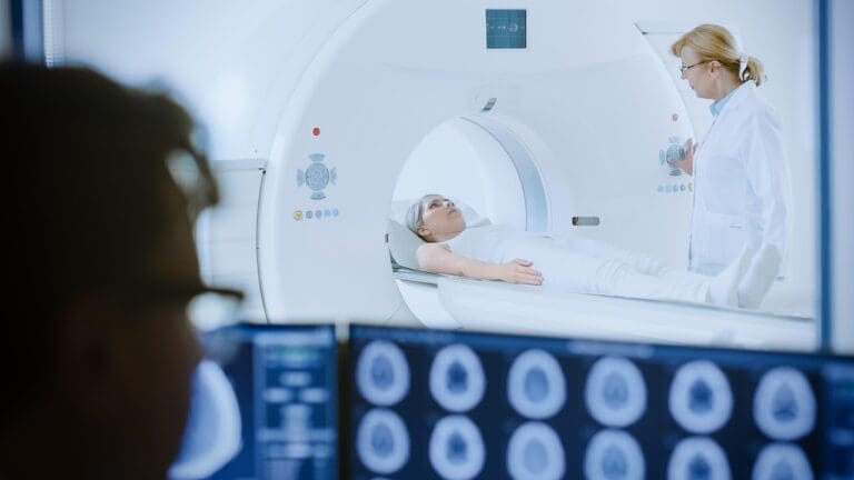
At Liv Hospital, we use kidney scintigraphy, also known as renal scintigraphy. It’s a test that checks how well your kidneys work. We use tiny amounts of radioactive materials called radiopharmaceuticals for this.
This test helps us understand your kidney health. It lets us give you the right treatment. With a nuclear renal scan, we can see how your kidneys are doing and find any problems.
Key Takeaways
- Kidney scintigraphy is a nuclear medicine imaging test.
- It evaluates the structure and function of the kidneys.
- Radiopharmaceuticals are used to provide insights into kidney health.
- Liv Hospital uses this diagnostic tool for accurate diagnoses.
- Personalized treatment plans are developed based on scan results.
The Fundamentals of Kidney Scintigraphy

Kidney scintigraphy is a way to see how well the kidneys work. It uses special images made by a small amount of radioactive material. This helps doctors understand kidney function and structure.
Definition and Basic Principles
Kidney scintigraphy, or renal scintigraphy, is a nuclear medicine test. It uses a tiny bit of radioactive material that the kidneys take up. A gamma camera then detects this radiation to create images of the kidneys.
This method helps doctors see how well the kidneys are working. It shows things like blood flow and how well the kidneys drain. This is key to diagnosing kidney problems.
Key aspects of kidney scintigraphy include:
- Assessment of renal function
- Evaluation of kidney structure
- Measurement of renal blood flow
- Detection of obstruction or other pathologies
Historical Development of Renal Nuclear Medicine
The first kidney imaging radiopharmaceuticals were made in the mid-20th century. Over time, better radiopharmaceuticals and technology have made kidney scintigraphy more accurate.
“The development of new radiopharmaceuticals has been instrumental in expanding the applications of renal nuclear medicine, enabling more precise and functional imaging of the kidneys.” – a Nuclear Medicine Specialist
Important milestones include the use of Technetium-99m. It’s a key part of kidney imaging today because of its good properties and how it works in the body.
| Year | Milestone | Significance |
| 1950s | Introduction of the first radiopharmaceuticals for renal imaging | Initial development of renal nuclear medicine |
| 1970s | Development of Technetium-99 m-labeled compounds | Improved image quality and expanded diagnostic capabilities |
| 2000s | Advancements in gamma camera technology and image processing | Enhanced sensitivity and specificity of kidney scintigraphy |
Key Terminology in Nuclear Renal Imaging
Knowing the terms used in nuclear renal imaging helps us understand kidney scintigraphy results. Key terms include:
- Radiopharmaceutical: A compound containing a radioactive element used for diagnostic or therapeutic purposes.
- Gamma camera: A device used to detect and image the radiation emitted by radiopharmaceuticals.
- Time-activity curve: A graphical representation of the activity of a radiopharmaceutical in a region of interest over time.
By learning these terms, we can better grasp kidney scintigraphy. This helps us diagnose and manage kidney issues more effectively.
The Science Behind Nuclear Renal Scans

To understand nuclear renal scans, we need to look at radiopharmaceuticals and imaging methods. These scans use special compounds and advanced tech to see and check how well the kidneys work.
Radiopharmaceuticals Used in Kidney Imaging
Radiopharmaceuticals are substances with tiny amounts of radioactive material. They help diagnose and treat diseases. For kidney imaging, these compounds go to the kidneys, helping to check their function and structure.
Common ones used are Technetium-99m (Tc-99m) labeled compounds like Tc-99m DTPA and Tc-99m MAG3. The National Center for Biotechnology Information says the right one depends on what the doctor wants to know.
How Radioactive Tracers Work in the Body
After being given, radioactive tracers move through the blood and into the kidneys. How fast and how they spread tells doctors about kidney health. For example, Tc-99m MAG3 shows how well the kidney tubules work.
A gamma camera tracks the tracer’s path in the kidneys. It catches the gamma rays from the tracer.
Gamma Camera Technology and Image Formation
The gamma camera is key in nuclear renal scans. It finds the gamma rays from the tracer. This info makes images that show where and how much tracer is in the kidneys.
The camera’s tech uses special detectors and computers to make detailed kidney images. These images help doctors understand kidney function and spot problems.
Types of Kidney Scintigraphy Procedures
Kidney scintigraphy includes different procedures for various needs. The choice of scan depends on what information is needed for a correct diagnosis.
Static Renal Scintigraphy
Static renal scintigraphy gives a snapshot of the kidney’s structure. It helps check the size, shape, and position of the kidneys. It also finds any structural issues.
Dynamic Renal Scintigraphy
Dynamic renal scintigraphy takes images over time. It shows how well the kidney works. This is key to seeing how the kidney takes in and gets rid of the radiopharmaceutical.
Captopril Renography
Captopril renography is a special dynamic renal scintigraphy. It’s used to find renovascular hypertension. Captopril, an ACE inhibitor, is given to see its effect on kidney function and blood pressure.
Diuretic Renography
Diuretic renography checks for obstructive uropathies. A diuretic is given to make more urine. This helps tell if there’s an obstruction or not.
Each procedure has its own strengths for diagnosis. Healthcare providers pick the best test for each patient’s needs.
Clinical Applications and Indications
Kidney scintigraphy is used in many ways, from finding renal vascular diseases to checking how well transplanted kidneys work. It’s a key tool in nephrology, helping us understand kidney function and problems.
Detecting Renal Vascular Diseases
Renal vascular diseases, like renal artery stenosis, can harm kidney function and health. Kidney scintigraphy, with captopril renography, shows how blood flows and kidneys work. It helps find patients with renovascular hypertension, so they can get the right treatment.
Evaluating Obstructive Uropathies
Obstructive uropathies are another area where kidney scintigraphy is very helpful. It shows how blockages affect kidney drainage. This helps doctors see how bad the problem is and if treatments are working.
An expert found that diuretic renography is very good at spotting these blockages. It helps doctors make better treatment decisions.
Assessing Transplant Function
For people with kidney transplants, scintigraphy is key. It checks if the new kidney is working properly and finds problems early. This helps doctors act fast to fix any issues.
Diagnosing Congenital Anomalies
Kidney scintigraphy also helps find birth defects of the kidneys and urinary tract. It gives clear pictures of the kidneys, helping doctors spot problems like ectopic kidneys. This info is vital for planning treatment.
| Clinical Application | Diagnostic Utility | Key Benefits |
| Detecting Renal Vascular Diseases | High sensitivity for renovascular hypertension | Guides therapeutic interventions |
| Evaluating Obstructive Uropathies | Assesses the functional impact of obstruction | Monitors the effectiveness of interventions |
| Assessing Transplant Function | Evaluates graft perfusion, function, and drainage | Detects complications early |
| Diagnosing Congenital Anomalies | Provides detailed renal anatomy | Informs management strategies |
In conclusion, kidney scintigraphy is a powerful tool with many uses. It gives important information about kidney function. This makes it essential for diagnosing and treating many kidney problems.
Preparing for a Nuclear Kidney Scan
Getting ready for a nuclear kidney scan is important. This test checks how well your kidneys work and finds problems. To make sure the scan works well and is safe, follow these steps.
Pre-Procedure Guidelines
Before the scan, there are important things to do. Tell your doctor about all the medicines you take. Some might need to be changed or stopped. Also, let them know if you have any allergies to contrast agents or other medicines.
Medication Considerations
Some medicines can mess with the scan or cause problems. Your doctor will tell you which medicines to keep taking or stop. It’s very important to listen to them to stay safe and get accurate results.
Hydration Requirements
Drinking water is key before, during, and after the scan. Drink lots of water as your doctor tells you to. This helps your kidneys work properly and gets rid of the medicine quickly.
What to Bring and Wear
On the day of the scan, wear comfy, loose clothes. Don’t wear metal things like jewelry or clothes with metal parts. Bring all the papers you need, like your doctor’s note, insurance, and a list of medicines. Being ready will make the process easier.
The Kidney Scintigraphy Procedure: Step by Step
Let’s go through the kidney scintigraphy procedure step by step. This will help us understand what patients can expect. The process includes several key stages, from the administration of a radiopharmaceutical to post-procedure care.
Administration of the Radiopharmaceutical
The first step is the administration of a radiopharmaceutical. This is done through an intravenous injection. The radiopharmaceutical used is designed to be taken up by the kidneys, allowing for the assessment of renal function.
The choice of radiopharmaceutical depends on the specific clinical question being addressed. For example, Technetium-99m DTPA is commonly used for assessing renal function and drainage.
Positioning and Image Acquisition
After the radiopharmaceutical is administered, the patient is positioned under a gamma camera. The gamma camera detects the gamma rays emitted by the radiopharmaceutical. This allows for the creation of images that reflect kidney function.
Patients are typically asked to lie on a table under the gamma camera. The imaging process may involve multiple stages. This includes dynamic imaging to assess the flow of the radiopharmaceutical through the kidneys.
Duration and Patient Experience
The duration of a kidney scintigraphy procedure can vary. It typically lasts between 1 to 3 hours. Patients may be asked to wait between different stages of the imaging process.
During the procedure, patients are generally able to remain fully clothed. They may be asked to remove any jewelry or other items that could interfere with the imaging.
| Procedure Stage | Duration | Patient Experience |
| Radiopharmaceutical Administration | 5-10 minutes | Mild discomfort from the injection |
| Imaging | 1-3 hours | Lying under the gamma camera |
| Post-Procedure | Varies | Normal activities can be resumed |
Post-Procedure Care
After the procedure is complete, patients can generally resume their normal activities. The radiopharmaceutical is cleared from the body through urine. Patients are encouraged to stay hydrated to help eliminate the radioactive tracer.
It’s also recommended that patients follow any specific instructions provided by their healthcare provider regarding post-procedure care.
Interpreting Renal Scan Results
Renal scan results give important insights into kidney health. They help doctors make better decisions for patient care. The scan shows how well the kidneys work, their size, and shape. This information is key to diagnosing kidney problems.
Normal vs. Abnormal Findings
It’s important to know the difference between normal and abnormal scan results. Normal results mean the kidneys are working properly and look good. But, abnormal results might show problems like poor kidney function, blockages, or scarring.
A normal scan shows that both kidneys take up and release the radiopharmaceutical evenly. Any difference could mean there’s a problem.
Quantitative Analysis Methods
Quantitative analysis of renal scan results involves measuring several things to check kidney function. Some common methods include:
- Calculating the differential renal function
- Assessing the rate of radiopharmaceutical uptake and excretion
- Evaluating the time-activity curves
These measurements give important information about kidney function. They help doctors diagnose certain conditions.
Common Diagnostic Patterns
Some patterns in renal scan results can point to specific conditions. For example:
| Pattern Observed | Possible Diagnosis |
| Delayed uptake and excretion | Renal obstruction or hydronephrosis |
| Reduced or absent uptake | Kidney damage or a non-functioning kidney |
| Asymmetrical uptake | Renal vascular disease or congenital anomalies |
Time-Activity Curves and Their Significance
Time-activity curves show how the radiopharmaceutical moves in the kidney over time. They give info on kidney function, like how fast it takes up and releases the radiopharmaceutical.
A normal curve shows quick uptake and slow release as the radiopharmaceutical is removed. But, abnormal curves might show blockages, poor function, or other issues.
“The use of time-activity curves in renal scan interpretation has significantly improved our ability to diagnose and manage kidney diseases.” – A Renowned Nephrologist
By studying renal scan results, including time-activity curves, doctors can better understand kidney function. This helps them make more accurate diagnoses.
Comparing Kidney Scintigraphy to Other Imaging Modalities
Different imaging techniques have their own strengths in showing kidney health. We’ll look at how kidney scintigraphy compares to ultrasound, CT, and MRI. Each has its own benefits and drawbacks.
Ultrasound vs. Nuclear Renal Scan
Ultrasound uses sound waves to show kidney images. It’s good for finding structural issues like cysts. But it doesn’t tell much about kidney function.
A nuclear renal scan, or kidney scintigraphy, shows how well the kidneys work. It checks their filtering and drainage. A study on the National Center for Biotechnology Information website says scintigraphy is great for kidney function tests.
Ultrasound is often first because it’s safe and easy. But, for detailed function info, scintigraphy is better.
CT and MRI Alternatives
CT and MRI scans also check kidney health. CT gives detailed images and is good for finding stones and tumors. MRI shows high-quality images without harmful radiation.
CT and MRI are great for body structure. But, they don’t give the function info scintigraphy does. The choice depends on what the doctor needs to know.
When Scintigraphy Is the Preferred Choice
Scintigraphy is key when you need to know how the kidneys work. It’s useful for checking on kidney transplants or suspected artery problems.
Key Scenarios for Kidney Scintigraphy:
- Evaluating renal function in transplant patients
- Assessing for renal artery stenosis
- Diagnosing obstructive uropathy
Complementary Use of Multiple Imaging Techniques
Often, doctors use more than one imaging method. They might start with ultrasound, then use scintigraphy for function tests. CT or MRI might follow for detailed body images.
| Imaging Modality | Primary Use | Functional Information |
| Ultrasound | Structural abnormalities | Limited |
| Kidney Scintigraphy | Renal function and drainage | Detailed |
| CT Scan | Detailed anatomical imaging | Limited |
| MRI | High-resolution anatomical imaging | Limited |
Knowing what each imaging method does helps doctors choose the best test for patients.
Radiation Safety and Concerns
When we use radiopharmaceuticals in kidney scintigraphy, we must think about radiation safety. We use small amounts of radioactive materials to check how well the kidneys work. It’s important to know about the radiation exposure and how we keep everyone safe.
Typical Radiation Exposure Levels
The radiation from a kidney scintigraphy is usually low. The dose from the radiopharmaceutical is a few millisieverts (mSv). For comparison, a chest X-ray is about 0.1 mSv, and the yearly background radiation is 2.4 mSv in the U.S. We use the least amount of radiopharmaceutical needed to get good images, keeping exposure low.
Safety Protocols and Precautions
We follow strict safety rules to protect everyone. These include:
- Using the least amount of radiopharmaceutical needed.
- Having trained professionals handle and give the radiopharmaceutical.
- Checking the gamma camera and other equipment regularly.
- Telling patients about radiation safety and what to do after the test.
Special Considerations for Children and Pregnant Women
For kids and pregnant women, we carefully think about the risks and benefits of kidney scintigraphy. Kids get smaller doses because of their size. Pregnant women usually avoid it unless it’s really needed, and we look for other imaging options. When it’s necessary, we take extra steps to reduce radiation.
Minimizing Radiation Exposure
We follow the ALARA principle to reduce radiation. This means:
- Adjusting the radiopharmaceutical dose for each patient.
- Using the latest technology to lower doses.
- Training all staff on radiation safety.
By sticking to these rules, we make sure kidney scintigraphy is safe and effective. We get important information while keeping radiation exposure low.
Advantages and Limitations of Nuclear Medicine Scans for the Kidneys
It’s important to know the good and bad of nuclear medicine scans for kidney health. These scans give both functional and anatomical info. This is key to diagnosing and managing kidney issues.
Unique Benefits of Functional Imaging
Nuclear medicine scans give functional information about the kidneys. This info isn’t always available with other scans, like ultrasound or CT. It helps doctors see how kidneys work and spot problems early.
One big plus is that they can give exact numbers on kidney function. For example, dynamic renal scintigraphy can show the GFR of each kidney. This is super helpful for patients with kidney disease or before surgery.
“Nuclear medicine techniques offer a unique window into the functional status of the kidneys, allowing for early detection and management of renal diseases.”
Renal Nuclear Medicine Expert
Diagnostic Limitations
Nuclear medicine scans have some downsides. They don’t show as much detail as MRI or CT scans. They’re great for function but not for anatomy.
Another issue is radiation exposure. While doses are usually safe, there’s a risk, mainly for those needing many scans.
| Imaging Modality | Functional Information | Anatomical Detail | Radiation Exposure |
| Nuclear Medicine Scan | High | Low | Yes |
| CT Scan | Low | High | Yes |
| MRI | Moderate | High | No |
Cost and Availability Factors
The cost and where you can get nuclear medicine scans vary a lot. They’re seen as a specialized diagnostic tool and might not be as common as other scans.
Also, the price can be high. They need specialized equipment and trained personnel, which adds to the cost.
Patient Selection Criteria
Choosing the right patients for nuclear medicine scans is key. Doctors must think about the clinical question and the patient’s overall health status before deciding.
In summary, nuclear medicine scans have both good and bad points for kidney checks. Knowing these helps doctors decide when to use them.
Recent Advances in Renal Nuclear Medicine
Renal nuclear medicine has grown a lot. This is thanks to new imaging methods and radiopharmaceuticals. These changes help us better diagnose and treat kidney diseases.
Hybrid Imaging Techniques
Hybrid imaging is a big step forward. It combines nuclear medicine with CT or MRI. This gives us detailed information on kidney function and structure. Hybrid imaging improves how well we can diagnose and plan treatments.
“Hybrid imaging is a huge leap in renal nuclear medicine,” says an expert. “It lets us see how kidneys work and their structure together. This gives us a full view of kidney health.”
New Radiopharmaceuticals
New radiopharmaceuticals are also making progress. These agents are more specific and sensitive to kidney issues. For example, some tracers target specific kidney functions. This makes assessing kidney health more precise.
- Improved specificity for kidney function assessment
- Enhanced sensitivity for detecting renal diseases
- Better safety profiles for patients
Artificial Intelligence in Image Interpretation
Artificial intelligence (AI) is being used more in renal nuclear medicine. AI helps analyze complex images. It spots patterns that humans might miss. This leads to quicker and more accurate diagnoses.
AI in renal nuclear medicine is a game-changer. It boosts how well we can read images. This lets doctors focus more on patient care.
Personalized Medicine Applications
Recent advances also support personalized medicine. We use specific radiopharmaceuticals and advanced imaging. This way, we can tailor treatments to each patient. This approach can lead to better treatment plans and outcomes.
As we keep moving forward in renal nuclear medicine, these new developments are changing patient care. By using these innovations, we can give more accurate diagnoses and treatments. This leads to better care for our patients.
Conclusion
Kidney scintigraphy, or a nuclear renal scan, is a key tool for checking kidney health. We’ve looked at how it works and its uses in this article. It’s important for spotting and treating kidney problems.
At Liv Hospital, we aim to offer top-notch healthcare. We use kidney scintigraphy to find the right treatments for our patients. This helps them get the best care possible.
In short, kidney scintigraphy is essential for kidney disease diagnosis and treatment. Knowing how it works helps doctors give better care to patients. We keep improving our diagnostic tools to ensure our patients get the best care.
FAQ
What is kidney scintigraphy?
Kidney scintigraphy is a test that uses special medicines to see how kidneys work. It looks at both the structure and function of the kidneys.
How does a nuclear renal scan work?
A nuclear renal scan uses a special medicine that the kidneys absorb. Then, a camera captures images. This shows how well the kidneys are working.
What are the different types of kidney scintigraphy procedures?
There are several types of kidney scintigraphy. These include static, dynamic, captopril, and diuretic renography. Each has its own use and benefits.
What is the purpose of a kidney scintigraphy?
A kidney scintigraphy helps find and manage kidney problems. This includes diseases, blockages, and birth defects.
How do I prepare for a nuclear kidney scan?
To get ready for a scan, follow certain steps. This includes taking medicine as told, drinking water, and wearing comfy clothes. This helps the scan go smoothly.
What happens during a kidney scintigraphy procedure?
During the scan, a special medicine is given. Then, a camera takes pictures. This usually takes about 30 minutes to a few hours.
How are renal scan results interpreted?
Results are checked by looking at the pictures and data from the scan. This helps find normal or abnormal findings. It also helps measure kidney function.
What are the advantages of kidney scintigraphy compared to other imaging modalities?
Kidney scintigraphy shows how the kidneys work. This makes it a great tool for diagnosis. It’s better than ultrasound, CT, and MRI for this purpose.
Are there any radiation safety concerns with kidney scintigraphy?
Yes, there are safety concerns about radiation. But the levels are low. Safety steps are taken to protect everyone, including those who are more sensitive.
What are the latest advances in renal nuclear medicine?
New things in renal nuclear medicine include hybrid imaging and new medicines. Artificial intelligence and personalized medicine are also being used. These advancements help improve kidney care and diagnosis.
What is the difference between a renal scan and a kidney scan?
Renal scan and kidney scan mean the same thing. They are tests that check how the kidneys work and look.
How is a nuclear medicine kidney scan different from other kidney imaging tests?
A nuclear medicine kidney scan is unique. It uses special medicines to see how the kidneys function. This is different from ultrasound, CT, and MRI, which don’t show function as well.
References
- Taylor, A. T. (2014). Radionuclides in nephrourology, Part 1: Radiopharmaceuticals, quality control, and quantitative indices. Journal of Nuclear Medicine, 55(4), 608-615. https://jnm.snmjournals.org/content/55/4/608
- Procedure guidelines for dynamic renal scintigraphy with 99mTc-MAG3, 123I-hippuran, and 99mTc-DTPA. (2008). Nuklearmedizin, 46(5), 203-205. https://pubmed.ncbi.nlm.nih.gov/15480507/


