Last Updated on November 25, 2025 by Ugurkan Demir

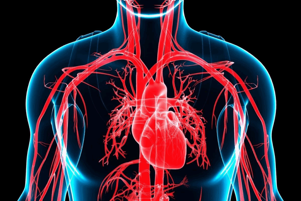
Knowing how an layers of artery works is key to keeping your heart healthy. An artery is a blood vessel that carries vital stuff like blood, oxygen, and nutrients. It helps keep your body running smoothly.
A healthy arterial wall structure is vital. The three main layers work together to keep blood flowing right. Learning about these layers helps us understand our heart and blood system better.

Layers of Artery are more than just blood vessels. They are key paths that keep us alive by bringing oxygen and nutrients. The arterial system is vital for our health, making sure every part of our body gets what it needs.
We count on our arteries to keep our body working right. If they get damaged, it can lead to heart problems. So, knowing how arteries work is key to staying healthy.
Arteries are built strong to handle the blood pressure from the heart. They have a three-layered wall that’s both strong and flexible. This design lets them do their job well.
| Layer | Composition | Function |
| Tunica Intima | Endothelium | Regulates blood flow and blood pressure |
| Tunica Media | Smooth muscle and elastic fibers | Controls vessel diameter and blood pressure |
| Tunica Externa | Collagen fibers | Provides structural support |
Understanding arteries is key to seeing how our body works. By knowing their role, we can take better care of them. This means living a healthy life and getting regular check-ups.

The arterial wall is made up of three layers. Each layer has its own role and features.
The innermost layer is the tunica intima. It has a single layer of endothelial cells. These cells line the artery’s inside.
The middle layer is the tunica media. It’s mostly smooth muscle cells and elastic fibers. These help control blood pressure.
The outermost layer is the tunica externa. It’s made of connective tissue. This layer supports and protects the artery.
| Layer | Composition | Function |
| Tunica Intima | Endothelial cells | Lines the lumen, reducing friction |
| Tunica Media | Smooth muscle cells and elastic fibers | Regulates blood pressure |
| Tunica Externa | Connective tissue | Provides support and protection |
The Tunica Intima is more than a simple lining. It’s a dynamic interface between the bloodstream and the vessel wall. It has a single layer of endothelial cells.
The endothelium is a key part of the Tunica Intima. It acts as a selective barrier. This barrier controls the exchange of materials between the blood and tissues.
It regulates blood flow, immune responses, and inflammation. This makes it vital for maintaining vascular homeostasis.
The Tunica Intima’s functions include:
Dysfunction of the Tunica Intima is linked to cardiovascular diseases. Understanding its role is key for developing treatments.
The tunica media is a key part of artery walls. It helps keep blood pressure right and the vessel size just right. We’ll look into what makes it up and why it’s so important for blood flow.
The tunica media has smooth muscle cells and elastic fibers. These work together to control blood pressure and the size of the vessel. The smooth muscle cells are arranged in a special way. This lets them tighten or loosen the vessel as needed.
The elastic fibers in the tunica media are also important. They make the artery wall stretch and snap back with each heartbeat. This helps keep blood pressure steady and ensures tissues get enough blood.
The structure and function of the tunica media are vital for the circulatory system’s health. By knowing about its makeup and how it works, we can understand how blood flow and pressure are managed.
The Tunica Externa is the outermost layer of the artery. It keeps the artery strong and flexible. It’s made mostly of collagen and elastic fibers.
This layer has collagen fibers for strength and durability. It also has elastic fibers. These fibers let the artery stretch and recoil with each heartbeat. This ensures blood keeps flowing.
The Tunica Externa’s collagen is vital for its support. It keeps the artery from expanding too much or bursting under pressure.
To sum up, the Tunica Externa is essential for the artery’s strength and flexibility. Its mix of collagen and elastic fibers is key to keeping arteries working right.
The thickness of smooth muscle in blood vessels is very important. It affects how they work. Let’s look at how different blood vessels have different amounts of smooth muscle.
Smooth muscle thickness changes a lot in arteries, veins, and capillaries. Arteries, like muscular arteries, have a lot more smooth muscle than veins. This is because arteries face higher pressures to keep blood flowing.
Muscular arteries have a thick tunica media layer with smooth muscle cells. This layer helps control blood pressure and flow. Veins, on the other hand, have less smooth muscle because they handle lower pressures.
Looking at histological sections of different blood vessels shows the difference.
In summary, the thickness of smooth muscle varies among blood vessels. Muscular arteries have the most to meet their needs. Knowing this helps us understand vascular health and disease better.
The structure of blood vessels changes a lot between arteries, veins, and capillaries. This change shows their different roles in the circulatory system. We will look at these differences, focusing on wall thickness and lumen size.
Arteries carry blood away from the heart and have thicker walls than veins. Veins return blood to the heart and have thinner walls. Arteries need to handle the heart’s high blood pressure.
Let’s examine the differences in a structured format:
| Blood Vessel Type | Wall Thickness | Lumen Size |
| Arteries | Thick walls | Smaller lumen |
| Veins | Thinner walls | Larger lumen |
| Capillaries | Very thin walls (single layer of endothelium) | Very small lumen |
As shown in the table, arteries have thicker walls and smaller lumens to handle high blood pressure and ensure continuous blood flow. In contrast, veins have thinner walls and larger lumens, accommodating lower pressure and blood volume. Capillaries, with their extremely thin walls, facilitate the exchange of nutrients and waste products.
Understanding these structural differences is key to seeing how the circulatory system works. Each type of blood vessel is vital for keeping the heart and blood vessels healthy.
Looking at the artery wall under a microscope shows its detailed structure. It has three main layers. Knowing about these layers helps us understand the artery’s health and how it works.
The three layers of the artery wall are clear when we look closely. The Tunica Intima, the innermost layer, has a single layer of endothelial cells. The Tunica Media, in the middle, is mostly smooth muscle cells and elastic fibers. These help control blood pressure and flow.
The Tunica Externa, the outermost layer, is made of connective tissue. It gives the artery the support it needs.
| Layer | Composition | Function |
| Tunica Intima | Endothelial cells | Regulates blood flow and blood pressure |
| Tunica Media | Smooth muscle cells and elastic fibers | Controls vessel diameter and blood pressure |
| Tunica Externa | Connective tissue | Provides structural support |
Elastic and muscular arteries have unique structures that fit their roles in the circulatory system. Their differences are not just about how they look. They are also linked to their functions.
Elastic arteries, like the aorta and its main branches, have lots of elastic fibers. This lets them stretch and snap back with each heartbeat. This keeps blood pressure steady throughout the arteries.
The elastic nature of these arteries is key during the heart’s contraction. The aorta expands to handle the blood surge. Then, it snaps back, pushing blood forward and keeping the flow smooth.
The aorta and big arteries are mostly elastic. Their elasticity is essential for smoothing out the pressure wave from the heart. This ensures blood flows smoothly to the rest of the body.
| Characteristics | Elastic Arteries | Muscular Arteries |
| Main Composition | High elastic fiber content | High smooth muscle content |
| Function | Windkessel effect, maintaining blood pressure | Regulation of blood flow to specific areas |
| Examples | Aorta, large arteries | Smaller distributing arteries |
Muscular arteries, on the other hand, have more smooth muscle cells. This lets them control blood flow and pressure better in certain areas.
Knowing how elastic and muscular arteries work helps us understand heart health. It shows how complex and vital the circulatory system is.
It’s key to understand how the structure of arterial walls relates to their function. The walls are made to handle the high pressures from the heart. They also keep blood flowing to our body’s tissues.
The arterial wall has three main layers: the tunica intima, tunica media, and tunica externa. Each layer is special and helps the artery work well.
The tunica intima, closest to the blood, has cells that keep blood flowing smoothly. These cells also help control how wide the artery is. This layer’s smooth surface helps blood move without resistance.
The tunica media is in the middle. It’s made of smooth muscle cells and elastic fibers. This layer can change the artery’s size to control blood pressure and flow. The muscle cells are arranged in a circle to control the artery’s width.
The tunica externa is the outer layer. It’s strong and elastic, thanks to collagen fibers. It also has nerves and blood vessels to support the artery itself.
In short, the structure and function of arterial walls work together. Each layer’s unique features help keep blood flowing right and our heart healthy.
“The detailed structure of arterial walls shows how our bodies adapt to different needs.”
The layers of blood vessel walls can change in unhealthy ways. A big problem is atherosclerosis. We’ll look at how these changes affect blood vessels’ structure and function.
Atherosclerosis causes plaque to build up in artery walls. This makes them hard and narrow. It starts in the endothelium, the innermost layer, where damage lets lipids and inflammatory cells get in.
As it gets worse, it also affects the tunica media. This leads to a loss of flexibility in the artery wall. The smooth muscle cells in the tunica media are key in this process.
Atherosclerosis has many effects on arteries. It reduces blood flow to important organs, raises the risk of blood clots, and can cause serious problems like heart attacks or strokes.
It’s important to understand atherosclerosis to find ways to prevent and treat it. We need to know how each part of the artery wall contributes to the disease.
Looking at the unhealthy changes in blood vessel walls, like in atherosclerosis, helps us understand how different layers work together. This knowledge is key to improving heart health.
We’ve looked into the details of the arterial wall, which has three layers: the tunica intima, tunica media, and tunica externa. The innermost layer of blood vessels, the tunica intima, is key to keeping blood vessels healthy. The tunica media, the thickest layer in arteries, helps with elasticity and contraction.
The structure of arteries and veins and capillaries is quite different. Arteries have a thicker wall to handle high blood pressure. The 3 layers of an artery work together for good blood flow. Arteries, mainly muscular arteries, have the most smooth muscle.
Knowing the microscopic structure of the blood vessels and their layers helps us understand the circulatory system better. The wall of artery is built to handle blood flow constantly. Its design shows how adaptable the body is.
In wrapping up our look at arterial structure, it’s clear that the arteries structure is key to heart health. By understanding the layers of an artery and their role, we see how complex life-sustaining mechanisms work.
The arterial wall has three layers: the Tunica Intima, Tunica Media, and Tunica Externa. The Tunica Intima is the innermost layer. The Tunica Media is in the middle, and the Tunica Externa is the outermost layer.
The Tunica Media has the thickest layer of smooth muscle. It’s made of smooth muscle cells and elastic fibers. These help control blood pressure and flow.
The endothelium is a single layer of cells in the Tunica Intima. It’s key to keeping the blood vessels healthy. It helps control blood flow, pressure, and clotting.
Arteries have thick walls with three layers. Veins are thinner and have valves to stop backflow. Capillaries are the smallest and have a single layer of cells. They help exchange oxygen and nutrients with tissues.
The Tunica Externa is the outermost layer. It gives structural support and protection. It’s made of collagen fibers and other connective tissue.
Atherosclerosis causes plaque buildup in the arterial walls. This leads to inflammation and damage to the endothelial layer. The walls become thicker and less flexible, raising the risk of heart disease.
Elastic arteries, like the aorta, have lots of elastic fibers. This lets them stretch and recoil with each heartbeat. Muscular arteries have more smooth muscle cells. This lets them constrict and dilate with blood pressure changes.
Subscribe to our e-newsletter to stay informed about the latest innovations in the world of health and exclusive offers!