
We’re here to explain Epiretinal Membrane (ERM), a common eye issue affecting millions globally. ERM, or macular pucker, is when cells grow on the retina’s inside. This can lead to vision problems.Learn to read a macula epiretinal membrane OCT. This guide explains what ERM is and how it appears on an OCT scan.
But, the good news is most cases are mild and don’t need treatment. Yet, for those with vision issues, catching it early and getting expert help is key.
Key Takeaways
- ERM is a common eye condition affecting the retina.
- It is also known as macular pucker or cellophane maculopathy.
- Most cases are mild and do not require treatment.
- Early detection is critical for those with vision problems.
- Getting an expert ophthalmologist’s opinion is vital for the right treatment.
Understanding Epiretinal Membrane (ERM)

ERM, or epiretinal membrane, is a layer of cells on the retina’s surface. It can cause vision problems. This layer affects the macula, key for clear vision. Knowing about ERM helps both patients and doctors manage it better.
Definition and Basic Anatomy
An epiretinal membrane forms on the retina’s inner surface. It looks like a greyish, semi-transparent layer. The macula, in the retina’s center, is vital for tasks like reading and driving.
When an ERM forms, it can make the macula pucker. This leads to vision distortion. In erm in ophthalmology, this is important because it can cause serious issues like macular edema or retinal detachment.
Alternative Names: Macular Pucker and Cellophane Maculopathy
ERM is also called macular pucker and cellophane maculopathy. These names describe how it affects the macula. Macular pucker means the macula gets wrinkled. Cellophane maculopathy describes the membrane’s thin, translucent look.
People with ERM might see blurry or distorted images. A detailed eye exam can spot ERM. This exam might use Optical Coherence Tomography (OCT) to see the erm eyes and how severe it is.
To sum up, knowing about ERM means understanding its definition, how it affects the retina, and its other names. This knowledge is key for diagnosing and treating ERM. It helps ensure patients get the right care, whether it’s just watching it or needing surgery.
The Structure and Function of the Macula

The macula helps us see clearly in the center, making everyday tasks easy. It’s a key part of the retina at the back of the eye. Knowing how the macula works is key to understanding vision problems and why treatment is so important.
Normal Macular Anatomy
The macula is a special area of the retina that lets us see details clearly. It has lots of photoreceptor cells. These cells turn light into signals for the brain.
Key components of the macula include:
- The fovea, a depression in the innermost layer of the retina, responsible for high-acuity vision.
- A high concentration of cone photoreceptors, which are sensitive to color and detail.
- A complex network of retinal layers that work together to facilitate visual processing.
How the Macula Affects Vision
The macula is key for seeing small details, faces, and reading. If it gets damaged, like by an epiretinal membrane, vision can get worse. Its health is vital for doing daily tasks that need clear vision.
Macular Function | Impact on Vision |
Sharp, central vision | Enables reading, face recognition, and detailed tasks |
High-acuity vision | Facilitates activities like driving and watching TV |
Color perception | Enhances overall visual experience |
In conclusion, the macula’s detailed structure and function are essential for clear vision. Understanding its role helps us see why vision problems, like epiretinal membrane, need treatment.
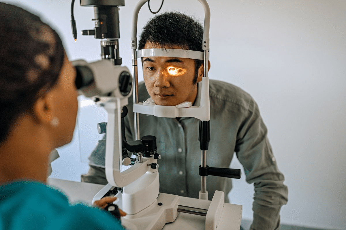
Macula Epiretinal Membrane OCT: Diagnosis and Imaging
Optical Coherence Tomography (OCT) has changed how we diagnose macular conditions, like epiretinal membrane (ERM). This non-invasive method gives us clear images of the retina. It helps us diagnose and track ERM accurately.
What is Optical Coherence Tomography (OCT)?
OCT is a test that uses low-coherence interferometry to create detailed images of the retina. It’s like a high-tech camera that looks inside the eye. OCT helps us see the retinal layers and spot any problems, like ERM.
We use OCT to get detailed images of the retina. These images are key for diagnosing and measuring ERM’s severity. They let us see the retina’s thickness, any distortions, and if there’s an ERM.
How OCT Detects Epiretinal Membrane
OCT spots ERM by showing us the retinal surface and what’s under it. An ERM looks like a bright line on the retina’s surface. It’s often linked to thicker retina and distortions.
OCT helps us find key signs of ERM, like:
- Thickening of the retinal surface
- A hyperreflective membrane
- Retinal distortion and folds
- Cystoid macular edema linked to ERM
These signs are important for diagnosing ERM and seeing how it affects the retina.
Other Diagnostic Methods
While OCT is a main tool for ERM diagnosis, other methods are used too. These include:
- Fundus Fluorescein Angiography (FFA): Shows vascular leakage and retinal ischemia.
- Fundus Autofluorescence: Tells us about the retinal pigment epithelium’s health.
- Visual Acuity Tests: Check how ERM affects vision.
Using these methods together gives us a full picture of ERM. It helps us plan the best treatment.
Types of Epiretinal Membrane
Epiretinal membrane (ERM) is not just one thing. It’s a condition with many forms and levels of severity. This matters a lot for how it affects your vision and what treatment you need. We’ll look at the different kinds of ERM, what they are like, and what they mean for your health.
Cellophane Macular Reflex (CMR)
Cellophane Macular Reflex (CMR) is a type of ERM. It’s a thin, clear membrane on the retina. It often looks shiny when doctors check your eyes. CMR is usually an early sign of ERM and might not hurt your vision much at first. But, it can get worse if not watched closely.
Preretinal Macular Fibrosis (PMF)
Preretinal Macular Fibrosis (PMF) is a more serious form of ERM. The membrane gets thicker and more fibrous. This can cause big problems with your vision. People with PMF might see things in a weird way or have trouble seeing clearly. How fast CMR turns into PMF can vary, so regular check-ups are important.
Differences in Severity and Presentation
How bad ERM is can really vary. Some people might not notice it much, while others could have big vision problems. The type of ERM you have can affect how bad it is. For example, PMF is usually worse than CMR. Knowing this helps doctors give you the right treatment.
ERM can happen for no reason or because of something else like eye surgery or injury. Knowing why you have ERM helps doctors figure out the best way to treat it.
In short, knowing the different types of ERM is key to treating it right. Doctors can give better care by understanding CMR and PMF. This helps improve your life and how well you can see.
Causes and Risk Factors of Epiretinal Membrane
Epiretinal Membrane (ERM) can develop due to several factors. These can be divided into idiopathic and secondary types. Knowing what causes ERM helps in managing and treating it effectively.
Idiopathic Epiretinal Membrane
Idiopathic ERM happens without a known cause. It’s the most common type of ERM. Age is a big risk factor, with more cases in older adults. We’ll look at how age affects idiopathic ERM.
“The exact reasons for idiopathic ERM are not fully understood,” says a leading ophthalmologist. “But age-related changes in the retina are thought to play a key role.”
Secondary Epiretinal Membrane
Secondary ERM is caused by other retinal diseases or injuries. These include retinal detachment, diabetic retinopathy, or eye trauma. The underlying condition leads to the formation of the epiretinal membrane.
- Retinal detachment
- Diabetic retinopathy
- Ocular trauma
These conditions can cause inflammation or scarring in the retina. This leads to the development of ERM.
Age and Gender as Risk Factors
Age is a major risk factor for both idiopathic and secondary ERM. The risk increases after 50. While some studies suggest a gender difference, the evidence is not clear.
As we age, the retina changes, raising the risk of ERM. Knowing these risk factors helps in early detection and management.
By understanding the causes and risk factors of Epiretinal Membrane, healthcare providers can offer better treatments. This improves patient outcomes.
Symptoms of Epiretinal Membrane
The symptoms of Epiretinal Membrane can vary. Recognizing them early is key to keeping your vision. Understanding these symptoms is important for daily life.
Early Signs and Symptoms
In the early stages, Epiretinal Membrane may cause subtle visual issues. Some people see metamorphopsia, where straight lines look wavy. Others might see blurred vision or struggle with fine details, like reading or threading a needle.
“The early detection of ERM is key for effective management,” experts say. Early signs are often mild but can get worse if not treated.
Progressive Vision Changes
As ERM gets worse, symptoms get more noticeable. Vision can become very blurry, and distortion can get worse. Some people may see a big drop in central vision, making daily tasks hard.
Patients often struggle with recognizing faces, driving, or simple tasks like cooking or watching TV. The way ERM progresses can vary. But regular monitoring is key to managing its effects.
When Symptoms Require Medical Attention
If your vision suddenly changes, like it gets much blurrier or more distorted, see a doctor fast. While ERM isn’t usually an emergency, quick evaluation by an eye doctor is important.
See an ophthalmologist if you have persistent or worsening vision problems, flashes of light, or more floaters. Early treatment can greatly improve epiretinal membrane treatment outcomes.
How Serious Is Epiretinal Membrane?
It’s important to know how serious epiretinal membrane (ERM) is. This condition affects the retina and can impact people differently. Knowing this helps decide the best treatment and what to expect.
Severity Spectrum of ERM
ERM can be mild or severe. Mild cases might not bother patients much, but severe cases can really affect vision. Doctors can tell how severe it is and choose the right treatment.
Some people with ERM don’t notice anything wrong, while others have blurry or distorted vision. This can make everyday tasks hard, like reading or driving. In some cases, vision problems can be quite severe.
Impact on Daily Activities and Quality of Life
ERM can really change how people live their daily lives. Those with severe ERM might struggle with things that need clear vision, like reading or driving. This can make their life and independence worse.
ERM affects not just the person but also their family and caregivers. It’s key to understand this to give the best care and support.
Progression Rate and Long-term Outlook
How fast ERM gets worse can vary a lot. Some people might not see much change for years, while others might see it get worse quickly. Seeing a doctor regularly is important to keep vision good.
The future for people with ERM depends on several things. These include how bad the ERM is, how well treatment works, and any other health issues. With the right care, many people can keep their vision and quality of life good.
Treatment Options for Epiretinal Membrane
There are several ways to treat epiretinal membrane, from watching it closely to surgery. The right choice depends on how bad the symptoms are and how they affect daily life.
Conservative Management Approaches
For those with mild symptoms, watching it closely might be enough. This means regular checks with optical coherence tomography (OCT) to see if the membrane is changing. Conservative management works well for those with little vision trouble.
Some might need to make lifestyle changes to cope better. This could mean wearing glasses or contact lenses to fix vision issues. They might also avoid eye-straining activities.
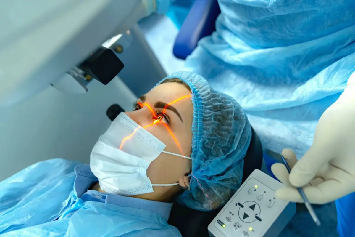
Surgical Intervention: Vitrectomy and Membrane Peeling
For serious cases, surgery is often needed. The most common surgery is vitrectomy with membrane peeling. This involves removing the vitreous gel and carefully peeling the membrane off the retina.
Vitrectomy and membrane peeling can greatly improve vision. The surgery is done under local anesthesia, and recovery is quick.
Recovery and Post-Treatment Care
After surgery, following a care plan is key for healing. This includes using eye drops to prevent infection and attending follow-up visits. It helps track how well the eye is healing.
Patients should avoid hard work and heavy lifting for a few weeks. Most can get back to normal activities within a few weeks.
Treatment Success Rates
Vitrectomy and membrane peeling have a high success rate. Many patients see big improvements in their vision. Studies show most patients get better vision and fewer symptoms after surgery.
But, like any surgery, there are risks. These include retinal detachment, infection, and cataracts. It’s important to talk about these risks and benefits with an eye doctor before deciding.
Living with Epiretinal Membrane
Living with epiretinal membrane (ERM) means finding ways to manage its effects on daily life. It’s important to understand how ERM affects vision and eye health.
Adapting to Vision Changes
Adapting to vision changes caused by ERM is a big challenge. People may see things differently, have blind spots, or notice other visual issues. To cope, they can make some lifestyle changes:
- Use magnifying glasses or other visual aids to enhance vision.
- Modify home environments to reduce fall risks and improve navigation.
- Employ technology, such as text-to-speech software, to assist with reading and other tasks.
These changes can help individuals with ERM manage their daily lives better.
Regular Monitoring and Follow-up Care
Regular check-ups with an eye care professional are key to managing ERM. This includes:
- Scheduled eye exams to track the progression of ERM.
- Utilizing diagnostic tools like Optical Coherence Tomography (OCT) to monitor changes in the retina.
- Discussing treatment options with an ophthalmologist, including the possibility of surgery.
Monitoring Aspect | Description | Frequency |
Scheduled Eye Exams | Comprehensive eye exams to assess ERM progression. | Every 6-12 months |
OCT Imaging | Use of OCT to monitor retinal changes. | As recommended by ophthalmologist |
Treatment Discussions | Consultations to discuss possible treatments, including surgery. | As needed based on ERM progression |
Support Resources and Low Vision Aids
Living with ERM can be tough, but there are many resources and aids available. These include:
- Low vision support groups, where individuals can share experiences and advice.
- Rehabilitation services, such as occupational therapy, to help adapt to vision loss.
- Technological aids, including apps and devices designed to assist with daily tasks.
By using these resources and keeping up with regular care, people with ERM can live fulfilling lives despite the challenges.
Prevention Strategies and Eye Health
To prevent epiretinal membrane (ERM), manage health conditions and keep your eyes healthy. Some risks, like age, can’t be changed. But, there are steps to lower your risk and protect your vision.
Managing Underlying Conditions
Health issues like diabetes can raise your risk for ERM. Managing these conditions through lifestyle changes and treatment can help. Keeping blood sugar levels in check can lower the risk of diabetic retinopathy, linked to ERM.
Regular Eye Examinations
Regular eye exams are key for catching ERM early. Comprehensive eye exams can spot early signs of ERM and other eye problems. The right exam schedule depends on age and risk factors. Adults should get their eyes checked every two years, or more often if they have risk factors.
Eye Examination Frequency | Age Group | Risk Factors |
Every 2 years | Adults under 40 | No known risk factors |
Annually | Adults over 40 | Presence of risk factors (e.g., diabetes, family history) |
Lifestyle Factors for Eye Health
Lifestyle choices are important for eye health. A balanced diet with fruits, veggies, and omega-3s supports your eyes. Also, protecting your eyes from UV radiation by wearing UV-protected sunglasses and avoiding smoking can help prevent eye conditions, including ERM.
Knowing the risks and taking care of your eyes can lower your chance of getting ERM. Regular eye exams and a healthy lifestyle are essential for preventing ERM and other eye problems.
Conclusion
Understanding epiretinal membrane (ERM) is key for those with vision changes. ERM affects the macula, causing big vision problems. Accurate diagnosis with Optical Coherence Tomography (OCT) is important for treatment.
ERM needs careful management in ophthalmology. Some cases might not need surgery, but others might. The treatment choice depends on how bad the symptoms are.
Knowing about ERM’s causes, symptoms, and treatments helps in eye care decisions. Regular eye checks are vital for catching ERM changes and adjusting treatments.
It’s important to see an eye doctor if you think you have ERM. With the right care, you can lessen ERM’s effects and keep your vision.
FAQ
What is Epiretinal Membrane (ERM) in ophthalmology?
Epiretinal Membrane (ERM) is a condition where a layer of cells forms on the retina. It happens over the macula, which can cause vision problems.
What are the alternative names for Epiretinal Membrane?
ERM is also known as macular pucker and cellophane maculopathy. These names describe its impact on the macula.
How is Epiretinal Membrane diagnosed?
Doctors use Optical Coherence Tomography (OCT) to diagnose ERM. This test is non-invasive and gives detailed images of the retina.
What are the symptoms of Epiretinal Membrane?
Symptoms include distorted vision and blurred vision. It can also make reading or other daily activities hard.
How serious is Epiretinal Membrane?
ERM’s seriousness varies. It can cause mild vision problems or serious visual impairment, depending on the condition.
What are the treatment options for Epiretinal Membrane?
Treatment options include watching the condition or surgery. Surgery might involve vitrectomy and membrane peeling, based on symptoms.
Can Epiretinal Membrane be prevented?
While ERM can’t be fully prevented, managing health and regular eye exams help. A healthy lifestyle also supports eye health.
What is the role of age in the development of Epiretinal Membrane?
Age is a big risk factor for ERM. It’s more common in older adults, suggesting age-related changes play a role.
How does Epiretinal Membrane affect daily life?
ERM can make daily activities like reading or driving harder. It can affect quality of life.
What support resources are available for individuals with Epiretinal Membrane?
People with ERM can use low vision aids and join support groups. Regular eye care helps manage the condition and adapt to vision changes.
What is the long-term outlook for someone with Epiretinal Membrane?
The outlook for ERM varies. Some people may have stable vision, while others may see vision changes over time. Regular monitoring is key.
References
National Health Service (NHS). Evidence-Based Medical Guidance. Retrieved from https://www.nhs.uk/conditions/macular-pucker/


