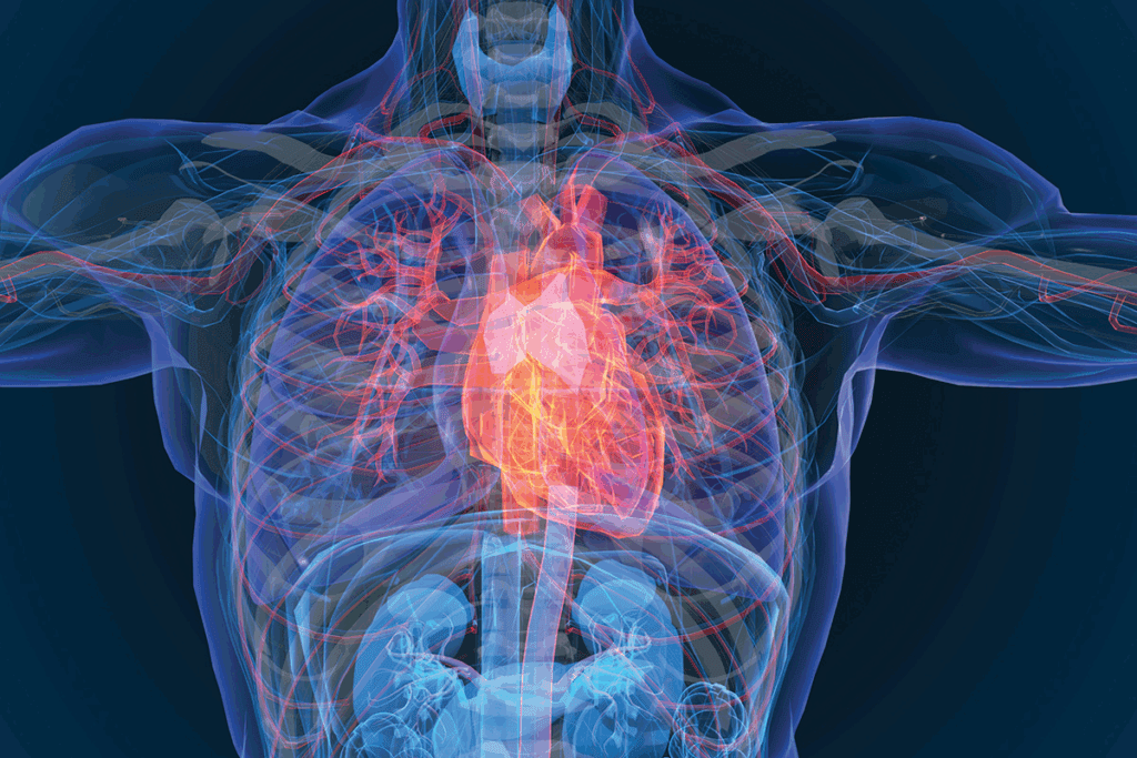
Knowing the arterial system is key to understanding how oxygenated blood spreads across the human body. At Liv Hospital, we stress the need to grasp the major arteries and their role in health. See major arteries of the body with labeled diagrams, charts, and an easy-to-understand anatomy guide.
The aorta, the biggest artery, starts from the heart. It branches out to key areas like the head, neck, and upper limbs. This guide will show you the artery map of the body. It includes labeled diagrams and charts to make the complex network clear.
We’ll look at the biggest artery in body and its branches. We’ll see how they are essential for delivering oxygenated blood to different organs and tissues.
Key Takeaways
- Understanding the arterial system’s role in distributing oxygenated blood.
- The aorta is the largest artery, originating from the heart.
- Critical branches of the aorta supply blood to the head, neck, and upper limbs.
- Labeled diagrams and charts help visualize the complex network of vessels.
- The importance of knowing the major arteries for overall health.
Understanding the Arterial System

The arterial system is key to our circulatory system. It sends oxygen-rich blood from the heart to the body. Knowing how it works and what it’s made of helps us understand heart health.
Function and Structure of Arteries
Arteries carry blood away from the heart to the body. They have strong walls to handle the heart’s high pressure.
The walls of arteries have three layers. The innermost layer, the tunica intima, is covered with cells that control blood flow and pressure.
Arterial Wall Structure
The arterial wall’s structure is vital for its job. The elastic properties of these walls help them stretch and recoil with each heartbeat. This keeps blood pressure steady.
This elasticity is key in the bigger arteries, which face high pressures.
Difference Between Arteries and Veins
Arteries and veins differ in their roles and blood flow direction. Arteries send oxygen-rich blood to the body, while veins bring deoxygenated blood back to the heart.
Arteries have thicker walls than veins to handle the heart’s blood pressure.
Understanding the arterial system’s role, structure, and how it differs from veins helps us see the circulatory system’s complexity. It also highlights the need to keep our heart health in check.
The Aorta: The Largest Human Artery

The aorta starts in the left ventricle of the heart. It is the biggest artery and very important for the body’s blood flow.
Anatomy and Structure of the Aorta
The aorta has three main parts: the ascending aorta, the aortic arch, and the descending aorta. The ascending aorta comes from the left ventricle. The aortic arch then curves back and to the left, supplying blood to the head and upper limbs. The descending aorta goes down through the chest and into the belly, splitting into the common iliac arteries.
The aorta’s wall has three layers: the tunica intima, the tunica media, and the tunica externa. The tunica intima is the innermost layer, made of endothelial cells. The tunica media is in the middle, with smooth muscle and elastic fibers. The tunica externa is the outermost, made of connective tissue.
Branches of the Aorta
The aorta has important branches that supply blood to different areas. The aortic arch branches include the brachiocephalic trunk, the left common carotid artery, and the left subclavian artery. These are key for blood to the head, neck, and upper limbs.
| Branch | Supply Area |
| Brachiocephalic Trunk | Right arm and head |
| Left Common Carotid Artery | Left side of the head and neck |
| Left Subclavian Artery | Left arm |
A leading cardiovascular specialist says, “The aorta’s structure and its branches are key to understanding how oxygenated blood is spread throughout the body.”
In summary, the aorta is not just the largest artery but also essential for the body’s blood flow. It makes sure oxygenated blood reaches all parts of the body efficiently.
Major Arteries of the Head and Neck
It’s key to know the major arteries in the head and neck for diagnosing and treating vascular issues. These arteries are vital for blood flow to the brain and other important areas.
Common Carotid Arteries
The common carotid arteries start from the brachiocephalic trunk on the right and the aortic arch on the left. They go up the neck, in front of the prevertebral fascia. They join the carotid sheath with the internal jugular vein and vagus nerve.
At the top of the thyroid cartilage, they split into the external carotid artery and the internal carotid artery. This split is a key spot.
External and Internal Carotid Branches
The external carotid artery sends blood to the neck and face. It branches into several arteries, like the superior thyroid and maxillary arteries.
The internal carotid artery mainly goes to the brain. It enters the skull through the carotid canal. It branches into the ophthalmic and anterior cerebral arteries.
Vertebral Arteries
The vertebral arteries start from the subclavian arteries and go up the neck. They enter the skull through the foramen magnum. There, they merge to form the basilar artery.
This artery supplies blood to the brainstem, cerebellum, and posterior inferior cerebellar arteries.
Branches of the Vertebral Arteries
The vertebral arteries have important branches. These include the anterior spinal artery and the posterior inferior cerebellar arteries. These branches are essential for the spinal cord and cerebellum.
In summary, the major arteries in the head and neck are vital for brain health. Knowing their anatomy and function is critical for doctors.
Major Arteries of the Upper Limbs
The upper limbs have a network of arteries that start from the subclavian arteries. We will look at the main arteries that bring blood to the upper limbs. We will cover their anatomy, function, and why they are important.
Subclavian Arteries
The subclavian arteries start from the brachiocephalic trunk on the right and the aortic arch on the left. They carry blood to the upper limbs, head, and neck. These arteries are key, providing oxygen-rich blood to the upper body.
Axillary and Brachial Arteries
When the subclavian artery reaches the first rib, it turns into the axillary artery. The axillary artery then moves into the arm, becoming the brachial artery. The brachial artery is a major artery that supplies blood to the arm and forearm. It splits into the radial and ulnar arteries.
Radial and Ulnar Arteries
The radial and ulnar arteries are the main arteries for the forearm and hand. The radial artery is on the outside of the forearm, and the ulnar artery is on the inside. These arteries are vital for the muscles and tissues in the forearm and hand.
Branches of the Radial and Ulnar Arteries
The radial and ulnar arteries have several branches. These include the radial recurrent artery, the ulnar recurrent artery, and the interosseous arteries. They also have branches for the wrist and hand.
“The arterial supply to the upper limb is a complex network that is essential for proper limb function,” says Medical Expert, a leading expert in vascular anatomy. “Understanding the anatomy of these arteries is critical for diagnosing and treating vascular disorders.”
In conclusion, the major arteries of the upper limbs are vital for blood supply to the arm, forearm, and hand. Knowing their anatomy and function is key for diagnosing and treating vascular disorders.
Major Arteries of the Thorax
Understanding the major arteries of the thorax is key to knowing how the circulatory system works. The thorax has vital arteries that help supply blood to the heart and other chest structures.
Coronary Arteries
The coronary arteries send blood straight to the heart muscle. They start from the aortic root and split into smaller arteries that cover the heart.
These arteries are vital for the heart’s health. They bring oxygen and nutrients. Any problems with these arteries can cause serious heart issues.
Brachiocephalic Trunk
The brachiocephalic trunk, or brachiocephalic artery, comes from the aortic arch. It sends blood to the right arm and head through its branches.
This artery is important because it’s the first big branch of the aortic arch. It plays a key role in blood supply to the upper body.
Intercostal Arteries
The intercostal arteries supply blood to the intercostal muscles and other thoracic structures. They start from the aorta and run between the ribs.
Branches of the Intercostal Arteries
The intercostal arteries have branches that supply blood to various thoracic structures. These include:
- Anterior intercostal arteries
- Posterior intercostal arteries
These branches are vital for blood supply to the muscles and tissues between the ribs.
| Artery | Origin | Supply |
| Coronary Arteries | Aortic Root | Heart Muscle |
| Brachiocephalic Trunk | Aortic Arch | Right Arm and Head |
| Intercostal Arteries | Aorta | Intercostal Muscles and Thoracic Structures |
Major Arteries of the Body: A Complete Systemic Overview
The body’s arterial system is a complex network. It starts with the aorta and ends with the smallest arteries. Each part plays a vital role in keeping blood flowing.
The aorta is the largest artery, carrying blood from the heart. It splits into two main arteries: the brachiocephalic trunk and the left common carotid artery. These arteries then branch into smaller ones, like the subclavian arteries.
These arteries lead to the arteries of the upper body. The subclavian arteries give rise to the vertebral arteries and the internal thoracic arteries. The vertebral arteries go to the brain, while the internal thoracic arteries supply blood to the chest wall.
The arteries of the lower body branch from the aorta’s left side. The left common iliac artery splits into the common femoral arteries. These arteries then branch into the femoral arteries, which supply blood to the legs.
The arteries of the lower body also include the arteries of the abdominal wall. These arteries are vital for blood circulation in the abdominal area.
Understanding the major arteries is key to grasping the body’s systemic arterial circulation. Each artery has its own role, ensuring blood reaches every part of the body.
Arterial junctions and branches are essential for this process. They allow blood to flow to different areas, supporting the body’s functions.
By studying the major arteries, we can appreciate the complexity and importance of the body’s arterial system.
Major Arteries of the Abdomen
Knowing how blood flows to the abdomen is key for treating stomach problems. The abdominal aorta splits into important branches. These branches help the digestive system and other parts of the abdomen.
Celiac Trunk and Its Branches
The celiac trunk starts just below the diaphragm. It quickly splits into three main parts: the left gastric artery, the common hepatic artery, and the splenic artery.
- The left gastric artery feeds the stomach.
- The common hepatic artery leads to the liver and the stomach and duodenum through the gastroduodenal artery.
- The splenic artery supplies the spleen, pancreas, and stomach.
Superior and Inferior Mesenteric Arteries
The superior mesenteric artery comes from the abdominal aorta below the celiac trunk. It goes to the small intestine and the right colon.
The inferior mesenteric artery comes from the abdominal aorta lower down. It feeds the left colon, rectum, and upper anal canal.
Branches of the Mesenteric Arteries
The superior mesenteric artery has branches like the inferior pancreaticoduodenal artery and the ileocolic artery. The inferior mesenteric artery has the left colic artery and the sigmoid branches.
| Artery | Origin | Supply |
| Celiac Trunk | Abdominal Aorta | Stomach, Liver, Pancreas, Spleen |
| Superior Mesenteric Artery | Abdominal Aorta | Small intestine, Right half of the colon |
| Inferior Mesenteric Artery | Abdominal Aorta | Left half of the colon, Rectum, Upper anal canal |
In conclusion, the major arteries of the abdomen are vital for the digestive system. Knowing their anatomy helps in diagnosing and treating stomach issues.
Major Arteries of the Lower Limbs
It’s important to know how blood flows to the lower limbs. This knowledge helps in treating vascular problems. The lower limbs have a complex system of arteries. These arteries make sure blood reaches the thigh, leg, and foot.
Femoral Artery
The femoral artery is a key artery for the thigh. It starts from the external iliac artery and goes down through the femoral triangle. Here, it branches out to the thigh muscles.
The femoral artery is vital. If it gets blocked, it can cause serious problems.
Popliteal Artery
The popliteal artery comes from the femoral artery at the adductor hiatus. It carries blood to the knee and leg. It splits into the anterior and posterior tibial arteries.
The popliteal artery is in the popliteal fossa. It’s at risk for injury and disease.
Anterior and Posterior Tibial Arteries
The anterior tibial artery and posterior tibial artery branch off from the popliteal artery. They supply blood to the leg and foot. The anterior tibial artery goes to the front of the leg. The posterior tibial artery goes to the back.
Branches of the Tibial Arteries
The tibial arteries have branches for the leg and foot. The anterior tibial artery has the anterior medial and lateral malleolar arteries. The posterior tibial artery has the peroneal artery and nutrient arteries for the tibia and fibula.
Knowing about these branches is key for treating vascular issues in the lower limbs.
Advanced Imaging and Mapping of Arteries
Advanced imaging has changed vascular medicine a lot. It lets us see the arteries clearly. Angiography and computed tomography angiography (CTA) help map arteries well. This is key for finding and treating artery diseases.
Arterial imaging is now a must in medicine. It lets doctors see the arteries in great detail. This helps in diagnosing and planning surgeries.
Using CTA and magnetic resonance angiography (MRA) has made diagnosing artery diseases better. These methods let doctors see the arteries from many sides. This boosts their confidence in making diagnoses.
These imaging tools also help in making detailed maps of arteries. This is great for spotting blockages, aneurysms, and other problems that need treatment.
Also, these advanced imaging methods help track how diseases progress and how treatments work. They give a clear view of the arteries. This helps doctors make better choices for patient care.
To wrap it up, advanced imaging and artery mapping are vital for treating vascular diseases. As technology gets better, these methods will get even more accurate. This will lead to better care for patients.
Conclusion
Knowing about the arterial system is key to keeping our hearts healthy. The major arteries carry oxygen-rich blood to our organs and tissues. In this article, we’ve looked at the aorta, carotid arteries, and other important ones.
These arteries are vital for getting blood to our head, neck, and upper limbs. Knowing how they work helps doctors diagnose and treat heart diseases.
Healthcare experts can manage heart-related conditions better by understanding the arterial system. As medical science and technology improve, knowing about the arterial system will become even more important.
FAQ
What is the largest artery in the human body?
The aorta is the largest artery. It starts from the left ventricle of the heart. It carries oxygenated blood to the rest of the body.
What are the major arteries of the head and neck?
The head and neck have several major arteries. These include the common carotid arteries and their branches. Also, the vertebral arteries supply blood to the brain and other vital areas.
What is the function of the coronary arteries?
The coronary arteries give blood to the heart muscle. They provide the heart with the oxygen and nutrients it needs to work.
How do arteries differ from veins?
Arteries carry oxygen-rich blood away from the heart. Veins bring deoxygenated blood back to the heart. Arteries have thicker walls because they handle higher pressure.
What is the significance of understanding the arterial system?
Knowing the arterial system is key for diagnosing and treating heart diseases. It also helps in keeping the heart and blood vessels healthy.
What are the major arteries of the lower limbs?
The lower limbs have important arteries. These include the femoral artery and the popliteal artery. The anterior and posterior tibial arteries also play a role in supplying blood to the legs.
How are arterial diseases diagnosed?
Doctors use advanced imaging to diagnose arterial diseases. Techniques like angiography, ultrasound, and CT scans help in identifying these conditions.
What is the role of the aorta in the arterial system?
The aorta is the main artery from the heart. It branches out to supply oxygenated blood to the body.
What are the branches of the aorta?
The aorta has several branches. These include the coronary arteries and the brachiocephalic trunk. It also has intercostal arteries, among others.
Why is it important to understand the arterial system?
Understanding the arterial system is vital for heart health. It helps in diagnosing and treating diseases. It also prevents serious heart problems.
References:
- StatPearls. (2023). Anatomy, Head and Neck: Carotid Arteries. In StatPearls https://www.ncbi.nlm.nih.gov/books/NBK545238/
- Jones, O. (2023, February). Major arteries of the head and neck. TeachMeAnatomy. https://teachmeanatomy.info/neck/vessels/arterial-supply/
- Foran, P., & Co-authors. (2023). Clinical basis for the knowledge of anatomy of the carotid artery: A review article. Yenagoa Medical Journal, 5(2), 24-29. https://yenagoamedicaljournal.net/clinical-basis-for-the-knowledge-of-anatomy-of-the-carotid-artery-a-review-article/
- Omotoso, B. R., et al. (2021). Radiological anatomy of the intracranial vertebral artery in [population studied]. Scientific Reports. https://www.nature.com/articles/s41598-021-91744-9/


