Heart health is key to feeling good overall. Nuclear cardiology is a big help in finding and treating heart problems.
Learning nuclear cardiology well is important. It helps find and fix heart issues. This leads to better heart health.

Nuclear cardiology is a big step forward in treating heart diseases. It mixes nuclear medicine and cardiology for better heart care.
Nuclear cardiology uses small amounts of radioactive tracers to evaluate heart function. It looks at how these materials spread in the heart. This shows how well the heart is working.
This method lets see the heart’s shape and how it moves. They can spot heart problems, see if heart muscle is alive, and check how well the heart pumps.
Cardiac nuclear medicine has grown a lot in recent years. At first, it was mainly for finding heart artery disease with special images.
With new tech and medicines, it now checks heart function and if heart muscle is alive. Today, it’s key in managing heart disease, helping make better treatment plans.
| Year | Development | Impact |
| 1970s | Introduction of Thallium-201 | Enabled myocardial perfusion imaging |
| 1980s | Development of Technetium-99m sestamibi | Improved image quality and reduced radiation exposure |
| 2000s | Advancements in SPECT and PET technology | Enhanced diagnostic accuracy and expanded applications |
Nuclear cardiology uses special materials to see the heart. It’s a field that helps diagnose and manage heart diseases. This is done with tiny amounts of radioactive materials.
Radiopharmaceuticals are key in nuclear cardiology. They help see how the heart works and spot heart problems. These substances focus on certain heart areas, giving important health info.
Common Radiopharmaceuticals:
Radiopharmaceuticals are vital but also involve radiation. Keeping everyone safe from radiation is a big deal.
Important things to think about include:
| Consideration | Description |
| Dosage | Using the least amount of radiopharmaceuticals needed |
| Shielding | Protecting against radiation with the right shields |
| Patient Education | Telling patients about radiation risks and benefits |
By managing these carefully, nuclear cardiology’s benefits can be high while risks are low.
“The safe use of radiopharmaceuticals is key in nuclear cardiology. It’s about finding the right balance between getting good results and keeping radiation exposure low.”
Nuclear cardiology uses several key procedures to check the heart’s function. These tests are vital for understanding heart health and managing heart diseases.
Myocardial perfusion imaging (MPI) is a common test in nuclear cardiology. It checks blood flow to the heart muscle. It finds areas where blood flow is low, which might mean blockages or damage.
MPI is great for spotting coronary artery disease and seeing if treatments work.
Key aspects of MPI include:
Cardiac blood pool imaging, or MUGA scan, checks how well the heart pumps. It uses a tiny amount of radioactive tracer in the blood. This lets see how well the heart’s ventricles pump blood.
“MUGA scans provide valuable information on cardiac function, particularlly in patients with heart failure or those undergoing chemotherapy.” –
Cardiology Expert
PET cardiac imaging is a detailed test in nuclear cardiology. It shows how well the heart’s tissue works and blood flow. It’s great for checking if heart tissue can recover and helping decide treatment for heart disease or failure.
| Procedure | Purpose | Benefits |
| Myocardial Perfusion Imaging | Assess coronary artery disease | Identifies areas of reduced blood flow |
| Cardiac Blood Pool Imaging (MUGA) | Evaluate heart’s pumping function | Assesses ventricular function |
| PET Cardiac Imaging | Assess myocardial viability | Guides treatment decisions |
These nuclear cardiology procedures are key for diagnosing and managing heart conditions. They give important insights into the heart’s function and help guide treatment plans.
Nuclear medicine has changed how we find and treat heart diseases. It gives both functional and anatomical details. This helps diagnose heart conditions well.
Nuclear medicine is key in finding coronary artery disease. This disease is a big cause of illness and death. Myocardial perfusion imaging (MPI) spots areas where blood flow to the heart is low.
This shows if coronary artery disease is present. can then decide how serious it is and what treatment is needed.
Nuclear medicine also helps a lot with heart failure. It uses cardiac blood pool imaging (MUGA) and PET cardiac imaging. These methods show how well the heart pumps and if the heart muscle is working.
This info is key for figuring out why heart failure happens. It helps choose the right treatments and check if they work.
With nuclear medicine, can make plans that fit each patient’s needs. This makes heart failure treatment better.
Nuclear cardiology is key in checking how well the heart works and helping decide on treatments. It’s a big help for to spot and treat heart problems well.
Choosing the right patients for nuclear cardiology tests is very important. look for those who might really need these tests, like those with heart disease or heart failure.
They consider the patient’s symptoms, medical history, and early test results. This makes sure the tests give useful info for treatment plans.
Nuclear cardiology works best when used with other heart tests. This combo gives a full picture of heart health. It helps make better treatment plans.
For example, mixing nuclear cardiology with echocardiography or MRI gives insights into heart structure and function. Here’s how different tests work together:
| Diagnostic Test | Information Provided | Utility |
| Nuclear Cardiology | Assesses cardiac function and perfusion | Diagnoses coronary artery disease, heart failure |
| Echocardiography | Provides structural and functional heart information | Assesses valve function, cardiac chambers |
| Cardiac MRI | Detailed images of heart structure and function | Evaluates cardiac morphology, viability |
Using nuclear cardiology with other tests helps make better choices. This leads to better care for patients.
The patient experience in nuclear cardiac testing includes several stages. These stages range from preparation to post-procedure care. Knowing about these stages can help reduce anxiety and make the process smoother.
Getting ready for a nuclear cardiac test is important. Patients are usually told to avoid caffeine and certain medications that might affect the test results. They should also wear comfy clothes and remove any jewelry that could get in the way of the imaging.
It’s also important to tell the healthcare provider about any allergies or medical conditions. This includes any past reactions to contrast agents or radiopharmaceuticals used in these tests.
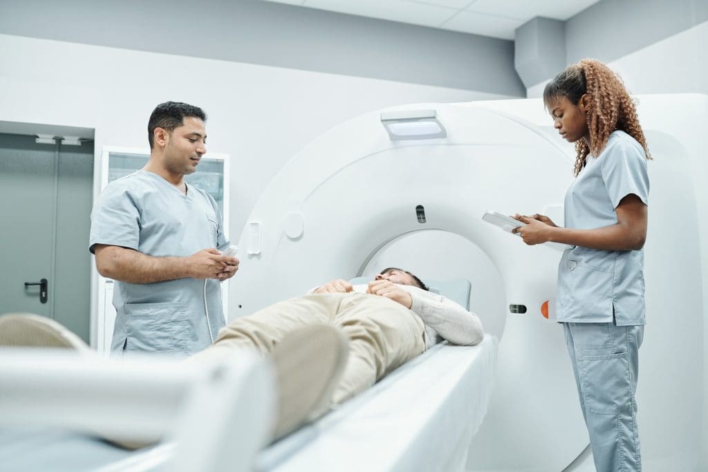
During the test, a small amount of radiopharmaceutical is given. This lets the heart be imaged. The test happens in a special department, where you might lie on a table while a gamma camera takes pictures of your heart.
After the test, you can usually go back to your normal activities unless your doctor says not to. The radiopharmaceutical is removed from your body through urine or feces. It’s key to follow any instructions from the healthcare team to stay safe and get accurate test results.
It’s key to understand nuclear cardiology results well to spot heart issues and find the right treatment. Tests like myocardial perfusion imaging show the heart’s details. This helps diagnose and manage heart disease.
Nuclear cardiology test reports talk about the heart’s blood flow, function, and health. Knowing cardiac anatomy and physiology is important to get these reports. The reports have a summary, image description, and conclusion.
should focus on the summary. It outlines the main findings and their impact on care. Standardized reporting makes reports clear and consistent. This helps understand the results better.
Tests often show perfusion defects, meaning less blood flow to the heart. Reversible defects might mean ischemia, while fixed defects suggest scar tissue. Other findings include ventricular function issues, like a low ejection fraction.
“The accurate interpretation of nuclear cardiology results is key for treatment decisions and better patient outcomes.”
Knowing what these findings mean helps create treatment plans. This might include lifestyle changes, medication, or more tests and treatments.
Nuclear cardiology has both good points and downsides. It’s a complex tool for checking heart health.
Nuclear cardiology has some big advantages. It’s very good at finding heart disease and seeing how well the heart works. It looks at both blood flow and heart function.
The benefits of nuclear cardiology include being non-invasive. It gives both diagnosis and future health outlook. It’s also widely available, which makes it a top choice for .
But, nuclear cardiology also has some limitations. One big worry is radiation exposure. While it’s mostly safe, it’s something to think about, mainly for younger people or those needing many tests.
Other limitations of nuclear cardiology include the . It also needs special equipment and trained people. Plus, there can be issues with the images that might affect how they’re read. Knowing these limits helps choose and understand tests better.
There are many ways to check the heart for problems. Nuclear cardiology is one method that shows the heart’s function and structure well. It’s getting more attention for its detailed images.
Echocardiography uses sound waves to see the heart. It’s good for looking at the heart’s structure and how it works. But, it might not show as much as nuclear cardiology about blood flow and how well the heart works.
Nuclear cardiology is better at finding heart disease and checking how well the heart works and gets blood.
Cardiac CT gives clear pictures of the heart’s shape. It’s mainly used to see if there’s calcium in the heart’s arteries. Nuclear cardiology looks more at how the heart works and gets blood.
Nuclear cardiology is better for checking blood flow and how well the heart works. Cardiac CT is better for looking at the heart’s shape and finding calcium in arteries.
| Imaging Modality | Primary Use | Diagnostic Strengths |
| Nuclear Cardiology | Myocardial perfusion, viability | Functional assessment, disease severity |
| Cardiac CT | Coronary artery calcium, stenosis | Anatomical detail, calcium scoring |
Cardiac MRI can look at the heart’s shape and how it works. It’s great for many things but not as common as nuclear cardiology. It can be harder to do.
Nuclear cardiology is more common and easy to use. It’s good for finding and managing heart problems.
In summary, each way to look at the heart has its own good points. Nuclear cardiology is special because it shows how the heart works. It’s very useful for diagnosing and treating heart disease.
Women and the elderly greatly benefit from nuclear cardiology. It offers detailed heart images. This is key for these groups.
Heart disease is a big killer for women. Nuclear cardiology is key in finding and treating it. It spots coronary artery disease and future heart risks.
The American Heart Association says heart disease kills 1 in 5 women in the U.S. Early detection and treatment with nuclear cardiology can save lives.
Elderly people often have complex heart issues. Nuclear cardiology helps by checking heart function without surgery. It finds problems like scar tissue.
“The use of nuclear cardiology in elderly patients allows for a more accurate diagnosis and tailored treatment plans, improving outcomes in this high-risk population.”
People with diabetes or high blood pressure face higher heart disease risks. Nuclear cardiology helps by showing how well the heart works and how blood flows.
| Comorbidity | Nuclear Cardiology Application | Benefit |
| Diabetes | Myocardial Perfusion Imaging (MPI) | Early detection of coronary artery disease |
| Hypertension | Cardiac Blood Pool Imaging (MUGA) | Assessment of left ventricular function |
Nuclear cardiology gives detailed heart images. It’s vital for managing heart disease in different groups.
Nuclear cardiology has seen big changes with new cameras and software. These updates have made it better at diagnosing heart problems. Now, can see the heart more clearly than before.
The newest cameras and tools in nuclear cardiology are top-notch. Cameras like the Cadmium Zinc Telluride (CZT) detectors make images better and faster. This helps find heart diseases more easily.
New software has also improved nuclear cardiology a lot. It can handle complex data quickly, giving important insights. Techniques like attenuation correction and resolution recovery make images clearer, helping make better diagnoses.
These new technologies help diagnose and treat heart problems more accurately.
It’s key for healthcare providers and patients in the US to grasp nuclear cardiology. This field is a major tool for diagnosing and treating heart disease. Yet, it’s not without its practical challenges.
Nuclear cardiology tests are usually covered by big insurance companies in the US, like and Medicaid. But, how much they cover can change based on the test and the patient’s plan.
The of nuclear cardiology services can differ, affecting what patients pay out of pocket. It’s important for patients to know what their insurance covers and any extra they might face.
Finding a skilled nuclear cardiology specialist is vital for those needing these tests. Here’s how to find them:
When picking a nuclear cardiology specialist, look at their experience, qualifications, and the technology at their place.
Knowing about insurance and finding the right specialist helps patients get the most from nuclear cardiology for their heart health.
Nuclear medicine in cardiac care is on the verge of a big change. New technologies and methods are coming along. These advancements are making diagnosis and treatment better.
New cameras and software are making images clearer in nuclear cardiology. Advanced SPECT and PET technologies help spot coronary artery disease and check heart health. Hybrid imaging mixes nuclear medicine with CT or MRI for a full view of the heart.
| Technology | Application | Benefit |
| Advanced SPECT | Improved diagnostic accuracy | Better detection of CAD |
| PET Imaging | Assessment of myocardial viability | Enhanced prognostic value |
| Hybrid Imaging | Combination with CT or MRI | Comprehensive cardiac assessment |
Personalized medicine is key in cardiac care, with nuclear medicine leading the way. It means treatments fit each patient’s needs. This approach can lead to better results and lower . Genomic and biomarker-based strategies are being used with nuclear cardiology for more precise treatments.
The mix of new technologies and personalized medicine will change nuclear cardiology. It brings hope for those with heart disease.
Nuclear cardiology is key in improving heart health. It helps make accurate diagnoses and choose the right treatments. This leads to better care for patients.
There are many nuclear cardiology tests, like myocardial perfusion imaging. These tests show how well the heart works and blood flows. They help find problems like coronary artery disease and heart failure.
Nuclear cardiology is getting better with new technology and medicines. Keeping up with these changes helps give better care. This improves heart health for patients.
To make the most of nuclear cardiology, need to understand it well. Using it in their work helps them make better choices. This leads to more personalized care for patients with heart issues.
The future looks bright with new technologies and techniques. These advancements will likely improve diagnosis and patient care.
Yes, insurance usually covers it. But, coverage and payment details vary. Always check with your insurance provider.
Ask your doctor for a referral or check your insurance for specialists. You can also search online for certified nuclear cardiologists in your area.
Yes, it can be used for women and elderly patients. The test might need to be adjusted for their specific needs.
Yes, it’s generally safe with a low risk of bad reactions. But, like any test, there are risks like radiation. Talk to your doctor about these.
Nuclear cardiology is one of several imaging options, like echocardiography and MRI. Each has its own benefits and drawbacks. The right test depends on the patient’s needs and the doctor’s question.
To prepare, avoid certain foods and medications. Wear comfy clothes and remove metal jewelry. Your healthcare provider will give you specific instructions.
Common tests include myocardial perfusion imaging and cardiac blood pool imaging (MUGA). PET cardiac imaging is also used. These tests help find and manage heart conditions like coronary artery disease and heart failure.
Nuclear cardiology is a field that uses tiny amounts of radioactive tracers. It helps diagnose and manage heart disease. This method uses nuclear medicine to see the heart and its blood vessels.
Subscribe to our e-newsletter to stay informed about the latest innovations in the world of health and exclusive offers!