
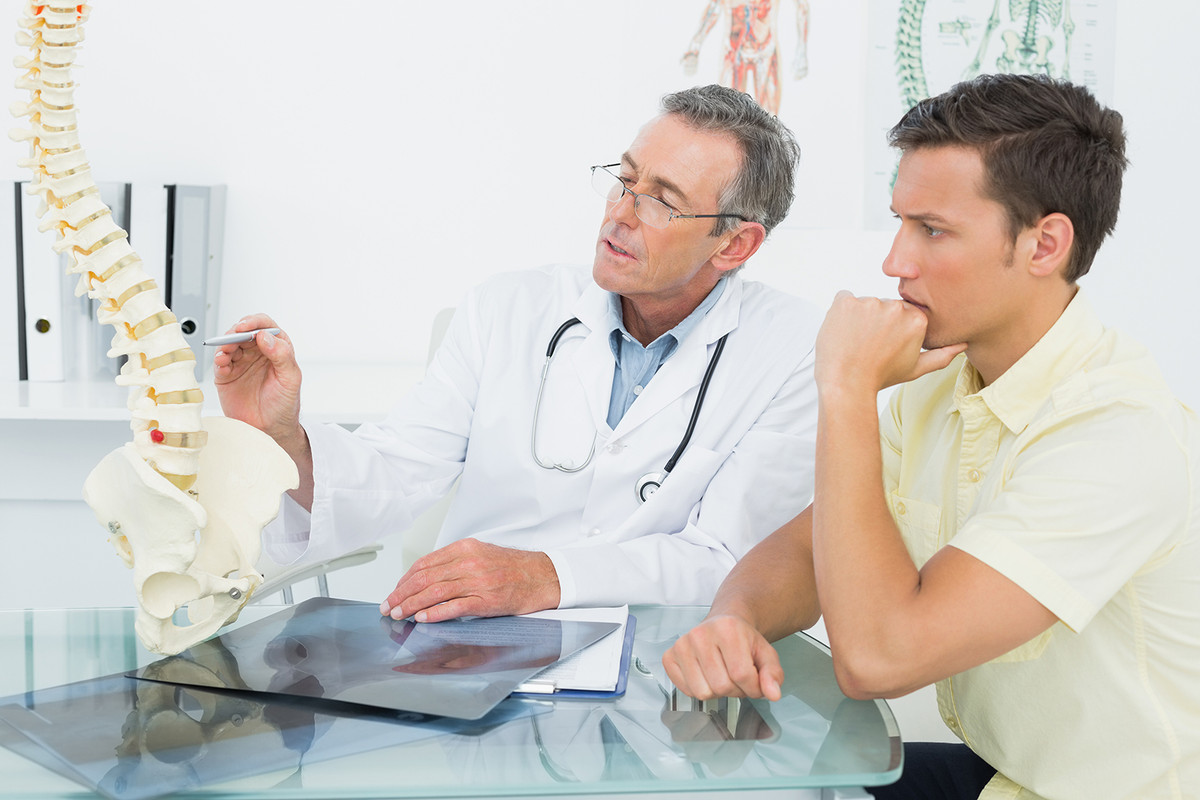
Knowing the 12 cranial nerves is key for doctors and students. These nerves link the brain to the head, face, neck, and upper body. They control many functions, like seeing, tasting, and moving our face.match the cranial nerve number with its name7 Key Facts About Arnold Chiari Malformation Type 2 and Its Management
We’ll look at where these nerves are and what they do. They are numbered from I to XII. The first two come from the cerebrum, and the rest from the brainstem. This guide will make it easier to know each nerve’s role.
Key Takeaways
- The 12 cranial nerves play a vital role in controlling various critical functions.
- These nerves are directly connected to the brain.
- Understanding cranial nerves is essential for medical professionals.
- The nerves are numbered using Roman numerals for identification.
- Knowledge of cranial nerve anatomy is key for diagnosis and treatment.
The Fundamentals of Cranial Nerves

To understand the nervous system, we must start with the basics of cranial nerves. The 12 cranial nerves are key to the nervous system. Knowing their origin and how they work is important for medical professionals.
Origin and Organization in the Brain and Brainstem
The cranial nerves come from the brain and brainstem. The olfactory nerve (CN I) and optic nerve (CN II) start in the cerebrum. The rest (III – XII) come from the brainstem. This shows how complex the cranial nerves are in the central nervous system.
The brainstem, made up of the midbrain, pons, and medulla oblongata, is where most cranial nerves start. Each nerve has its own spot of origin in these parts. This shows the detailed anatomy of the brainstem.
Functional Classification: Sensory, Motor, and Mixed Nerves
Cranial nerves are divided into sensory, motor, and mixed nerves. Sensory nerves send information to the brain. Motor nerves send signals to muscles and glands, causing movement or responses. Mixed nerves do both.
Knowing this classification helps in diagnosing and treating nerve disorders. For example, knowing a nerve’s type helps find where a problem is and what symptoms it might cause.
By understanding the different roles of cranial nerves, we can see how complex the nervous system is. This helps us match each cranial nerve with its number and function better.
How to Match the Cranial Nerve Number with Its Name

Learning to match the cranial nerve number with its name is more than just memorizing. It’s about knowing their roles and importance in health. This skill is key for students and doctors to diagnose and treat brain and nerve issues well.
We use two main ways to learn this: mnemonic devices and an anatomical approach. Both help us understand the cranial nerves fully.
Effective Mnemonic Devices for Medical Students
Mnemonic devices are tools that link new info to something familiar, like a word or image. For cranial nerves, a common mnemonic is “O O O To Touch And Feel Very Good Velvet AH.” Each word starts with the first letter of each nerve: Olfactory, Optic, Oculomotor, Trochlear, Trigeminal, Abducens, Facial, Vestibulocochlear, Glossopharyngeal, Vagus, Spinal Accessory, and Hypoglossal.
Using mnemonics can make learning the cranial nerves faster and more fun. It lets students get creative with their own sentences or phrases.
Anatomical Approach to Learning Cranial Nerves
Mnemonics aid in memorization, but the anatomical approach gives deeper insights. It involves studying each nerve’s origin, path, and role, as well as its clinical importance.
Visualizing the nerves’ paths and their functions helps in remembering them. For example, knowing the olfactory nerve’s role in smell makes it easier to recall its name and function.
Combining mnemonics with the anatomical approach is a strong learning method. It helps in memorizing and understanding the cranial nerves’ clinical significance. This makes it simpler to match the cranial nerve number with its name in school and practice.
Cranial Nerve I: Olfactory Nerve
Cranial Nerve I, or the olfactory nerve, is key for smell. It sends sensory info from the nose to the brain. This nerve is the first of the 12 cranial nerves and helps us smell different odors.
Anatomical Pathway from Nasal Cavity to Brain
The olfactory nerve starts in the nasal cavity. Here, special olfactory receptor neurons are found. These neurons detect odor molecules in the air we breathe.
The nerve fibers carry signals from these receptors to the olfactory bulb in the forebrain. Then, the info goes to the brain’s olfactory cortex. This is where we process smells.
Function: Processing Olfactory Sensations
The main job of the olfactory nerve is to handle smell. When odor molecules meet olfactory receptors in the nose, it sends a signal to the brain. The brain then figures out what we’re smelling.
This process is key for enjoying food, detecting danger, and experiencing our surroundings.
Clinical Testing and Associated Disorders
Testing the olfactory nerve checks how well someone can smell. Doctors use smell tests to see if patients can identify different odors. Problems with the olfactory nerve include anosmia (not being able to smell), hyposmia (smelling less well), and dysosmia (smelling things wrong).
These issues can come from infections, head injuries, or neurological problems.
Condition | Description | Common Causes |
Anosmia | Loss of the ability to smell | Infections, head trauma, neurological disorders |
Hyposmia | Reduced ability to smell | Aging, nasal congestion, certain medications |
Dysosmia | Distorted sense of smell | Infections, head trauma, certain psychiatric conditions |
Cranial Nerve II: Optic Nerve
Cranial Nerve II, also known as the optic nerve, carries visual signals from the retina to the brain. It’s key for our vision. Damage to it can lead to serious vision problems.
Visual Pathway from Retina to Visual Cortex
The optic nerve starts in the retina, where light turns into electrical signals. These signals go to the optic disc and then through the optic canal into the brain. At the optic chiasm, the nerves from both eyes partially cross over, helping us see in 3D.
After crossing, the signals move to the lateral geniculate nucleus and then to the visual cortex. This is where our brain interprets what we see.
Function: Transmitting Visual Information
The optic nerve’s main job is to send visual information from the retina to the brain. It carries details about light, color, and where things are. This nerve is essential for us to see and understand our world.
Clinical Assessment and Visual Field Defects
Doctors use several tests to check the optic nerve, like visual acuity tests and visual field exams. They also use ophthalmoscopy to look at the optic disc. Damage to the optic nerve can cause blindness or other vision problems, depending on where and how much damage there is.
Clinical Test | Purpose | Significance |
Visual Acuity Test | Measures the sharpness of vision | Helps diagnose optic nerve dysfunction |
Visual Field Examination | Assesses the extent of the visual field | Detects defects such as hemianopia |
Ophthalmoscopy | Examines the optic disc and retina | Reveals signs of optic nerve damage or disease |
Knowing about the optic nerve’s anatomy, function, and how doctors check it is key for treating vision problems. By spotting optic nerve damage early, doctors can help keep our vision sharp.
Cranial Nerve III: Oculomotor Nerve
Cranial nerve III, or the oculomotor nerve, controls eye movements and pupil size. It’s key for eye functions like moving the eyeball and controlling the eyelid. It also helps in constricting the pupil.
Anatomical Course Through the Cavernous Sinus
The oculomotor nerve starts in the midbrain. It goes between the superior cerebellar and posterior cerebral arteries. Then, it passes through the cavernous sinus, a space near the brain base, before entering the orbit.
In the cavernous sinus, the nerve is close to important structures. This includes the internal carotid artery and other cranial nerves. This close proximity makes it vulnerable to damage from various conditions.
Functions: Eye Movement, Pupil Constriction, and Accommodation
The oculomotor nerve has several roles:
- It controls eye movement by working with the medial rectus, superior rectus, inferior rectus, and inferior oblique muscles.
- It manages pupil constriction by controlling the sphincter pupillae muscle.
- It helps with accommodation by coordinating the ciliary muscles for focusing on near objects.
These roles are essential for everyday tasks like reading and driving. They require precise eye movements.
Clinical Signs of Oculomotor Nerve Palsy
Oculomotor nerve palsy can be caused by trauma, aneurysms, or diabetes. Signs include:
Signs | Description |
Ptosis | Drooping of the eyelid due to weakness of the levator palpebrae superioris muscle. |
Dilated Pupil | The pupil may be dilated and non-reactive due to loss of parasympathetic innervation. |
Eye Movement Limitation | Limitation in moving the eye in various directions due to paralysis of the extraocular muscles. |
Knowing the anatomy and functions of the oculomotor nerve is key for diagnosis and treatment. Clinical assessment and imaging studies help find the cause and guide treatment.
Cranial Nerve IV: Trochlear Nerve
The trochlear nerve is the thinnest cranial nerve and is key for eye movement. It is the fourth cranial nerve (CN IV). It has unique features that set it apart from other nerves.
Unique Anatomical Features and Course
The trochlear nerve has the longest path inside the brain. It starts in the midbrain, crosses over in the superior medullary velum, and comes out from the back of the brainstem. Because of its long path and thin structure, it is very vulnerable.
Decussation is a special feature of the trochlear nerve. It is the only nerve that crosses over before leaving the brainstem. This means it controls the opposite side’s superior oblique muscle.
Function: Superior Oblique Muscle Innervation
The trochlear nerve mainly controls the superior oblique muscle of the eye. This muscle helps the eye move down, inwards, and outwards. It’s important for looking down correctly.
Diagnosing Trochlear Nerve Dysfunction
Trochlear nerve problems can cause trochlear nerve palsy. This leads to double vision, mainly when looking down. Doctors check eye movements to diagnose it. People with this problem often tilt their head to see better.
Doctors use the Parks-Bielschowsky three-step test to find the cause of double vision. This test helps figure out which eye and muscle are affected.
Cranial Nerve V: Trigeminal Nerve
The trigeminal nerve, or cranial nerve V, is complex. It has sensory and motor functions. It’s key for facial sensation and controlling actions like chewing.
Three Major Divisions: Ophthalmic, Maxillary, and Mandibular
The trigeminal nerve splits into three main parts: the ophthalmic, maxillary, and mandibular divisions. Each part has its own role and area it covers.
- Ophthalmic Division (V1): It handles sensory input from the eye and its surroundings.
- Maxillary Division (V2): It’s in charge of feeling in the middle face, like the maxillary sinus and nasal area.
- Mandibular Division (V3): This part has both sensory and motor fibers. It deals with feeling in the lower face and controls chewing muscles.
Clinical Assessment and Trigeminal Neuralgia
Checking the trigeminal nerve involves looking at its sensory and motor abilities. This includes testing facial touch, corneal reflexes, and chewing muscles.
Clinical Test | Description | Function Assessed |
Facial Sensation Testing | Checking how well the patient feels touch, pain, and temperature on their face. | Sensory Function |
Corneal Reflex Test | Touching the cornea to see if the patient blinks. | Sensory and Motor Function |
Muscles of Mastication Strength | Testing the strength of chewing muscles. | Motor Function |
Trigeminal neuralgia causes sharp, stabbing pain in the face. It happens when the trigeminal nerve gets compressed or irritated. Treatment can include medicine or surgery.
“Trigeminal neuralgia is a debilitating condition that affects the quality of life. Accurate diagnosis and appropriate management are critical for symptom relief.”
We know how tough trigeminal nerve disorders can be. We’re dedicated to giving our patients the best care possible.
Cranial Nerve VI: Abducens Nerve
Understanding the abducens nerve is key to diagnosing and treating eye movement disorders. The abducens nerve, or cranial nerve VI, controls the lateral rectus muscle. This muscle is vital for moving the eye outward.
Anatomical Course and Vulnerability
The abducens nerve has a long path inside the brain, making it prone to damage. It starts in the pons, exits the brainstem, and goes through several areas before reaching the eye. This long journey makes it vulnerable to injury.
Its long path means the abducens nerve can be hurt by trauma, high brain pressure, or blood vessel pressure. This often leads to abducens nerve palsy, causing trouble with moving the eye laterally.
Function: Lateral Gaze Control
The abducens nerve mainly controls the lateral rectus muscle. This muscle helps the eye move outward, which is important for seeing in 3D and moving around.
Damage to the abducens nerve can cause double vision. This is because the eyes can’t line up right. Double vision can make everyday tasks hard, like driving or reading.
Identifying Abducens Nerve Palsy
To diagnose abducens nerve palsy, doctors do a detailed check. They look at how the eyes move, how well you see, and the pupils. Signs of this nerve problem include:
- Double vision that gets worse when looking to the side
- The eye can’t move as far outward as it should
- People might move their head to avoid double vision
Clinical Feature | Description |
Diplopia | Double vision, mainly when looking to the side |
Limited Abduction | The eye can’t move as far outward as it should |
Compensatory Head Movement | People might turn their head to make up for eye movement issues |
Knowing about the abducens nerve’s role in eye movement is vital for doctors. It helps them give the right treatment and improve patient care. By understanding this nerve, we can better handle eye movement problems.
Cranial Nerve VII: Facial Nerve
The facial nerve is one of the twelve cranial nerves. It has a complex path and many roles. It controls the muscles of facial expression, among other things.
Complex Anatomical Course and Branches
The facial nerve starts in the brainstem and goes through the temporal bone. It ends up in different places. It has branches like the temporal, zygomatic, buccal, marginal mandibular, and cervical branches. These branches help the muscles of facial expression work.
Here’s how the nerve travels:
- It starts in the brainstem, between the pons and medulla oblongata.
- Then, it goes through the internal auditory meatus into the facial canal in the temporal bone.
- It comes out of the skull through the stylomastoid foramen.
- It branches out to the muscles of facial expression.
Bell’s Palsy and Other Facial Nerve Disorders
Bell’s palsy is a common issue with the facial nerve. It causes sudden weakness or paralysis on one side of the face. The cause is often a viral infection.
Other problems with the facial nerve include:
Condition | Description |
Ramsay Hunt Syndrome | A complication of shingles that affects the facial nerve, causing pain, hearing loss, and facial paralysis. |
Hemifacial Spasm | A condition characterized by irregular, involuntary muscle contractions on one side of the face. |
Knowing about the facial nerve’s anatomy and functions is key. It helps in diagnosing and treating these conditions.
“The facial nerve is a complex structure with a wide range of functions, making it a critical component of our neurological makeup.” –
A renowned neurologist
Cranial Nerve VIII: Vestibulocochlear Nerve
Cranial Nerve VIII, also known as the vestibulocochlear nerve, is key for hearing and balance. It has two parts: the vestibular and cochlear divisions. Each part has its own job, helping us sense our surroundings.
Vestibular and Cochlear Divisions
The vestibulocochlear nerve has two main parts: the vestibular and cochlear divisions. The vestibular division helps us stay balanced. The cochlear division is for hearing.
The vestibular division works with the inner ear’s vestibular apparatus. It detects changes in head position and movement. This is vital for balance and spatial awareness.
The cochlear division, on the other hand, deals with sound. It converts sound vibrations into electrical signals for the brain to understand as sound.
Functions: Hearing Perception and Balance Maintenance
The vestibulocochlear nerve is essential for hearing and balance. The cochlear division lets us hear different sounds. The vestibular division helps us stay balanced and oriented.
A neurologist notes, “The vestibulocochlear nerve is vital for our senses. Its problems can cause big issues with hearing and balance.”
“Dysfunction of the vestibulocochlear nerve can result in vertigo, hearing loss, and tinnitus, significantly impacting a person’s quality of life.”
Vertigo, Hearing Loss, and Diagnostic Tests
Problems with the vestibulocochlear nerve can cause vertigo, hearing loss, and tinnitus. Vertigo makes you feel like you’re spinning. Hearing loss can be mild or severe, affecting how well you hear.
Condition | Description | Diagnostic Tests |
Vertigo | Sensation of spinning or feeling like the environment is spinning | Romberg test, Electronystagmography (ENG) |
Hearing Loss | Partial or complete loss of hearing | Pure Tone Audiometry, Speech Audiometry |
Tinnitus | Perception of noise or ringing in the ears | Tinnitus matching, Tympanometry |
Tests for vestibulocochlear nerve issues include audiology tests like Pure Tone Audiometry. Vestibular tests like Electronystagmography (ENG) are also used.
Cranial Nerve IX: Glossopharyngeal Nerve
Cranial nerve IX, also known as the glossopharyngeal nerve, plays a key role in our body. It helps with taste, swallowing, and sensing the carotid body.
Anatomical Distribution in the Throat and Tongue
The glossopharyngeal nerve spreads out in the throat and the back third of the tongue. It starts in the medulla oblongata and goes out through the jugular foramen. Its branches help the stylopharyngeus muscle and the parotid gland.
Key Anatomical Features:
- Originates from the medulla oblongata
- Exits through the jugular foramen
- Innervates the stylopharyngeus muscle
- Provides sensation to the posterior one-third of the tongue
Functions: Taste, Swallowing, and Carotid Body Sensation
The glossopharyngeal nerve has important roles:
- Taste Sensation: It carries taste from the back third of the tongue, helping us taste different flavors.
- Swallowing: It helps the stylopharyngeus muscle, which is important for swallowing.
- Carotid Body Sensation: It senses changes in blood oxygen levels through the carotid body.
Glossopharyngeal Neuralgia and Gag Reflex Testing
Glossopharyngeal neuralgia causes severe pain in the tongue, throat, or ear. It can be triggered by swallowing or talking. Doctors might check the gag reflex to diagnose it.
Condition | Symptoms | Diagnostic Test |
Glossopharyngeal Neuralgia | Severe pain in tongue, throat, or ear | Gag reflex testing |
Gag Reflex Abnormality | Absent or diminished gag reflex | Gag reflex testing |
Knowing about the glossopharyngeal nerve’s functions and related conditions is key. It helps doctors diagnose and treat related disorders.
Cranial Nerve X: Vagus Nerve
The vagus nerve is key to the parasympathetic nervous system. It controls many bodily functions. It’s the longest cranial nerve, reaching many parts of the body.
Extensive Anatomical Distribution Throughout the Body
The vagus nerve starts in the medulla oblongata and goes down to the abdomen. It connects to the heart, lungs, and stomach. This lets it control many things like heart rate and digestion.
Key areas innervated by the vagus nerve include:
- The pharynx and larynx, influencing swallowing and vocalization
- The heart, affecting heart rate and cardiac function
- The lungs, regulating respiration and bronchial tone
- The gastrointestinal tract, modulating digestion and gut motility
Vagal Tone and Clinical Implications of Dysfunction
Vagal tone is how active the vagus nerve is. It’s important for balance and calm. A strong vagal tone means better heart health and stress handling.
But, if the vagus nerve doesn’t work right, it can cause problems. These include:
Condition | Description |
Gastroparesis | Delayed gastric emptying due to impaired gastric motility |
Vocal cord paralysis | Impaired vocal cord function leading to voice changes or breathing difficulties |
Heart rate variability disorders | Abnormal heart rate responses to stress or physical activity |
Experts say the vagus nerve is vital for our health. Its problems can affect us a lot.
“The vagus nerve is often referred to as the ‘wanderer’ due to its extensive distribution throughout the body, highlighting its complex and multifaceted role in maintaining physiological homeostasis.”
Knowing about the vagus nerve helps doctors diagnose and treat problems. They use tests like heart rate checks and studies on digestion.
Cranial Nerve XI: Accessory Nerve
The accessory nerve, also known as Cranial Nerve XI, is key for neck and shoulder muscle control. It’s special because it comes from both the brain and the spinal cord.
Cranial and Spinal Roots and Their Pathways
The accessory nerve has two roots: the cranial and the spinal. The cranial root starts in the brainstem, at the medulla oblongata. It briefly joins the spinal root before splitting off to join the vagus nerve (Cranial Nerve X).
The spinal root comes from the upper spinal cord (C1-C5/C6). It goes up through the foramen magnum into the brain. There, it meets the cranial root. Together, they leave the brain through the jugular foramen.
Function: Sternocleidomastoid and Trapezius Muscle Control
The accessory nerve controls two main muscles: the sternocleidomastoid and the trapezius. The sternocleidomastoid helps with head rotation and flexion. The trapezius is important for shoulder movements like elevation, depression, and rotation of the scapula.
Damage to this nerve can cause muscle weakness or paralysis. This leads to shoulder problems.
Clinical Testing and Shoulder Dysfunction
Doctors test the accessory nerve by checking the sternocleidomastoid and trapezius muscles. They do this through manual muscle testing. The patient must rotate their head or shrug against resistance.
Weakness or unevenness in these actions suggests nerve damage. Shoulder issues from nerve injury include trouble lifting the arm, drooping shoulders, or scapular winging.
Cranial Nerve XII: Hypoglossal Nerve
The hypoglossal nerve is key for tongue movement. It’s part of the cranial nerve family. It mainly controls the tongue’s movements.
Course from Medulla Oblongata to Tongue
The hypoglossal nerve starts in the medulla oblongata. It goes through the neck, between the carotid artery and jugular vein. Its path is important for understanding its role and possible damage sites.
When it reaches the tongue, it helps the intrinsic and extrinsic muscles. These muscles are vital for speech, swallowing, and food manipulation.
Function: Intrinsic and Extrinsic Tongue Muscle Control
The hypoglossal nerve controls the tongue’s muscles. The intrinsic muscles change the tongue’s shape. The extrinsic muscles control its position and movement. Together, they allow for precise tongue movements needed for speech and swallowing.
Muscle | Function |
Genioglossus | Protrudes the tongue |
Hyoglossus | Depresses and retracts the tongue |
Styloglossus | Retracts and elevates the tongue |
Tongue Deviation and Other Signs of Hypoglossal Nerve Damage
Damage to the hypoglossal nerve can cause tongue deviation. If one side is damaged, the tongue will deviate towards it. Other signs include muscle atrophy and fasciculations.
Knowing the hypoglossal nerve’s anatomy and function is key. It helps in diagnosing and treating tongue mobility issues and neurological problems.
Conclusion: Mastering Cranial Nerve Identification
Knowing the 12 cranial nerves is key for doctors. We’ve covered the basics of cranial nerves, where they start, and what they do. We’ve looked at each nerve, from the olfactory to the hypoglossal, explaining their roles and importance.
Being able to link a nerve number with its name is essential. It helps doctors understand the body better and improve care. Using memory aids and knowing how nerves work can make doctors better at diagnosing and treating patients.
In conclusion, knowing cranial nerves is more than just book knowledge. It’s a skill that helps doctors do their job well. We urge doctors to keep learning and practicing, as it’s vital for great patient care.
FAQ
What are the 12 cranial nerves and their functions?
The 12 cranial nerves are key parts of our nervous system. They help us see, smell, taste, and hear. They also help us balance and control our facial expressions and swallowing.
How can I effectively match cranial nerve numbers with their names?
To match cranial nerve numbers with their names, use mnemonic devices. They make it easier to remember. Learning about their paths and functions can also help.
What is the function of the olfactory nerve (CN I)?
The olfactory nerve is important for smell. It sends smell information from our nose to our brain.
What is the role of the optic nerve (CN II) in vision?
The optic nerve is key for our vision. It carries visual information from our eyes to our brain.
How do I identify the different cranial nerves?
To identify cranial nerves, learn about their paths, functions, and how to test them. Knowing the 12 cranial nerves is a must for doctors.
What is the significance of understanding the functional classification of cranial nerves?
Knowing how cranial nerves work is important. They are divided into sensory, motor, and mixed types. This helps us understand their roles in our body.
How can I learn the names and order of the 12 cranial nerves?
To learn the names and order, use mnemonic devices and learn about their paths and functions. This makes it easier to remember.
What are some common disorders associated with cranial nerve dysfunction?
Disorders like trigeminal neuralgia, Bell’s palsy, vertigo, and hearing loss are common. They affect cranial nerves.
Why is it important to master the identification of cranial nerves?
Mastering cranial nerve identification is key for doctors. It helps them understand human anatomy and improve their practice.
References
National Center for Biotechnology Information. Evidence-Based Medical Guidance. Retrieved from https://www.ncbi.nlm.nih.gov/pmc/articles/PMC11523702/
