Thyroid disorders are common worldwide, with many people getting thyroid nodules. With up to 70% of the global population developing these nodules, it’s no surprise that their detection is the most common reason for imaging the thyroid.
Thyroid imaging is important for diagnosing and managing thyroid issues. It’s used to find nodules, check how well the thyroid works, and see if thyroid cancer has spread. Knowing thyroid uptake timing is also key for accurate diagnosis.
Key Takeaways
- Thyroid disorders are prevalent globally, affecting a significant portion of the population.
- Thyroid imaging is vital for diagnosing and managing thyroid-related issues.
- Common reasons for thyroid imaging include detecting nodules and assessing thyroid gland function.
- Understanding thyroid uptake timing is essential for accurate diagnosis.
- Thyroid imaging helps in evaluating the spread of thyroid cancer.
The Fundamentals of Thyroid Imaging
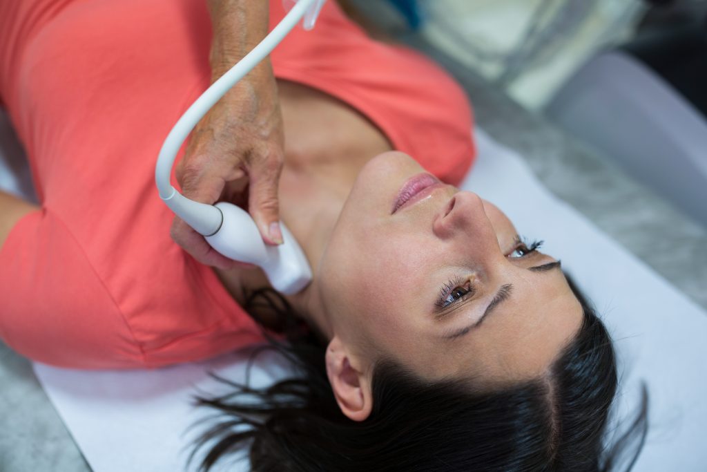
Understanding thyroid imaging is key for accurate diagnosis and treatment. It includes different methods, each with its own strengths and uses.
Common Thyroid Imaging Modalities
Thyroid imaging uses ultrasound, radioiodine scans, CT, MRI, and PET scans. Ultrasound is often the first choice because it’s non-invasive and shows detailed images of thyroid nodules. Radioiodine scans help check thyroid function and find thyroid cancer metastases.
| Modality | Primary Use | Key Benefits |
| Ultrasound | Nodule detection and characterization | Non-invasive, detailed images |
| Radioiodine Scan | Thyroid function assessment, cancer metastasis detection | Functional information, sensitive for metastases |
| CT/MRI | Evaluation of large goiters, substernal extension | Detailed anatomy, soft tissue characterization |
When Physicians Order Thyroid Imaging
Doctors order thyroid imaging for many reasons. This includes checking thyroid nodules, suspected thyroid cancer, and hyperthyroidism. The choice of imaging depends on the clinical question and patient factors.
For example, a patient with a noticeable thyroid nodule might get an ultrasound. This helps check the nodule’s characteristics and decide if a fine-needle aspiration is needed.
Thyroid Nodules: The Primary Reason for Thyroid Imaging
Thyroid imaging is often started because of nodules found. These nodules can be harmless or cancerous. Checking them is key to spotting cancer early.
Prevalence and Detection of Thyroid Nodules
Many people have thyroid nodules, and more women than men do. They are often found by accident during tests or exams.
Risk Stratification of Nodules
Not every nodule is at the same risk for cancer. Doctors look at several things to figure out the risk. Features seen on ultrasound help decide if a nodule is high risk.
Imaging Protocols for Nodule Evaluation
Ultrasound is the main tool for checking thyroid nodules. It shows details about the nodule’s size and what it’s made of. Sometimes, CT or MRI scans are used too, like for looking at deeper areas or lymph nodes.
| Imaging Modality | Primary Use | Advantages |
| Ultrasound | Initial evaluation of thyroid nodules | High resolution, non-invasive |
| CT Scan | Assessing substernal extension or lymph node involvement | Detailed cross-sectional imaging |
| MRI | Evaluating soft tissue involvement or when ultrasound is inconclusive | Excellent soft tissue differentiation |
Checking thyroid nodules involves looking at symptoms, imaging, and sometimes a biopsy. Knowing how common they are, what risks they have, and the best imaging methods is key to making the right diagnosis and treatment plan.
Suspected Thyroid Cancer: Urgent Imaging Indications
When thyroid cancer is suspected, quick and precise imaging is key. This ensures a fast diagnosis and treatment. Thyroid cancer, though rare, needs immediate action to help patients.
Clinical Red Flags That Necessitate Imaging
Certain signs point to the need for thyroid imaging right away. These include:
- A palpable thyroid nodule or mass
- Hoarseness or voice changes
- Difficulty swallowing or breathing
- Family history of thyroid cancer
- Previous radiation exposure
Anyone showing these symptoms should get a detailed check-up. This includes imaging to check for thyroid cancer.
Imaging Features Suggestive of Malignancy
Ultrasound, CT, and MRI are vital for spotting thyroid cancer signs. Key signs include:
- Nodule characteristics: Hypoechoic nodules with irregular margins and microcalcifications are suspicious for malignancy.
- Size and growth: Larger nodules or those showing significant growth over time warrant further investigation.
- Lymph node involvement: Enlarged or abnormal lymph nodes may indicate metastatic disease.
Spotting these signs is essential for diagnosing thyroid cancer. It helps guide the right treatment.
Hyperthyroidism Diagnosis Through Imaging
Imaging is key in diagnosing hyperthyroidism. It helps figure out the cause.
Hyperthyroidism means the thyroid gland makes too much thyroid hormone. Imaging helps find out why, like Graves’ disease or toxic nodular goiter.
Graves’ Disease Imaging Patterns
Graves’ disease is an autoimmune issue causing hyperthyroidism. Thyroid scans show a diffused increase in uptake. This is because autoantibodies stimulate the gland evenly.
“The thyroid gland in Graves’ disease is usually enlarged and has a characteristic ‘increased uptake’ pattern on scintigraphy.” – Endocrinology Expert
Toxic Nodular Goiter Identification
Toxic nodular goiter has nodules that make thyroid hormones on their own. Scans show a heterogeneous uptake pattern. This means areas of high uptake match the toxic nodules.
Distinguishing Between Hyperthyroid Causes
Graves’ disease and toxic nodular goiter are different based on scan patterns. Graves’ shows a uniform increase. Toxic nodular goiter has a patchy or nodular uptake pattern.
| Characteristics | Graves’ Disease | Toxic Nodular Goiter |
| Uptake Pattern | Diffuse, uniformly increased | Heterogeneous, patchy or nodular |
| Thyroid Gland Appearance | Enlarged, homogeneous | Nodular, heterogeneous |
Knowing these patterns is vital for correct diagnosis and treatment of hyperthyroidism.
Goiter Assessment and Management
Imaging is key in diagnosing and managing goiter, a thyroid gland issue. It’s vital to accurately assess goiter to choose the right treatment.
Diffuse vs. Multinodular Goiter Imaging
Goiter can be either diffuse or multinodular, based on imaging. Diffuse goiter means the thyroid gland is evenly enlarged. On the other hand, multinodular goiter has many nodules in the gland. Ultrasound and CT scans help tell these apart.
- Ultrasound is great for checking thyroid gland shape and finding nodules.
- CT scans show how big the goiter is and its effect on nearby areas.
Substernal Goiter Evaluation Techniques
Substernal goiter extends below the chest’s top. CT scans are best for this because they give clear images of the gland’s size and its position.
The right imaging choice depends on the goiter’s symptoms and size. Good imaging is key for planning surgery if needed.
Thyroiditis Detection and Characterization
Imaging techniques are key in spotting and figuring out different thyroiditis types. Thyroiditis is when the thyroid gland gets inflamed. This can happen for many reasons, like autoimmune diseases, infections, or radiation.
Acute and Subacute Thyroiditis Imaging
Acute thyroiditis starts suddenly because of infections. Ultrasound might show an enlarged thyroid with mixed textures. Subacute thyroiditis, or De Quervain thyroiditis, causes pain and high inflammation markers. It looks like a hypoechoic thyroid gland with less blood flow.
Hashimoto’s and Silent Thyroiditis Patterns
Hashimoto’s thyroiditis is an autoimmune disease causing long-term inflammation. It shows a diffusely hypoechoic thyroid gland with more blood flow on Doppler ultrasound. Silent thyroiditis looks similar but doesn’t cause pain. Using imaging to diagnose thyroiditis helps doctors make better treatment plans.
Radioiodine Uptake Test Methodology
The radioiodine uptake test is key for checking thyroid health. It sees how much radioactive iodine the thyroid takes in. This helps doctors spot and treat thyroid problems.
4 Hour Uptake Procedure and Significance
The 4-hour uptake test is a first look at thyroid health. A small amount of radioactive iodine is given. Then, after 4 hours, a gamma counter measures how much is taken up.
This test quickly shows how active the thyroid is. It helps doctors figure out if you have too much thyroid hormone.
24 Hour Uptake Protocol and Interpretation
The 24-hour uptake test gives a detailed look at thyroid health. It measures iodine uptake 24 hours after it’s given.
When looking at 24-hour uptake results, doctors check the percentage of iodine the thyroid takes in. A high uptake might mean Graves’ disease or toxic nodular goiter. A low uptake could point to thyroiditis or hypothyroidism.
Knowing when to do the thyroid uptake timing is key for right diagnosis. Both the 4-hour and 24-hour tests help doctors understand thyroid function and decide on treatment.
Thyroid Scan Procedure: Patient Experience
When you’re set for a thyroid scan, knowing what to expect can help calm your nerves. A thyroid scan is a test that checks how well your thyroid works and spots any issues.
Pre-scan Preparation Requirements
Before your thyroid scan, there are a few things to do. Patients are usually told to stay away from foods and meds with iodine to get accurate results. You should also wear comfy clothes and no jewelry that might get in the way. Always check with your doctor for any special instructions.
Here’s what you need to do before the scan:
- Avoid iodine-rich foods for a few days before the scan
- Inform your doctor about any medications or supplements you’re taking
- Wear comfortable, loose-fitting clothing
- Remove any jewelry that could interfere with the scan
Step-by-Step Procedure Walkthrough
The thyroid scan process has a few steps. First, a small amount of radioactive material is given through an IV. This material goes to the thyroid gland, letting the camera take clear pictures. The scan happens in a special room, and you’ll lie on a table while the camera moves around your neck.
Here’s what happens during the scan:
- Administration of the radioactive tracer
- Waiting period for the tracer to accumulate in the thyroid gland
- Positioning on the scan table
- Capturing images with a gamma camera
Result Turnaround Times for Different Thyroid Imaging Methods
How fast you get thyroid imaging results depends on the method used. This speed is key for patient care and treatment plans. Knowing these times helps doctors meet patient needs and make better choices.
Factors Affecting Scan Report Time
Many things can change how long it takes to get scan results. These include the imaging’s complexity, the radiology team’s workload, and the skill of those reading the images. Also, the technology and specific rules used play a role.
Key factors include:
- Complexity of the imaging procedure
- Radiology department workload
- Availability of skilled personnel
- Technology and protocols used
Typical Timeframes by Imaging Modality
Each imaging method has its own usual time for results. Ultrasound results are quick, often in a few hours. But CT or MRI scans take longer because they’re more complex and need detailed analysis.
| Imaging Modality | Typical Turnaround Time |
| Ultrasound | 2-4 hours |
| CT Scan | 4-24 hours |
| MRI | 4-48 hours |
Expedited Results in Urgent Scenarios
In emergencies like suspected thyroid cancer or acute thyroiditis, fast results are vital. Hospitals have plans to quickly handle these cases. Good communication between doctors and radiology is essential for quick results.
Understanding what affects scan report times and the usual times for each method helps doctors care for patients better. In emergencies, fast results can save lives. This shows how important quick communication and prioritizing is.
Interpreting Thyroid Uptake Percentage and Scan Results
Understanding thyroid uptake percentage and scan results is key to diagnosing thyroid issues. Thyroid scans show how well the thyroid is working. They help doctors spot where the thyroid is too active or too slow.
Normal Range Values and Variations
The normal range for thyroid uptake is between 10% and 30% at 24 hours. But, this can change due to iodine intake and some medicines. Knowing these changes is important for correct interpretation.
Hot Nodules vs. Cold Nodules: Clinical Significance
Thyroid nodules are called ‘hot’ or ‘cold’ based on how they take up radioactive iodine. Hot nodules work too well and are rarely cancerous. On the other hand, cold nodules don’t work well and might be cancerous. This helps doctors decide what to do next.
| Nodule Type | Uptake Characteristic | Malignancy Risk |
| Hot Nodule | Increased uptake | Low |
| Cold Nodule | Decreased uptake | Higher |
Pattern Recognition in Thyroid Disorders
Spotting patterns in thyroid scans is vital for diagnosing thyroid problems. For example, a big thyroid with high uptake might be Graves’ disease. A multinodular goiter shows up as multiple areas with different uptake levels.
Nuclear Medicine Reports for Thyroid Imaging
Getting a precise diagnosis and treatment plan depends on the details in nuclear medicine reports for thyroid imaging. These reports come from thyroid scan procedures. They give doctors the information they need to make smart choices.
Standard Components of a Thyroid Scan Report
A complete thyroid scan report has several important parts. These are:
- Patient Information: Basic details and medical history.
- Scan Details: The scan type and the radiopharmaceutical used.
- Findings: A detailed look at the thyroid gland’s appearance and any issues found.
- Interpretation: The radiologist’s analysis of the findings, matched with clinical data.
- Recommendations: Suggestions for what to do next or further tests.
Knowing these parts helps patients understand their diagnosis and treatment plan better.
How to Read and Understand Your Results
Understanding thyroid scan results needs a basic grasp of thyroid anatomy and function. Normal results mean the thyroid gland looks and works as it should. Abnormal results might show different thyroid sizes, nodules, or function changes.
A “hot nodule” shows a spot with more radioactive uptake, meaning it’s working too much. On the other hand, a “cold nodule” might be a non-working area, which could be harmless or cancerous.
Diagnostic Timeline: From Imaging to Treatment Decision
Knowing the diagnostic timeline is key for good thyroid care. It takes steps and teams from first imaging to treatment choice.
Typical Workflow in Thyroid Diagnosis
The thyroid diagnosis starts with a doctor’s check-up and imaging like ultrasound. Accurate imaging is key for making the next steps. The steps include:
- Clinical assessment and history taking
- Imaging studies (ultrasound, thyroid scan)
- Laboratory tests (thyroid function tests, fine-needle aspiration biopsy)
- Multidisciplinary team discussion for diagnosis and treatment planning
Factors That May Delay Diagnosis
Many things can slow down thyroid diagnosis and treatment. These include:
“Delays in diagnosis can lead to increased anxiety and uncertainty for patients, stressing the need for quick diagnostic paths.”
- Limited access to specialized care
- Complex cases needing more tests
- Patient issues like not following diagnostic advice
Knowing these delays can help make the diagnosis process faster. This ensures quicker treatment choices.
Ultrasound as the First-Line Imaging Tool
Ultrasound is a key tool in thyroid diagnostics. It’s chosen first because it gives clear images of the thyroid gland. Plus, it doesn’t use harmful radiation.
Ultrasound has many benefits. It’s a non-invasive way to check thyroid nodules. Doctors can see their size, number, and details. This helps decide if more tests are needed, like fine-needle aspiration.
Advantages of Thyroid Ultrasound
Ultrasound is safe and can be used on many patients. This includes those who are pregnant or have other health issues. It also lets doctors see the thyroid’s blood flow and guide needles for tests.
Ultrasound is fast and cheaper than MRI or CT scans. It’s a great first step for checking thyroid problems.
Ultrasound-Guided Fine Needle Aspiration
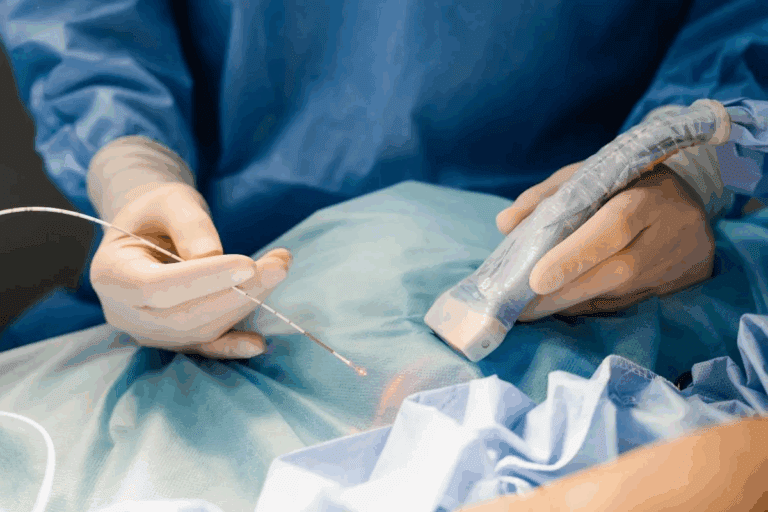
Ultrasound is great for guiding fine-needle aspiration (FNA) procedures. It makes sampling more accurate by showing the nodule in real-time. This is helpful for hard-to-reach or tricky-to-find nodules.
Using ultrasound and FNA together is key in diagnosing thyroid nodules. It gives doctors the info they need to decide on treatment.
Advanced Imaging Techniques: CT, MRI, and PET
Advanced imaging like CT, MRI, and PET is key in checking thyroid problems. They give detailed views of the thyroid’s shape and any issues. This helps doctors figure out what’s wrong and how to treat it.
When Advanced Imaging Is Necessary
Doctors often use advanced imaging when they think there might be complications. For example, CT scans help see if thyroid cancer has spread to lymph nodes. They’re also good for checking on goiters that are below the sternum.
Comparing Effectiveness Across Modalities
Each imaging method has its own benefits. CT scans show the thyroid’s shape and how it fits with other parts of the body. MRI is better at showing soft tissues, which is helpful for thyroid cancer checks. PET scans are great for spotting cancer cells because they show how active they are.
| Modality | Strengths | Common Uses |
| CT | Excellent anatomical detail | Substernal goiter, thyroid cancer staging |
| MRI | Superior soft tissue contrast | Thyroid cancer extent, soft tissue evaluation |
| PET | Metabolic activity assessment | Malignancy detection, cancer staging |
Special Populations and Thyroid Imaging Considerations
Thyroid imaging for special groups like kids and pregnant women is tricky. It needs careful planning to keep everyone safe and get accurate results.
Pediatric Protocols and Safety Measures
For kids, we adjust thyroid imaging to cut down on radiation. Ultrasound is often the best choice because it’s safe and doesn’t use harmful radiation. If CT scans are needed, we follow strict rules for kids.
| Imaging Modality | Pediatric Considerations | Safety Measures |
| Ultrasound | Preferred for its safety and efficacy | No radiation exposure |
| CT Scan | Use with caution; adjust dose | Use pediatric-specific protocols |
Pregnancy and Thyroid Imaging Options
Thyroid issues are big during pregnancy, and imaging is key. Ultrasound is the safest choice for checking thyroid health. If radioactive tests are needed, we plan them carefully to protect the baby.
Choosing the right imaging method and plan is vital for special groups. It helps meet diagnostic needs while keeping safety in mind.
Conclusion: Optimizing Thyroid Imaging for Accurate Diagnosis
Getting thyroid imaging right is key for diagnosing and treating thyroid issues. We’ve looked at the basics of thyroid imaging, its role in finding thyroid nodules and cancer, and the need for better imaging protocols.
Good thyroid imaging is vital for making accurate diagnoses. Knowing the strengths and weaknesses of imaging methods helps doctors choose the best tests for each patient. Making sure imaging protocols are top-notch is also important. This ensures images are clear, helping doctors plan the right treatment.
Modern imaging tools like ultrasound and nuclear medicine have changed thyroid imaging. Using these technologies well helps doctors give better care and avoid mistakes. Good thyroid imaging is essential for top-notch patient care.
FAQ
What is the typical turnaround time for thyroid scan results?
The time it takes to get thyroid scan results varies. It depends on the type of scan and the facility. Usually, you’ll get your results in a few hours to a few days. For urgent cases, you might get them faster.
How is the radioiodine uptake test performed?
For the radioiodine uptake test, you’ll get a small dose of radioactive iodine. Then, measurements are taken at 4 hours and/or 24 hours. This checks how well your thyroid is working.
What is the difference between a 4-hour and 24-hour uptake test?
The 4-hour test gives a quick look at thyroid iodine uptake. The 24-hour test shows more about thyroid function and iodine uptake over time.
How do I prepare for a thyroid scan?
Before a thyroid scan, avoid certain medicines and foods. Also, remove any jewelry or metal objects. They might affect the scan.
What is the significance of hot and cold nodules on a thyroid scan?
Hot nodules are usually benign and make too much thyroid hormone. Cold nodules don’t work well and might be cancerous.
How is thyroid uptake percentage interpreted?
Thyroid uptake percentage shows how much iodine your thyroid takes up. Normal ranges vary by lab and test.
What are the common imaging modalities used for thyroid assessment?
Common imaging methods include ultrasound, radioactive iodine scans, CT, MRI, and PET scans. Each has its own benefits and uses.
How long does it take to get results from a thyroid ultrasound?
Thyroid ultrasound results are usually quick. You might get them in a few hours or the same day.
What factors can delay thyroid diagnosis?
Delays in thyroid diagnosis can happen for several reasons. These include needing more tests, complex cases, or waiting for imaging or specialist appointments.
Are there special considerations for thyroid imaging in pregnant women?
Yes, pregnant women need special care for thyroid imaging. Avoid radioactive iodine scans and use safer alternatives when possible to protect the fetus.
How is thyroiditis detected using imaging?
Imaging like ultrasound and radioactive iodine scans can spot thyroiditis. They show specific patterns and changes in the thyroid gland.
What is the role of ultrasound-guided fine needle aspiration in thyroid assessment?
Ultrasound-guided fine needle aspiration is a procedure. It uses ultrasound to guide the sampling of thyroid nodules or lesions. This helps diagnose thyroid cancer or other conditions.


