Last Updated on November 27, 2025 by Bilal Hasdemir
When we talk about finding and treating medical issues, like cancer, two big tools are often used: PET scans and MRI scans. It’s important for both patients and doctors to know how these tools work and what they’re good for.
We’ll look at what makes these scans tick, how they’re used, and which one is better for finding and treating cancer. It gives a full breakdown of the differences.Compare MRI vs PET scan! Get 7 essential differences in their uses for cancer, technology, and what they reveal about the body.
Key Takeaways
- PET scans measure how active cells are, helping find diseases like cancer early.
- MRI scans use strong magnets and radio waves to show detailed body images.
- PET scans use a radioactive tracer, while MRI scans can be done with or without contrast.
- PET scans give patients a small dose of radiation, but MRI scans don’t.
- Choosing between PET scans and MRI scans depends on the medical issue and what the patient needs.
The Fundamentals of Medical Imaging
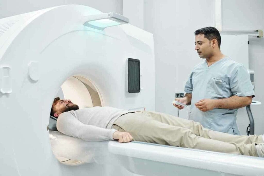
Medical imaging has changed how we diagnose diseases. It lets doctors see inside the body clearly. This has helped us understand and treat many health issues better.
Exploring medical imaging, we see how it has evolved. New technologies like PET scans and MRI scans have been developed. These tools help doctors see the body’s inner workings.
The Evolution of Diagnostic Technologies
Medical imaging has grown over time, with key milestones. Petroza et al. (2017) talked about how PET and MRI scans are used in medicine. Learn more about the differences between MRI and PET.
New imaging methods have made diagnosis more accurate. MRI shows soft tissues well, while PET scans reveal how the body works. This has opened up new areas in medicine.
| Imaging Modality | Primary Use | Key Benefits |
| MRI | Soft tissue visualization | High-resolution images, non-invasive |
| PET | Metabolic activity assessment | Early disease detection, functional insights |
The Importance of Selecting the Right Imaging Method
Choosing the right imaging method is key to good diagnosis and treatment. Doctors consider the patient’s condition, what they want to find, and each method’s strengths.
Understanding medical imaging and its tools helps doctors make better choices. This leads to better care for patients.
What Is an MRI Scan?
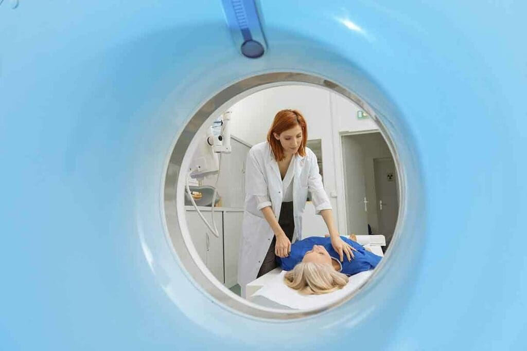
An MRI scan is a non-invasive way to see inside the body. It uses strong magnetic fields and radio waves. This method gives detailed images without surgery.
Basic Principles and Technology
MRI technology works by using nuclear magnetic resonance. When you have an MRI, you sit in a strong magnetic field. This field aligns hydrogen atoms in your body.
Then, radio waves disturb these atoms. The MRI machine picks up the signals. It uses these signals to make detailed images.
Key components of MRI technology include:
- A powerful magnet to align hydrogen atoms.
- Radiofrequency coils to transmit and receive signals.
- A computer system to reconstruct images from the received signals.
How Magnetic Resonance Imaging Works
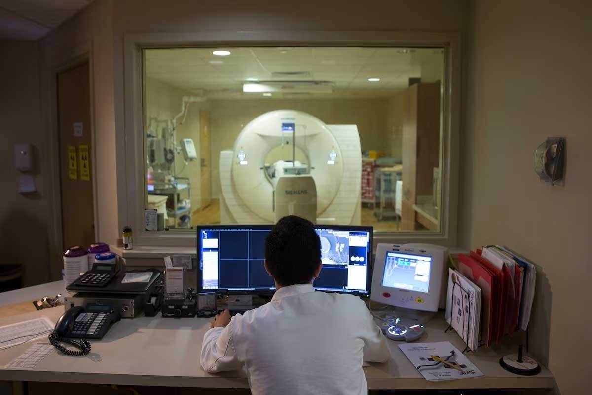
Getting an MRI involves several steps. First, you sit in the MRI scanner, a big machine. It has a strong magnet.
The scanner sends and receives radio waves. It captures signals from your body’s hydrogen atoms. Then, the MRI machine’s computer makes images of your body’s inside.
The MRI scan’s ability to distinguish between different types of soft tissues makes it very useful. It’s great for diagnosing conditions in the brain, spine, and joints.
Types of MRI Machines and Their Applications
There are many types of MRI machines. The most common is the closed MRI. It gives high-resolution images but can be scary for some.
Open MRI machines are more open. They can make people feel less anxious, but might not have the same quality as closed MRI images.
Special MRI machines, like functional MRI (fMRI), watch brain activity. They do this by seeing changes in blood flow. This makes fMRI very useful for brain research and diagnosis.
When we compare MRI scans to PET scans, we see they’re different. MRI shows detailed body structures. PET scans show how active the body’s cells are. So, MRI and PET scans are both important in medical diagnosis, but in different ways.
What Is a PET Scan?
Positron Emission Tomography (PET) is a top choice for diagnostic imaging. It shows how body tissues work by looking at their metabolic functions. This imaging test helps doctors see how different body parts function, focusing on metabolic activity.
Fundamentals of Positron Emission Tomography
PET scans use a radioactive drug, called a tracer, to highlight metabolic activity. This tracer goes to areas with high chemical activity, like growing cancer cells. This makes PET scans great for cancer diagnosis.
We use PET scans to check how body tissues and organs work. This is key to finding and managing diseases like cancer. Metabolic activity is a big clue for these diseases.
How Radioactive Tracers Function
The tracer in PET scans is like glucose, such as Fluorodeoxyglucose (FDG). Cancer cells, being more active than normal cells, take up more of this tracer. This makes them visible during the scan.
“The use of PET scans has revolutionized the field of oncology by providing a window into the metabolic activity of tumors, aiding in diagnosis, staging, and treatment monitoring.”
Researchers note.
The PET Scanner Technology and Process
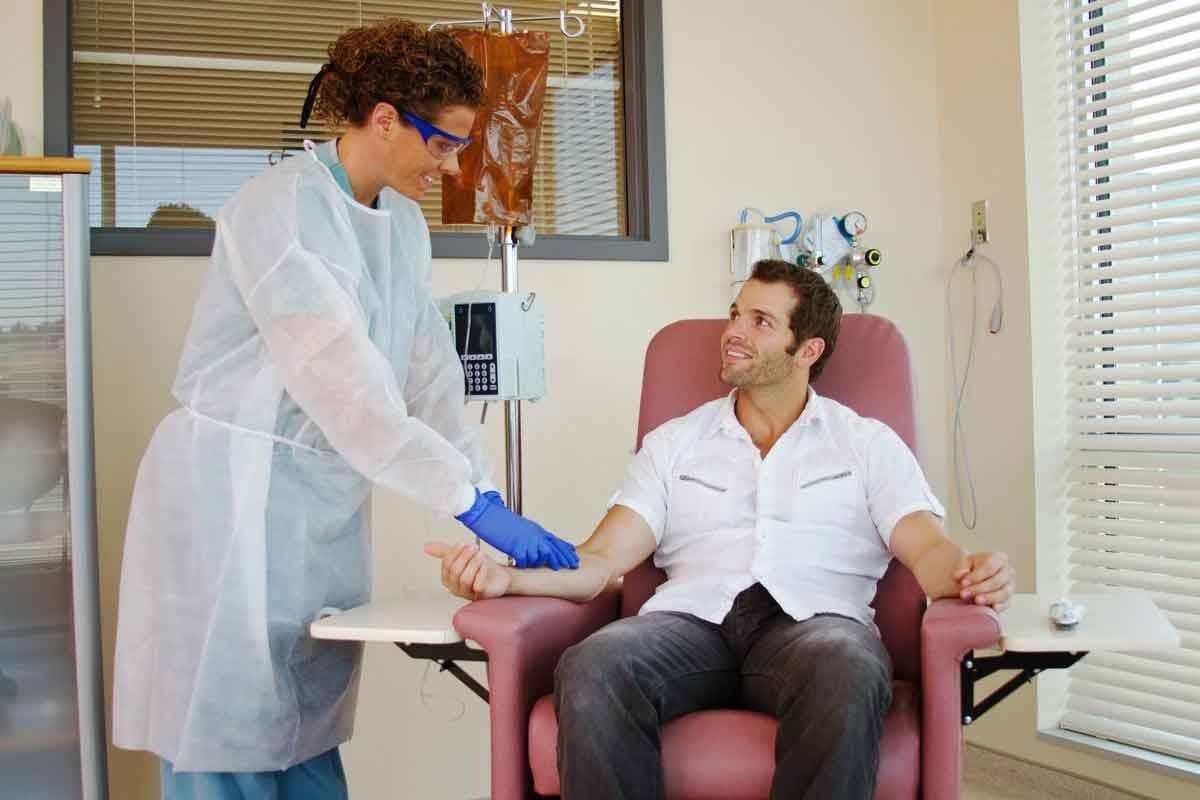
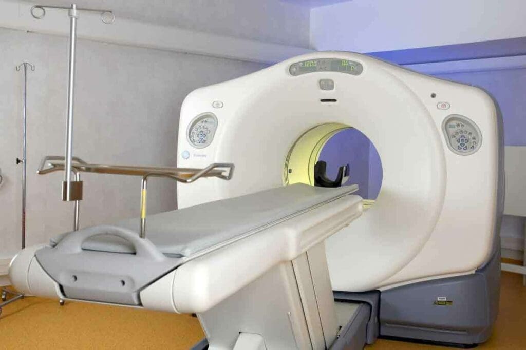
A PET scanner is a big, doughnut-shaped machine that picks up signals from the tracer. First, the tracer is injected into the patient. Then, the patient lies on a table that slides into the scanner. The scanner captures the tracer’s signals, and a computer makes images of metabolic activity.
| Aspect | Description |
| Tracer Injection | A radioactive tracer is injected into the patient. |
| Scanning Process | The patient lies on a table that slides into the PET scanner. |
| Image Reconstruction | A computer reconstructs images of the body’s metabolic activity based on the tracer’s signals. |
Understanding PET scans helps healthcare providers make better decisions for patient care. They can see the body’s metabolic functions clearly.
MRI vs PET: Underlying Technology Differences
MRI and PET scans are key tools in healthcare, but they work in different ways. Knowing how they differ helps us see their strengths and weaknesses.
Physics Behind Each Imaging Method
MRI uses nuclear magnetic resonance. It has a strong magnetic field and radio waves to show body structures. The magnetic field aligns hydrogen atoms, and radio waves disturb them, creating signals for images.
PET scans, on the other hand, detect gamma rays from a radioactive tracer in the body. This tracer goes to active areas like tumors. The PET scanner catches these gamma rays to show metabolic activity.
Image Generation Process Comparison
MRI images come from manipulating magnetic fields and radio waves. The quality depends on the magnetic field strength and radiofrequency coils.
PET images are based on the tracer’s distribution. The PET scanner catches gamma rays to show metabolic activity. The tracer type and scanner sensitivity affect image quality.
Technological Limitations of Each Method
MRI machines face limits like magnetic field strength and coil design. These affect image quality and detail.
PET scans are limited by tracer availability and scanner sensitivity. They might not show as much detail as an MRI.
Despite these limits, MRI and PET scans are essential in healthcare. They offer unique benefits that help in patient care. Understanding their differences helps healthcare providers use them wisely.
Key Difference #1: Anatomical vs Functional Imaging
MRI and PET scans are used for different things in medical care. MRI shows detailed pictures of body structures. PET scans look at how cells work, showing changes at the cellular level. Knowing these differences helps us understand their roles in treating patients.
Structural Visualization Capabilities of MRI
MRI is great at showing detailed pictures of the body’s inside. Doctors use it to see the brain, spine, joints, and more. It’s key to finding and treating many health issues.
For example, MRI is good at:
- Finding injuries like torn ligaments or cartilage
- Seeing lesions in the brain for conditions like multiple sclerosis
- Helping plan surgeries with clear images of the body’s parts
Metabolic Activity Detection by PET
PET scans, on the other hand, look at how body tissues work. They use special tracers to see how cells are working. This is very helpful in finding and tracking cancer.
PET scans are great for:
- Finding cancer and seeing how it responds to treatment
- Helping decide the best treatment by looking at tumor activity
- Finding unusual activity in the brain that might mean a neurological problem
Clinical Implications of This Fundamental Difference
The main difference between MRI and PET scans is important for doctors. MRI shows the body’s structure, while PET scans show how tissues work. This affects which scan is best for each situation.
In cancer care, MRI and PET scans work together. MRI shows the tumor’s shape, and PET scans show how active it is. This helps doctors understand the tumor’s behavior and how well it’s responding to treatment.
| Imaging Modality | Primary Use | Key Benefits |
| MRI | Anatomical Imaging | High-resolution images of structures, useful for diagnosing structural abnormalities |
| PET | Functional Imaging | Detection of metabolic activity, useful for assessing tissue function and identifying areas of abnormal activity |
Key Difference #2: Radiation Exposure and Safety Profiles
PET scans and MRI scans differ in how much radiation they use. This is important for both patients and doctors. Knowing these differences helps make better choices for medical tests.
Radiation Levels in PET Scanning
PET scans use radioactive tracers that give off radiation. The amount of radiation depends on the tracer and the dose. A PET scan’s radiation is usually between 2 to 7 millisieverts (mSv), similar to a CT scan.
“The use of PET scans requires careful consideration of the radiation dose to ensure that the benefits of the diagnostic information outweigh the risks,” experts say.
Factors influencing radiation exposure in PET scans include:
- The type and amount of radioactive tracer used
- The patient’s body size and composition
- The specific protocol followed for the scan
MRI’s Non-Ionizing Radiation Approach
MRI scans use a magnetic field and radio waves to create images. They don’t use ionizing radiation. This makes MRI safer for people who need many tests or are very sensitive to radiation, like pregnant women and kids.
“MRI is a valuable diagnostic tool that can provide critical information without exposing patients to ionizing radiation,” the American College of Radiology says.
The non-ionizing nature of MRI makes it safe for many patients. But it has its own safety issues, mainly because of the strong magnetic field.
Safety Considerations for Different Patient Populations
When choosing between PET scans and MRI, safety for different groups is key. Pregnant women and kids should avoid PET scans because of radiation. MRI is safer for them because it doesn’t use ionizing radiation.
Safety considerations include:
| Patient Group | PET Scan Considerations | MRI Considerations |
| Pregnant Women | Avoid unless abcessary due to radiation | Generally safe, but claustrophobia and contrast agents need consideration |
| Children | Used with caution, adjusting dose and tracer amount | Preferred due to lack of radiation, but may require sedation |
The right choice between PET and MRI scans depends on the patient’s needs and safety. Understanding the radiation and safety differences helps doctors make better decisions.
“The choice of imaging modality should be tailored to the individual patient, taking into account the diagnostic question, the patient’s condition, and the risks of the imaging technique.” – Expert in Diagnostic Imaging
Key Difference #3: Patient Experience and Procedure Requirements
When it comes to diagnostic imaging, PET and MRI scans have their own ways. Each scan has its own set of rules and things to consider. Knowing these differences helps manage patient expectations and makes the diagnostic process smoother.
Preparation Protocols for Each Scan
PET and MRI scans have different preparation steps. For a PET scan, patients usually need to fast for 4-6 hours before. This ensures the tracer works right. MRI scans don’t need fasting unless they use contrast agents. But patients must remove metal objects and may need to wear a gown for safety and image quality.
For PET scans, getting radioactive tracers is key. Patients might wait after getting the tracer before the scan. They might also drink a contrast agent to see the intestines better. Knowing the differences between PET, MRI, and other scans helps patients prepare better
Duration and Comfort Considerations
PET and MRI scans can last from 30 minutes to over an hour. MRI scans, in particular, can be long. They require patients to stay very quiet for a while, which can be hard for those with claustrophobia or discomfort.
Modern MRI machines have features like open designs to help with claustrophobia. Some places offer sedation or relaxation techniques to make patients more comfortable. PET scans are generally more comfortable, but patients need to stay very quiet to get good images.
Contraindications and Patient Restrictions
Both PET and MRI scans have things they can’t do with certain patients. MRI scans can’t be used on people with metal implants like pacemakers. Claustrophobia is also a big issue, but it can be managed.
PET scans involve a little radiation, so they’re not good for pregnant women unless it’s really needed. People with diabetes or on certain meds might need special prep for PET scans.
In summary, PET and MRI scans are both important for diagnosis, but they’re different. Understanding these differences helps healthcare providers prepare patients better. This makes the diagnostic process better for everyone involved.
Key Difference #4: Diagnostic Strengths and Clinical Applications
MRI and PET scans have different strengths for diagnosing diseases. Knowing these differences helps doctors choose the right test for each patient. This ensures the best care for each condition.
Conditions Best Diagnosed by MRI
MRI is great for soft tissues like the brain, spine, and muscles. It shows detailed images of these areas. This helps find injuries, infections, and tumors.
For example, MRI is key in diagnosing multiple sclerosis, herniated discs, and ligament sprains. It’s also used to check the blood flow in the body.
MRI is also used to look at the blood flow in the body. This is done through magnetic resonance angiography (MRA). It helps see how blood flows and find problems.
Conditions Best Diagnosed by PET
PET scans are best for finding metabolic activity in the body. They are very useful for cancer diagnosis and management. They show where cancer is and how it’s spreading.
PET scans help doctors see how well treatments are working. They are also used in neurology to diagnose and monitor Alzheimer’s disease. This is done by looking at glucose metabolism in the brain.
“PET scans have revolutionized the field of oncology by providing critical information about the metabolic activity of tumors, which is essential for developing effective treatment plans.”
As noted by an oncology imaging specialist.
Complementary Uses in Complex Diagnoses
Using MRI and PET scans together can give a clearer picture of a patient’s condition. This is important for complex cases. It shows both the structure and metabolic activity of the affected area.
| Condition | Preferred Imaging Modality | Complementary Imaging |
| Brain Tumor | MRI | PET for metabolic activity assessment |
| Cancer Staging | PET | MRI for detailed anatomical assessment |
| Musculoskeletal Injury | MRI | N/A |
Understanding MRI and PET scans helps doctors make better choices. This leads to better care and outcomes for patients.
PET Scan versus MRI for Cancer Detection and Monitoring
PET scans and MRI are key tools in fighting cancer. They serve different but important roles. The choice between them depends on the cancer type, its stage, and the needed information for diagnosis and treatment.
Early Cancer Detection Capabilities
Early cancer detection is key to effective treatment. PET scans are great at spotting metabolic changes in cancer cells early. MRI, on the other hand, gives detailed images of soft tissues and structural changes. MRI is better for seeing soft tissue tumors, but it might not catch cancer as early as PET scans.
“The use of PET scans in oncology has changed how we detect and manage cancer,” says a leading oncologist. “It offers functional information about tumors, which MRI’s detailed images can’t match.”
Tumor Characterization and Staging
After finding cancer, it’s important to understand the tumor and its stage. PET scans are great for this, showing how active tumors are. MRI gives detailed images of soft tissues, helping to see how big tumors are and where they are.
- PET scans show how active tumors are.
- MRI gives detailed images of soft tissues, aiding in local staging.
- Together, PET and MRI offer a complete view of the tumor, guiding treatment decisions.
Treatment Response Monitoring
It’s vital to check how well cancer responds to treatment. PET scans are excellent for this, spotting changes in tumor activity early. MRI can also monitor treatment response for tumors hard to see with other methods.
Effective treatment monitoring allows for timely therapy adjustments, which can improve patient outcomes. By combining PET’s functional info with MRI’s anatomical detail, doctors get a full picture of treatment response.
Key Difference #5: Accessibility, Cost, and Practical Considerations
It’s important to know the differences in accessibility, cost, and technology between PET and MRI scans. This helps make better choices in healthcare.
Comparative Costs and Insurance Coverage
PET scans are usually pricier because of the radioactive tracer. Insurance plans vary, sometimes covering one or both scans under specific conditions.
When looking at costs, consider:
- The price of the scan itself
- Any extra tests or procedures needed
- What insurance covers and what you’ll pay out-of-pocket
Insurance coverage for PET and MRI scans can be tricky. It often depends on whether the scan is medically necessary, as decided by a doctor.
Availability and Wait Times
PET scanners are less common than MRI machines, leading to longer waits. Wait times also depend on how busy scanning facilities are.
Important points to remember are:
- PET scanners are rarer, causing longer waits.
- How easy it is to get a scan can be critical in emergencies.
Technological Requirements for Healthcare Facilities
Healthcare places need specific tech to support PET and MRI scans. This includes setting up and keeping the equipment running, plus training staff.
Key tech points to think about are:
- The need for a cyclotron to make PET tracers
- High-field MRI machines for better images
- Keeping the equipment in good shape and checking quality
Understanding these practical aspects helps healthcare teams and patients make better choices about using PET and MRI scans.
Conclusion: Making the Right Choice Between PET and MRI
Choosing between a PET scan and an MRI depends on the situation and what you need to find out. We’ve looked at how each works, what they’re good at, and what they can’t do. This helps you pick the best tool for your health needs.
PET scans are great for seeing how diseases spread and finding cancers early. MRIs, on the other hand, show soft tissues well. They’re best for brain and muscle issues. For more on PET-CT vs MRI.
The right choice between PET and MRI depends on the patient’s health and other factors. Knowing the differences helps doctors and patients decide. The aim is to pick the best test for each person’s needs, ensuring they get the best care.
FAQ
What is the main difference between a PET scan and an MRI scan?
PET scans focus on how the body works, showing metabolic activity. MRI scans, on the other hand, show the body’s structure in detail.
Is a PET scan better than an MRI for cancer detection?
PET scans are great for finding cancer because they show metabolic activity. But whether to use a PET or an MRI scan depends on the situation.
What is the difference between a PET scan and an MRI in terms of radiation exposure?
PET scans use radioactive tracers, which emit radiation. MRI scans use non-ionizing radiation, making them safer for some, like pregnant women and kids.
How do I prepare for a PET scan versus an MRI scan?
For PET scans, you might need to fast or follow a diet. MRI scans require removing metal objects and possibly getting contrast agents.
Can I undergo both PET and MRI scans for cancer diagnosis?
Yes, you can have both scans to get a full picture of your health. MRI shows the body’s structure, while PET scans show metabolic activity.
What are the advantages of using PET scans over MRI for cancer monitoring?
PET scans are good at showing how well treatment is working. They’re useful for tracking cancer treatment progress.
Are there any contraindications for undergoing an MRI or PET scan?
Yes, some conditions, like claustrophobia, might prevent MRI scans. Certain metal implants or pregnancy also need special care. PET scans are not safe for pregnant women or breastfeeding mothers because of the radioactive tracers.
How do the costs of PET and MRI scans compare?
PET scans are usually more expensive than MRI scans. Costs depend on location, insurance, and the healthcare facility.
What is the difference between a PET scan and a PET/CT or PET/MRI scan?
PET/CT and PET/MRI scans combine PET’s metabolic imaging with CT or MRI’s structural imaging. This gives a more complete view of a patient’s health.
Can PET scans detect cancer at an early stage?
Yes, PET scans can spot cancer early by showing metabolic activity. This makes them valuable for early detection.
How do MRI and PET scans contribute to cancer staging?
MRI and PET scans help with cancer staging. MRI shows the body’s structure, and PET scans show metabolic activity, both indicating cancer spread.
Are PET scans and MRI scans available at all healthcare facilities?
No, not all places have PET and MRI scans. Availability depends on the facility’s technology and resources
References
- Mayerhoefer, M. E., Karanikas, G., Czernin, J., et al. (2020). PET/MRI versus PET/CT in oncology: a prospective, single-institution study. PubMed. https://pubmed.ncbi.nlm.nih.gov/31410538/
- Park, J., Hyun, S., Choi, Y., et al. (2020). Diagnostic accuracy and confidence of ^18F-FDG PET/MRI in comparison with PET or MRI alone in head and neck cancers. Scientific Reports. https:// www.nature.com/articles/s41598-020-66506-8


