Last Updated on November 27, 2025 by Bilal Hasdemir

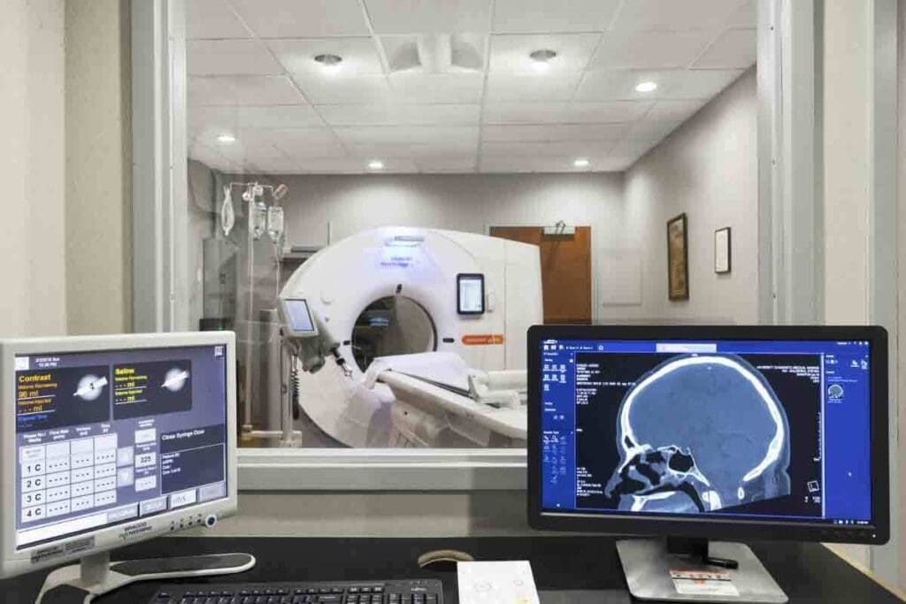
At LivHospital, we use myocardial SPECT imaging to check blood flow to the heart. This is key in finding coronary artery disease.
This method is non-invasive and common in myocardial perfusion imaging tests. It looks at the heart’s blood flow, mainly when stressed.
We focus on our patients and keep our methods up-to-date. This ensures our patients get the best care for heart diseases.
Key Takeaways
- Understanding the role of myocardial SPECT imaging in diagnosing coronary artery disease.
- Recognizing the importance of myocardial perfusion imaging tests in cardiac assessment.
- LivHospital’s commitment to patient-focused care and international medical standards.
- The non-invasive nature of myocardial SPECT imaging.
- The significance of evaluating blood flow to the heart muscle.
Understanding Myocardial SPECT Imaging Fundamentals

Learning about myocardial SPECT imaging is key to understanding MPI stress test results. Myocardial perfusion imaging (MPI) is vital for diagnosing and managing coronary artery disease (CAD).
What Is Myocardial SPECT Imaging?
Myocardial SPECT imaging is a nuclear medicine technique. It shows the heart’s function and structure. Small amounts of radioactive tracers, like Technetium-99m (Tc-99m) or Thallium-201 (Tl-201), are used. These tracers are taken up by the heart muscle based on blood flow.
The MPI stress test is often used with SPECT imaging. It checks how the heart responds to stress, usually through exercise or medicine. This test finds areas with less blood flow, which can mean CAD or other heart issues.
The Science Behind Nuclear Cardiac Imaging
Nuclear cardiac imaging, including SPECT, works on the idea that tracer distribution shows blood flow in the heart. By taking images of the heart under stress and at rest, doctors can see how blood flows. They can then spot any problems.
Key aspects of nuclear cardiac imaging include:
- Radiotracer uptake and distribution
- Image acquisition and processing
- Quantification of myocardial perfusion
Historical Development of SPECT in Cardiology
SPECT in cardiology has grown a lot over the years. It started with planar imaging and then got better with multi-head gamma cameras and new algorithms.
Now, SPECT imaging is a key tool for diagnosing and managing CAD. It’s very good at finding problems with blood flow.
| Aspect | Description | Clinical Significance |
| Radiotracer | Tc-99m or Tl-201 | Uptake reflects myocardial blood flow |
| Image Acquisition | Stress and rest imaging | Assesses perfusion under different conditions |
| Clinical Application | Diagnosis and management of CAD | Guides treatment decisions and risk stratification |
The Role of Myocardial Perfusion in Cardiac Health

Myocardial perfusion is key to keeping the heart healthy. It makes sure the heart muscle gets enough blood. Without it, the heart can’t work right.
Normal Myocardial Perfusion Patterns
In a healthy heart, blood flows evenly to all parts. This ensures the heart muscle gets the oxygen and nutrients it needs.
Healthy blood flow patterns are:
- Even uptake of radiotracers during imaging
- Blood flows the same across the heart muscle
- No areas with less or no blood flow
How Coronary Artery Disease Affects Heart Perfusion
Coronary artery disease (CAD) harms myocardial perfusion. CAD narrows or blocks coronary arteries, cutting off blood to the heart.
CAD’s effects on blood flow are:
| CAD Severity | Effect on Myocardial Perfusion |
| Mild Stenosis | Little impact on blood flow at rest; might cause stress-induced ischemia |
| Moderate Stenosis | Less blood flow during stress; might cause ischemia |
| Severe Stenosis or Occlusion | Big drop in blood flow at rest; high risk of heart attack |
Significance of Perfusion Abnormalities
Abnormal blood flow to the heart is a big warning sign. It can mean CAD, heart muscle disease, or other heart problems.
“Myocardial perfusion imaging is a critical tool in the diagnosis and management of coronary artery disease, allowing for the identification of areas of ischemia and infarction.” –
American Heart Association
Knowing about blood flow problems helps doctors:
- Find out how bad CAD is
- See the risk of heart problems later
- Make better treatment plans, like surgery
Myocardial perfusion imaging helps doctors make better choices for treatment. It’s useful for diagnosing and predicting heart problems. It helps with decisions about treatments and how well they work.
How MPI Stress Tests Evaluate Coronary Function
Myocardial perfusion imaging (MPI) stress tests are key in checking how well the heart’s blood flow works under stress. They help doctors find out if there’s any blockage in the heart’s arteries.
Exercise vs. Pharmacological Stress Testing
MPI stress tests can be done in two ways: through exercise or with medicine. Exercise stress tests use physical activity to make the heart work harder. This is best for people who can exercise.
For those who can’t exercise, pharmacological stress testing is used. This involves medicine like regadenoson, adenosine, and dipyridamole to make the heart think it’s working harder. This medicine makes the heart’s blood flow change, which MPI can detect.
Doctors choose between exercise and medicine stress tests based on the patient’s health and ability to exercise. Each method has its own benefits and is chosen for the best fit for each patient.
Physiological Changes During Stress Testing
Stress testing, whether through exercise or medicine, makes the heart rate and blood pressure go up. This means the heart needs more oxygen. In a healthy heart, blood flow increases to meet this need.
But in hearts with blocked arteries, blood flow can’t increase enough. This leads to changes that MPI can spot.
These changes help doctors find out if there’s heart disease. By comparing how the heart works at rest and under stress, MPI can find areas where blood flow is low. This could mean there’s a blockage or disease.
Interpreting Stress-Induced Perfusion Changes
When looking at MPI stress test results, doctors compare images from rest and stress. If an area of the heart takes up less radiotracer during stress but not at rest, it might be reversible ischemia. If it takes up less radiotracer both at rest and during stress, it could be scarred or infarcted.
Both rest and stress images help find ischemia. By comparing them, doctors can accurately diagnose heart disease. This helps them plan the best treatment for each patient.
- MPI stress tests evaluate coronary function by assessing blood flow under stress.
- Exercise and pharmacological stress testing are used based on patient capability and condition.
- Pharmacologic agents like regadenoson induce coronary vasodilation to simulate stress.
- Stress testing induces physiological changes that help diagnose coronary artery disease.
- Comparing rest and stress images helps identify reversible ischemia and scarred myocardium.
Radiotracers in Myocardial Perfusion Imaging
Radiotracers are key in heart perfusion studies through SPECT imaging. Myocardial perfusion scans help us see how the heart works and spot problems.
We use different radiotracers for top-notch images. Thallous chloride Tl-201 (201Tl) and technetium-based tracers like Tc-99m sestamibi and Tc-99m tetrofosmin are common.
Technetium-99m Based Agents
Technetium-99m agents are popular for their good physical traits and clear images. Tc-99m sestamibi and Tc-99m tetrofosmin are favorites. They help spot heart issues well.
These agents work for both rest and stress scans. Their stability and reliability make them a top choice in clinics.
Thallium-201 Applications
Thallium-201 has been used for heart imaging. It’s great for checking if heart muscle is alive. Tl-201 shows up in living heart muscle, making it good for viability studies.
But, Tl-201 has downsides. Its lower energy photons can cause more image problems than Tc-99m agents.
Comparing Radiotracer Effectiveness and Safety
When picking a radiotracer, we look at how well it works and if it’s safe. The table below shows key traits of common radiotracers.
| Radiotracer | Energy (keV) | Half-Life (hours) | Primary Use |
| Tc-99m Sestamibi | 140 | 6 | Perfusion Imaging |
| Tc-99m Tetrofosmin | 140 | 6 | Perfusion Imaging |
| Tl-201 | 69-83 | 73 | Viability, Perfusion Imaging |
The right radiotracer is key for heart perfusion studies and myocardial perfusion scans. Knowing each radiotracer’s strengths and weaknesses helps us improve patient care.
Rest vs. Stress Protocols in Myocardial Perfusion Studies
Choosing between rest, stress, or combined protocols is key in myocardial perfusion studies. These studies help diagnose and manage coronary artery disease (CAD). They show how well the heart’s blood flow changes under different conditions.
One-Day vs. Two-Day Imaging Protocols
Deciding between one-day and two-day imaging protocols depends on several factors. These include the patient’s health, the radiotracer used, and the clinical question. A one-day protocol does both rest and stress imaging on the same day. Rest imaging comes first, followed by stress imaging.
A two-day protocol separates these imaging sessions. This might improve image quality by reducing interference between rest and stress studies.
Table: Comparison of One-Day and Two-Day Imaging Protocols
| Protocol | Advantages | Disadvantages |
| One-Day | Convenience for patients, reduced overall study time | Potential for interference between rest and stress images |
| Two-Day | Improved image quality, reduced interference | Longer study duration, patient inconvenience |
When Rest-Only or Stress-Only Imaging Is Appropriate
Rest-only or stress-only imaging is used in certain cases. For example, a normal stress study can rule out significant CAD, making rest imaging unnecessary. On the other hand, rest-only imaging might be enough for patients at high risk of myocardial infarction. The choice depends on the patient’s specific situation and the doctor’s judgment.
Combined Protocol Benefits for Ischemia Detection
Combined rest and stress protocols are best for detecting ischemia. They allow doctors to see how blood flow changes between rest and stress. This helps identify ischemia more accurately. A study on PubMed Central shows these protocols improve CAD detection and provide important prognostic information.
In conclusion, the right protocol choice in myocardial perfusion studies is critical. It must match the patient’s needs. New technologies and protocols have made these studies more accurate and safe. Understanding the benefits and limitations of each protocol helps doctors provide better care.
Clinical Applications of Myocardial Perfusion Scans
Myocardial perfusion scans have many uses, from finding coronary artery disease to checking heart risk before surgery. They give vital info on heart function. This helps doctors make better choices for their patients.
Diagnosing Coronary Artery Disease
These scans are great for spotting coronary artery disease (CAD). They check blood flow to the heart muscle. If blood flow is low, it might mean blockages or narrow arteries.
Here’s what these scans can do:
- Spot patients with suspected CAD
- See how bad CAD is
- Help decide if more tests or treatments are needed
Risk Stratification After Cardiac Events
After a heart attack, these scans are key for figuring out risk. They look at damage and find areas that might not work well. This helps doctors know who’s at risk for more heart problems.
Here’s how they help with risk:
- Look at how bad the blood flow is
- Find out who’s at high risk for more heart issues
- Help make treatment plans that fit each patient
Preoperative Cardiac Risk Assessment
Before non-cardiac surgery, it’s important to check heart risk. Myocardial perfusion scans can spot who’s at higher risk. This helps doctors get patients ready for surgery and manage risks during it.
The good things about these scans for pre-surgery risk are:
- Find who needs to get ready before surgery
- Decide if more heart tests are needed
- Help plan how to keep heart risk low during surgery
Myocardial perfusion scans help a lot in managing heart disease. They help doctors make better plans for patients. As we learn more, it’s clear these scans are very important in cardiology today.
Viability Assessment Using Myocardial SPECT Imaging
Checking if heart muscle is alive is key for treating heart disease. Myocardial SPECT imaging helps by showing how well the heart works and gets blood.
Differentiating Viable from Non-Viable Myocardium
Myocardial perfusion imaging spots the difference between living and dead heart muscle. Living muscle can get better with the right treatment. Dead muscle, or scar tissue, won’t get better.
SPECT imaging helps doctors see how much living muscle there is. This helps decide if a patient needs surgery or other treatments.
Hibernating Myocardium Identification
Hibernating myocardium is heart muscle that’s not working well but can get better. SPECT imaging finds this by spotting areas that don’t get enough blood but can improve with treatment.
Implications for Revascularization Decisions
Knowing how much heart muscle is alive is very important for deciding on treatments. If a lot of muscle is alive, treatments like surgery can really help. This can make the heart work better and help patients live longer.
PET imaging is even better for some cases. It gives clearer pictures and helps measure blood flow to the heart. This is useful when you need to know exactly how well the heart is working.
Using SPECT imaging to check heart muscle helps doctors choose the best treatments. This leads to better care and outcomes for patients.
Technological Advances in Heart Perfusion Studies
New technologies are changing how we find and treat heart disease. These changes make heart scans more accurate and safe. This is key for heart health.
SPECT-CT Hybrid Systems
Hybrid systems mix SPECT and CT scans. This mix gives doctors both the heart’s function and its structure. It helps them make better diagnoses.
Benefits of SPECT-CT Hybrid Systems:
- Improved attenuation correction
- Enhanced anatomical localization
- Better diagnosis of complex cardiac conditions
A study showed that SPECT-CT systems cut down on unclear results by 50%.
Solid-State Detector Technology
New detector technology is boosting SPECT scans. Unlike old cameras, these detectors turn gamma rays into electrical signals. This makes scans clearer and more detailed.
Key benefits of these detectors include:
- Higher photon sensitivity
- Improved energy resolution
- Compact design allowing for more flexible imaging configurations
Software Innovations for Image Processing
New software is making heart scans better. It reduces noise and improves detail. This makes it easier to spot heart problems.
| Software Innovation | Description | Clinical Impact |
| Advanced Attenuation Correction | Algorithms that reduce artifacts caused by tissue attenuation | Improved specificity in diagnosing coronary artery disease |
| Resolution Recovery | Techniques that enhance image resolution | Better detection of small perfusion defects |
| Quantitative Analysis Tools | Software that provides objective measurements of perfusion and function | Enhanced risk stratification and treatment planning |
These advances, like SPECT-CT systems and better detectors, are improving heart scans. They help doctors diagnose and treat heart disease better. This might mean fewer invasive tests.
These technologies also work well with other tests, like PET scans. This gives doctors a full picture of the heart. For example, combining SPECT or PET with CT or MRI gives a detailed look at heart problems.
Comparing Myocardial Perfusion Testing Methods
There are many ways to check how well the heart is working. Myocardial perfusion imaging is the most common test used. We will look at the different methods, their good points, and what they can’t do.
SPECT vs. PET for Cardiac Assessment
Single Photon Emission Computed Tomography (SPECT) and Positron Emission Tomography (PET) are key in heart disease diagnosis. PET imaging has better detail and is more accurate than SPECT. It’s great for finding early heart disease and knowing how bad it is.
But, SPECT is more common because it’s cheaper and easier to find. It works well for checking how well the heart is working and if it’s alive. The choice between SPECT and PET depends on what the doctor needs to know, the patient’s situation, and what’s available.
Nuclear Imaging vs. Cardiac MRI
Nuclear imaging like SPECT and PET gives important info on heart blood flow and if it’s alive. Cardiac Magnetic Resonance Imaging (MRI) shows the heart’s shape and how it works without using radiation. Cardiac MRI is great for looking at the heart’s structure and how it moves.
- Nuclear imaging is better for checking blood flow and if the heart is alive.
- Cardiac MRI is best for looking at the heart’s shape and how it works.
- The right choice depends on what the doctor needs to know and the patient’s situation.
Complementary Role of Echocardiography
Echocardiography, or cardiac ultrasound, helps a lot in checking the heart. It shows how the heart is working right now, like how valves are doing and if the heart walls are moving right. It doesn’t check blood flow directly but is very useful for looking at how the heart is doing. It works well with nuclear imaging tests.
- Echocardiography is easy to get and doesn’t hurt.
- It’s good for looking at the heart’s structure and how it moves.
- Using echocardiography with nuclear imaging gives a full picture of the heart.
In summary, looking at different heart tests shows their good and bad sides. Knowing these helps doctors pick the best test for each patient. This makes diagnosing heart problems more accurate and helps patients get better care.
Patient Preparation and Safety Considerations
Keeping patients safe is key when doing myocardial perfusion imaging. This tool is great for checking heart health but needs careful thought. We must think about how to get accurate results and keep patients safe.
Pre-Test Instructions and Medication Management
Before a myocardial perfusion scan, patients get clear instructions. They learn about medication management, like what meds to stop or keep taking. It’s important to tell the doctor about all meds, including over-the-counter ones and supplements.
Patients also learn to avoid caffeine and certain foods that might mess with the test. Drinking plenty of water is encouraged to help the tracer spread right.
Radiation Exposure and Risk Minimization
One big safety issue is radiation exposure. We use the least amount of tracer needed for good images. We follow ALARA (As Low As Reasonably Achievable). Patients are told about radiation risks and how we lessen them.
We use new imaging tech and lower doses of tracer to cut down on radiation. Sometimes, we choose other imaging methods instead.
Managing Adverse Reactions to Stress Agents
Stress agents are used in myocardial perfusion imaging to mimic exercise. They’re usually safe but can cause adverse reactions in some. We watch patients closely and are ready to handle any problems quickly.
Common side effects are flushing, headaches, and dizziness. Rarely, more serious reactions can happen. We have plans for these situations too.
By preparing patients well and reducing risks, we make myocardial perfusion imaging safe and useful. It helps diagnose and manage heart issues effectively.
Conclusion: The Future of Myocardial Perfusion Imaging
Looking ahead, myocardial perfusion imaging will remain key in heart health checks. New tech makes mpi stress tests and myocardial spect imaging more accurate and safe. The use of SPECT-CT systems has greatly improved how we diagnose heart disease.
Myocardial spect imaging gives us vital info on heart blood flow. It helps doctors diagnose and manage heart disease better. With ongoing improvements in protocols and radiotracers, myocardial perfusion imaging will become even more useful in hospitals.
It’s important for healthcare workers to keep up with new developments in myocardial perfusion imaging. By doing so, we can offer better care and treatments for heart conditions. This helps improve patient outcomes and quality of life.
FAQ
What is myocardial SPECT imaging, and how does it work?
Myocardial SPECT imaging is a test that uses tiny amounts of radioactive material. It creates images of the heart. A radiotracer is injected into the blood, which the heart muscle absorbs. This lets doctors check blood flow and function.
What is the difference between a myocardial perfusion stress test and a regular stress test?
A myocardial perfusion stress test, or MPI stress test, uses nuclear medicine to check blood flow to the heart. It’s different from a regular stress test because it shows detailed images of the heart’s blood flow and function.
What are the benefits of using technetium-99m based agents in myocardial perfusion imaging?
Technetium-99m agents are popular in heart imaging because they provide high-quality images with low radiation. They accurately show blood flow and function in the heart.
How does coronary artery disease affect heart perfusion, and what are the implications for myocardial perfusion imaging?
Coronary artery disease reduces blood flow to the heart muscle. Myocardial perfusion imaging can spot these changes. This helps diagnose and manage coronary artery disease.
What is the difference between rest and stress protocols in myocardial perfusion studies?
Rest and stress protocols differ in when the imaging is done. Rest imaging is at rest, while stress imaging is after stress. The choice depends on the patient and the clinical question.
How do pharmacologic stress agents work, and what are their effects on coronary vasodilation?
Pharmacologic stress agents, like adenosine, widen blood vessels. This increases blood flow to the heart muscle. It helps detect perfusion problems.
What are the clinical applications of myocardial perfusion scans?
Myocardial perfusion scans help diagnose coronary artery disease and assess risk. They guide treatment and help plan surgeries. They’re key for making medical decisions.
How does myocardial SPECT imaging compare to other cardiac imaging modalities, such as PET and cardiac MRI?
Myocardial SPECT imaging is one of many heart imaging options. It’s widely available and relatively affordable. But, other methods like PET and MRI offer better sensitivity and detailed images.
What are the patient preparation and safety considerations for myocardial perfusion imaging?
Preparing for myocardial perfusion imaging includes following pre-test instructions and managing medications. It’s also important to minimize radiation exposure and handle stress agent reactions safely.
What is the role of radiotracers in myocardial perfusion imaging, and how are they used?
Radiotracers are key in myocardial perfusion imaging. They’re injected into the blood and absorbed by the heart. This lets doctors see blood flow and function.
What is viability assessment using myocardial SPECT imaging, and how is it used in clinical practice?
Viability assessment checks for living but dormant heart muscle. It helps decide if surgery is needed. This information improves treatment outcomes for heart disease patients.
What are the future directions for myocardial perfusion imaging, and how will it continue to evolve?
Myocardial perfusion imaging will get better with new technology. Advances include better radiotracers, SPECT-CT systems, and software. These will make imaging safer and more accurate.
References
- National Institute for Health and Care Excellence (NICE). (2016, November). Myocardial perfusion scintigraphy for the diagnosis and management of angina and myocardial infarction. NICE. https://www.nice.org.uk/guidance/dg17


