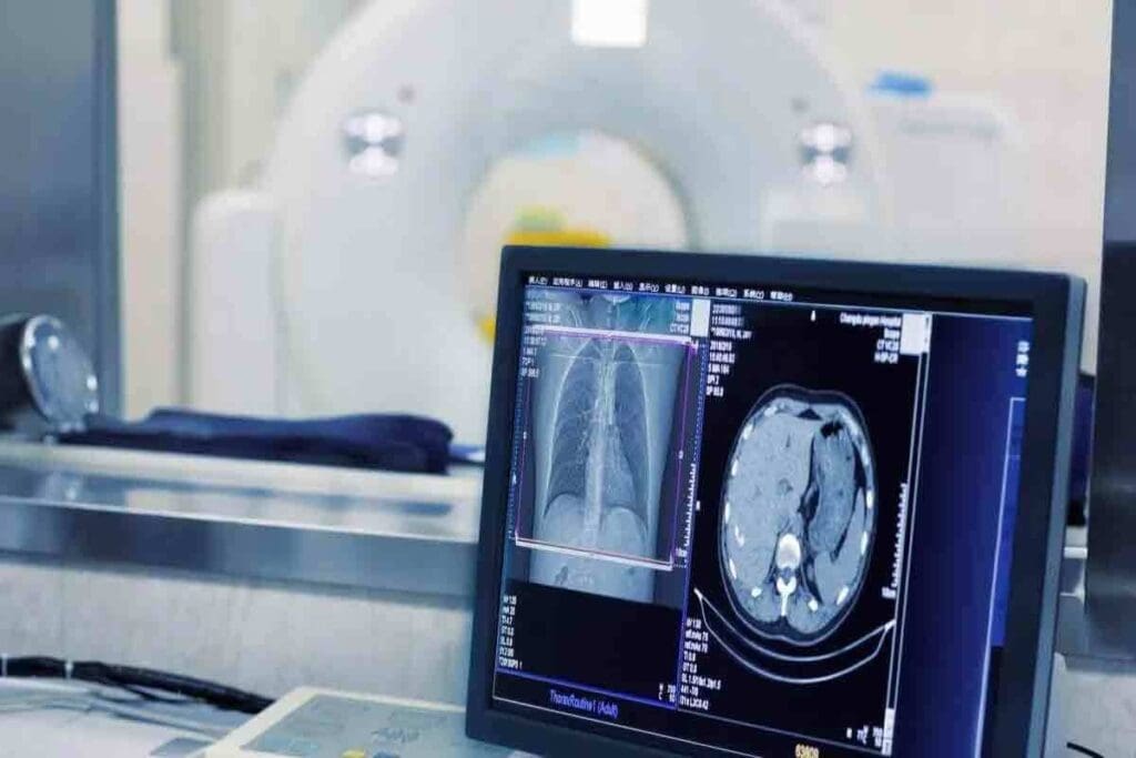
At Liv Hospital, we use advanced tests to check how well your kidneys work. The nm renal scan with Lasix is one of these tests. It helps us see how your kidneys handle urine and spot any problems.
The Lasix renal scan uses a tiny bit of radioactive material to see your kidneys. We give you Lasix, a medicine that helps us see how your kidneys work. This helps us find out if there are any blockages or issues.
Our team will help you through the 7 key steps of the scan. We make sure you’re comfortable and know what’s happening. We’ll also explain the results to you, so you understand your kidney health and what to do next.
Key Takeaways
- Understanding the purpose and significance of the nm renal scan with Lasix
- Preparation and procedure for the Lasix renal scan
- Interpretation of normal results and any possible issues
- The role of Lasix in checking kidney function
- The benefits of using advanced nuclear medicine tests at Liv Hospital
What is an nm Renal Scan with Lasix?
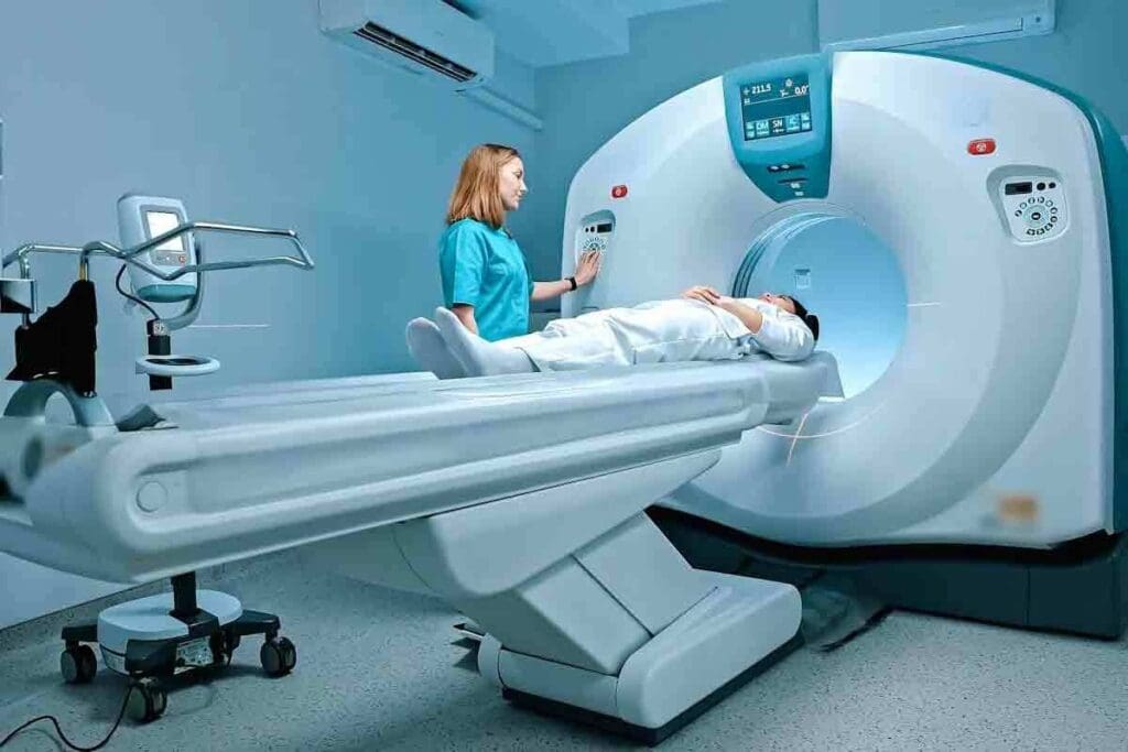
We use the nm renal scan with Lasix to understand kidney function and structure. This tool is key in nuclear medicine for checking kidney health and spotting problems.
Definition and Basic Principles
A nuclear medicine (nm) renal scan with Lasix is a test that looks at kidney function and shape. It uses tiny amounts of radioactive material. This material is injected into the blood and taken up by the kidneys, then excreted in urine.
The test uses a radiotracer that emits gamma rays. These rays are caught by a gamma camera, giving us images of the kidneys and how they work.
Lasix (furosemide) is added to the test to make it more useful. It makes the kidneys produce more urine. This helps us see how well the kidneys can handle the extra work, giving us important information about their health.
Difference Between Renal Scintigraphy and Renography
Renal scintigraphy and renography are often used together in nuclear medicine. But they’re not exactly the same. Renal scintigraphy is about imaging the kidneys with radioactive tracers, giving us both function and structure information. Renography, though, is about showing how the kidneys work over time, usually in a renogram curve.
- Renal scintigraphy gives us static and dynamic images of the kidneys.
- Renography focuses on the kidneys’ function, showing how they process the tracer over time.
Role in Kidney Function Assessment
The nm renal scan with Lasix is key for checking kidney function. It helps in:
- Checking how well the kidneys are working.
- Finding blockages in the urinary tract.
- Looking at how transplanted kidneys are doing.
- Telling the difference between blockages and other issues.
By giving us both function and structure info, the nm renal scan with Lasix is a top tool in nephrology and urology.
Clinical Indications for Lasix Renogram

A Lasix renogram is used for many kidney problems. It helps find blockages and check how well the kidneys work. This test is key for spotting issues and finding the right treatment.
Suspected Urinary Tract Obstruction
One main reason for a Lasix renogram is when there’s a blockage in the urinary tract. This blockage stops urine from flowing right. The test shows where and how bad the blockage is, helping doctors choose the best treatment.
Studies show it’s great for checking how serious the blockage is.
Evaluation of Hydronephrosis
Hydronephrosis means a kidney gets too big because of urine buildup. A Lasix renogram can tell if it’s because of a blockage or not. This helps doctors decide how to treat it.
It checks how well the kidney works and the flow of urine. This way, doctors can find the cause and fix it.
Assessment of Transplanted Kidneys
Lasix renography also checks on transplanted kidneys. It looks at how well the kidney is working and if there are any problems. This helps doctors make sure the transplant is okay and fix any issues fast.
Distinguishing Obstructive from Non-Obstructive Dilatation
Another big use of a Lasix renogram is to determine if a blockage is causing the problem or not. It shows how the kidney reacts to the diuretic. This helps doctors figure out what’s going on.
| Clinical Indication | Description | Diagnostic Utility of Lasix Renogram |
| Suspected Urinary Tract Obstruction | Blockage in the urinary tract | Diagnose the level and severity of obstruction |
| Evaluation of Hydronephrosis | Swelling of the kidney due to urine accumulation | Differentiates between obstructive and non-obstructive hydronephrosis |
| Assessment of Transplanted Kidneys | Monitoring kidney transplant function | Evaluates transplant function and detects complications |
| Distinguishing Obstructive from Non-Obstructive Dilatation | Dilatation of the urinary tract | Differentiates between obstructive and non-obstructive causes |
As said by an expert
“The Lasix renogram is a valuable diagnostic tool that provides critical information on kidney function and urinary tract dynamics, aiding in the management of various renal disorders.”
This shows how important Lasix renography is in medical care.
Preparation for Your nm Renal Scan with Lasix
Getting ready for your nm renal scan with Lasix is key. We’ll show you how to prepare. Being ready helps get accurate results and makes the test easier for you.
Pre-Procedure Instructions
Before your test, there are a few things to do:
- Tell your doctor about all medicines you take, including non-prescription ones and supplements.
- Let them know about any allergies, like medicines or contrast agents.
- Follow any special diet instructions from your healthcare team.
- Arrive a bit early to fill out any needed paperwork.
Hydration Requirements
Drinking lots of water is very important before your test. Drink plenty of water before your test to help your kidneys work well. This ensures clear images and accurate results.
Your doctor might give you specific water instructions. Make sure to follow them.
Medication Considerations
Some medicines can change how your test results come out. Tell your doctor about all medicines you’re taking. They’ll tell you if you should keep taking them, stop, or change them before the test.
It’s very important to mention any medicines that affect your kidneys or blood pressure.
What to Expect During the Test
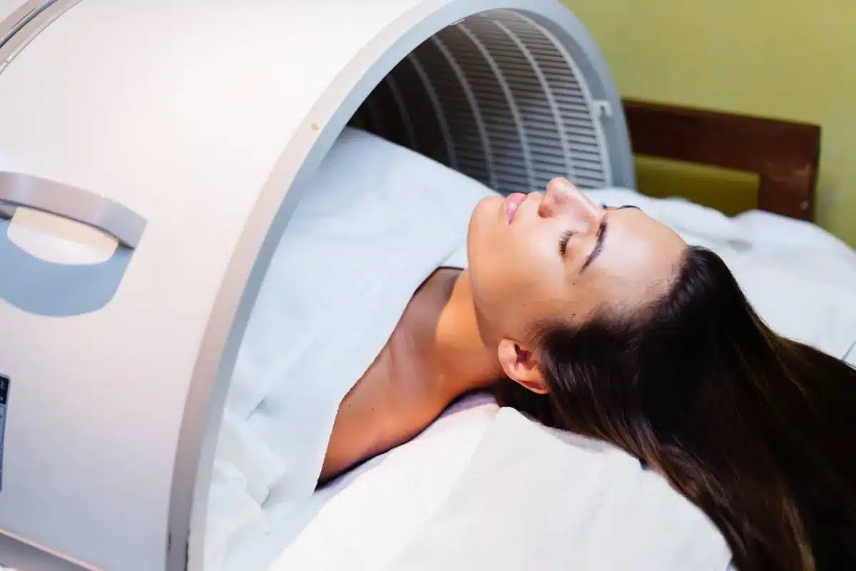
During the test, you’ll lie on a table. A radiotracer will be given to you through a vein. The scan will show how your kidneys work as the radiotracer moves through them. You might also get Lasix (furosemide) to see how your kidneys handle stress.
The test is usually easy to handle. Our medical team will be there to make sure you’re comfortable and safe.
The 7 Key Steps of an nm Renal Scan with Lasix
The nm renal scan with Lasix is a key test for checking kidney health. It helps find and treat many kidney problems.
Step 1: Patient Positioning and Initial Assessment
Getting the right images starts with how the patient is positioned. They lie on their back on a special table. A camera is placed over their kidneys. First, the team checks if everything is set up right and if there are any issues.
Step 2: Radiotracer Administration
Next, a radiotracer, a special dye, is given. This dye helps the camera see the kidneys and how well they work. The dye used is usually Technetium-99m DTPA or MAG3.
Step 3: Initial Dynamic Imaging
After the dye is given, the camera starts taking pictures. These pictures show how the dye moves through the kidneys. This helps understand renal blood flow, function, and drainage.
Step 4: Furosemide (Lasix) Administration
Furosemide, or Lasix, is then given to make the patient pee more. This helps tell if there’s a blockage in the urinary tract. The team watches how the kidneys react to the Lasix.
Step 5: Post-Furosemide Imaging
More pictures are taken after the Lasix is given. This shows how the kidneys handle the extra pee. It’s important to check how well the kidneys work.
Step 6: Data Analysis
The pictures and data are then looked at closely. This helps figure out how well the kidneys are working. It checks how the dye was taken up and how the kidneys reacted to the Lasix.
Step 7: Interpretation and Reporting
The last step is to understand what the results mean. Doctors look at the data to find out if there are any kidney problems. They then suggest what to do next.
Understanding DTPA Scan and Other Radiotracers
In nuclear medicine, different radiotracers help check kidney function. Technetium-99m DTPA is a common one. The right radiotracer depends on the medical question and what’s needed for diagnosis and treatment.
Technetium-99m DTPA: Properties and Applications
Technetium-99m DTPA is key for kidney imaging because of its good properties. It’s mainly filtered by the kidneys, making it great for checking glomerular filtration rate (GFR). Its quick removal and low secretion help show how well the kidneys work.
Alternative Radiotracers: MAG3 and DMSA
Other radiotracers like Technetium-99m MAG3 and Technetium-99m DMSA are also used for kidney imaging. Each has its own use.
Technetium-99m MAG3 is mainly secreted by the kidneys’ tubules. It’s best for patients with kidney problems. It gives clear images even when DTPA doesn’t work as well.
Technetium-99m DMSA is for static kidney images. It’s used to see renal cortical morphology and find pyelonephritis or renal scarring. It sticks to the kidney’s cortex, giving detailed views of the kidney tissue.
Selection Criteria for Different Radiotracers
Choosing a radiotracer depends on several things. These include the medical question, the patient’s condition, and what information is needed. Here’s a table that shows the main features and uses of the radiotracers we’ve talked about:
| Radiotracer | Primary Clearance Mechanism | Main Application |
| Technetium-99m DTPA | Glomerular Filtration | GFR Assessment, Dynamic Renal Imaging |
| Technetium-99m MAG3 | Tubular Secretion | Dynamic Renal Imaging, Even with Kidney Problems |
| Technetium-99m DMSA | Cortical Binding | Static Renal Imaging, For Kidney Shape and Damage |
DTPA Scanning Technique
The DTPA scan uses dynamic imaging after giving Technetium-99m DTPA. It takes quick pictures to see how the radiotracer moves through the kidneys. This helps check kidney function and calculate GFR.
Using Lasix (furosemide) during the scan helps tell if kidney problems are due to blockage or not. It checks how the kidneys react to the diuretic.
Normal Results of a Renal Scan with Lasix
A normal renal scan with Lasix shows quick tracer uptake, movement, and release. We’ll look at what makes results normal, like tracer patterns and renogram curves.
Expected Radiotracer Distribution Patterns
In a normal scan, the tracer spreads evenly in the kidney. It goes into the kidney tissue and then out into the collecting system. Prompt uptake and excretion show the kidney is working right.
A top nuclear medicine expert says, “A normal scan has even and quick tracer uptake in both kidneys. Then, it’s quickly excreted into the renal pelvis and drains well.”
“A normal renal scan shows symmetrical and prompt radiotracer uptake in both kidneys, followed by rapid excretion into the renal pelvis and subsequent drainage.”
Normal Renogram Curves
Renogram curves show the tracer’s activity over time. Normal curves quickly rise, peak, and then slowly fall as the tracer is released.
The curve has three parts:
- Phase 1: Quick tracer uptake
- Phase 2: Peak activity, showing maximum uptake
- Phase 3: Slow decline, as the tracer is released
Quantitative Parameters in Normal Studies
Several numbers help check if a scan is normal. These include:
| Parameter | Normal Value | Description |
| Tmax | 3-5 minutes | Time to maximum tracer uptake |
| T1/2 | Less than 10 minutes | Time for tracer activity to decrease by half after Lasix administration |
| Split Function | 45-55% | Relative function of each kidney |
What Constitutes a Normal Lasix Response
A normal Lasix response is a big increase in urine flow. This quickly removes the tracer from the renal pelvis. This is seen in the renogram curve as a sharp drop in activity after Lasix.
We’ve talked about what makes a renal scan with Lasix normal. This includes tracer patterns, renogram curves, and specific numbers. Knowing these is key to understanding scan results.
Renal Scan Results Interpretation
Understanding renal scan results means knowing about renogram patterns and their meanings. We’ll show you how to read time-activity curves and recognize different patterns.
Interpreting Time-Activity Curves (Renalgrams)
Time-activity curves, or renalgrams, show how a tracer moves through the kidneys. They tell us a lot about kidney function and how well they drain. We look at three main parts:
- Phase 1: This is when the tracer first goes into the kidneys, showing their blood flow and function.
- Phase 2: Here, the tracer builds up in the kidney tissue.
- Phase 3: This is when the tracer leaves the kidneys, showing how well they drain.
Any problems in these phases can point to kidney issues like blockages or poor function.
Obstructive Patterns
Obstructive patterns on a renogram show the tracer stays too long in the renal pelvis. Look for:
- A curve that goes up during the washout phase.
- It takes longer for the activity to reach its peak.
- The tracer takes a long time to wash out.
Non-Obstructive Dilatation Patterns
Non-obstructive dilatation patterns might show:
- Normal or slightly delayed washout.
- A good response to Lasix, meaning the dilatation isn’t due to a big blockage.
Telling apart obstructive and non-obstructive dilatation is key to the right treatment.
Abnormal Renogram Patterns
There are many abnormal renogram patterns, like:
| Pattern | Description |
| Poor renal function | Less initial uptake means the kidneys aren’t working well. |
| Severe obstruction | The tracer stays a lot longer with little Lasix response. |
Knowing these patterns helps us diagnose and treat kidney problems better.
Clinical Applications and Case Studies
The nm Renal Scan with Lasix is used in many ways. It helps check for urinary tract blockages and keeps an eye on kidney transplants. This tool gives doctors important information to help manage patient care.
Evaluation of Suspected Obstruction
This scan is great for finding urinary tract blockages. It looks at how well the kidneys work and if they’re draining properly. For example, it can tell if a blockage is causing kidney swelling.
A 45-year-old man had pain and thought he might have a blockage. The scan showed he did, and doctors fixed it. After surgery, the scan showed his kidneys were working better.
Post-Surgical Assessment
After surgeries like pyeloplasty, the scan checks if the blockage is gone. It shows if the kidneys are working better.
A 30-year-old man had pyeloplasty surgery. The scan after showed his kidneys were draining better, meaning the surgery was a success.
Renal Transplant Monitoring
People with kidney transplants get this scan to check on their new kidney. It helps find problems early, like blockages or damage to the kidney cells.
A 25-year-old with a kidney transplant had rising levels of waste in his blood. The scan showed his kidney wasn’t working as well, but it found no blockages. This helped doctors adjust his treatment.
Pediatric Applications
In kids, the scan helps find and treat problems like blockages and reflux. It’s good because it doesn’t need to be invasive.
A 6-year-old was checked for a blockage. The scan found one, and surgery fixed it. Later scans showed his kidneys were working better, proving the scan’s value.
Limitations, Risks, and Considerations
The nm renal scan with Lasix is a useful tool for doctors. But it has its own limits and risks. It’s important to know these to make smart choices.
Radiation Exposure
The test uses a small amount of radioactive material. This means patients get a little radiation. Even though the dose is safe, it’s key to think about the benefits and risks. This is true for pregnant women and kids. We try to use the least amount of radiation needed.
Contraindications
Some people should not get this test. Pregnant or breastfeeding women should tell their doctor about the radiation. Also, those allergic to the radiotracer or Lasix should let their doctor know. If you have kidney problems, it might not work well.
Factors Affecting Test Accuracy
Many things can change how accurate the test is. How well you’re hydrated, what meds you take, and when you get Lasix matter. Following the prep instructions is key for good results. We also look at your health and any other conditions.
Alternative Diagnostic Procedures
There are other tests you could have instead. Like ultrasound, CT scans, or MRI. The right test depends on what you need to know and your situation. We help decide the best test with you and your doctor.
Conclusion
We’ve looked into the nm renal scan with Lasix, a key tool for checking kidney health and finding blockages in the urinary tract. This test includes steps like setting up the patient, giving a radiotracer, and using Lasix.
This scan is very important in medical care. It helps doctors see how well the kidneys are working and spot any blockages. Knowing how to read these results helps doctors take better care of their patients.
To wrap it up, the nm renal scan with Lasix is essential for understanding kidney function. It’s used in many ways, like checking for blockages or watching over kidney transplants. This tool is vital for top-notch healthcare, helping us support patients worldwide.
FAQ
What is an nm renal scan with Lasix?
An nm renal scan with Lasix is a test that checks how well your kidneys work. It also looks for blockages in the urinary tract. It’s called a Lasix renogram or renal scintigraphy with furosemide.
What is the purpose of using Lasix in a renal scan?
Lasix helps in a renal scan by making more urine. This helps doctors see if there’s a blockage in the urinary tract. It shows how the kidneys respond to the increased urine production.
How should I prepare for an nm renal scan with Lasix?
To prepare, drink lots of water and avoid some medicines. Your doctor will give you specific instructions. This helps ensure the test works well.
What radiotracers are used in an nm renal scan with Lasix?
Technetium-99m DTPA and Technetium-99m MAG3 are common radiotracers. The choice depends on the patient’s needs and the question being asked.
What are the normal results of a renal scan with Lasix?
Normal results show both kidneys take up the radiotracer evenly. They also drain well after Lasix. The kidney function tests are within the normal range.
How is a Lasix renogram interpreted?
Doctors look at the time-activity curves and how the radiotracer moves. They check how the kidneys respond to Lasix. This helps find out if there’s a blockage or other issues.
What are the clinical applications of an nm renal scan with Lasix?
It’s used to check for blockages in the urinary tract. It helps see if there’s swelling in the kidneys. It’s also used for kidney transplants and to check kidney function in different situations.
Are there any risks or limitations associated with an nm renal scan with Lasix?
There’s a small risk of radiation exposure. Some people might have an allergic reaction to the radiotracer or Lasix. It’s not for everyone, and some things can make the test less accurate.
How does an nm renal scan with Lasix compare to other diagnostic tests?
It gives detailed information about how well the kidneys drain and if there’s a blockage. This is something other tests, like ultrasound or CT scans, can’t do. It’s very useful in certain cases.
What is the role of DTPA scanning in renal function assessment?
DTPA scanning helps check how well the kidneys work, focusing on the glomerular filtration rate (GFR). It also looks at kidney perfusion and drainage. This is key for diagnosing and managing kidney problems.
Can a renal scan with Lasix be used in pediatric patients?
Yes, it can be used in kids to check their kidney function and find urinary tract blockages. The test is adjusted for the child’s size and age.
What is renography, and how does it relate to an nm renal scan with Lasix?
Renography shows how kidney function changes over time during a nuclear medicine scan. An nm renal scan with Lasix includes renography to look at kidney function and drainage.
How does a Lasix renogram help in distinguishing obstructive from non-obstructive dilatation?
A Lasix renogram helps tell if swelling in the urinary tract is due to a blockage or not. It does this by seeing how the kidneys react to Lasix. If there’s a blockage, the kidneys drain slowly.
References
- Taylor, A. T., Nally, J., Aurell, M., et al. (2018). SNMMI Procedure Standard / EANM Practice Guideline for Diuretic Renal Scintigraphy in Adults With Suspected Upper Urinary Tract Obstruction. Seminars in Nuclear Medicine, 48(4), S1–S35. https://pmc.ncbi.nlm.nih.gov/articles/PMC6020824/
- Banks, K. P. (2022). Diuretic Renal Scintigraphy Protocol Considerations. Journal of Nuclear Medicine Technology, 50(4), 309–315. https://tech.snmjournals.org/content/50/4/309


