
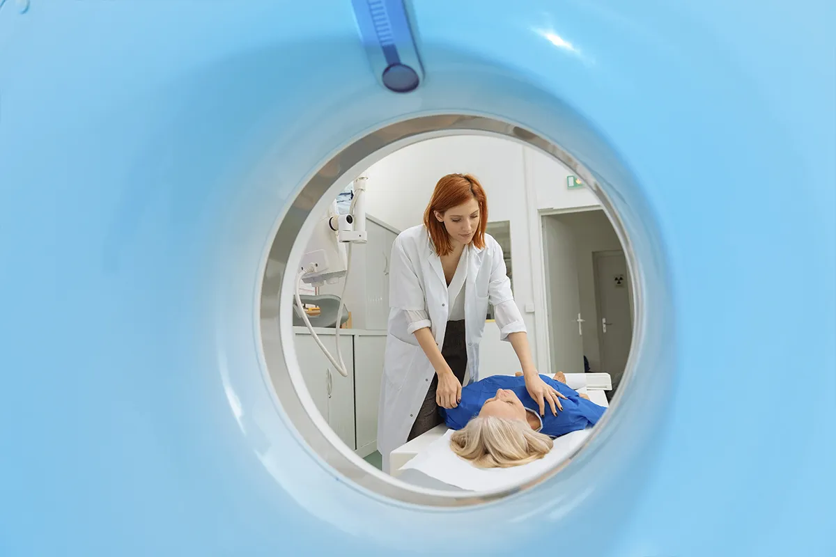
Medical imaging is key in finding and treating diseases. Radiology and nuclear medicine are two special areas that help us see inside the body. They use different methods to get images.
Radiology uses X-rays, CT scans, MRI, and ultrasound to see the body’s parts. It helps doctors find many health problems.
Nuclear medicine uses special drugs to see how organs work. It helps doctors understand how the body’s parts function.
Hospitals like Liv Hospital use both radiology and nuclear medicine. They work together to give the best care. This way, doctors can understand the body better.
Key Takeaways
- Radiology and nuclear medicine are distinct specialties in medical imaging.
- Radiology focuses on anatomical imaging using X-rays, CT, MRI, or ultrasound.
- Nuclear medicine employs radiopharmaceuticals to visualize organ function.
- Liv Hospital uses both specialties to deliver top-tier diagnostic care.
- The combination of radiology and nuclear medicine provides a more complete view of the body.
Understanding Medical Imaging Fundamentals

Advanced medical imaging technologies have greatly improved diagnosis. These tools are key in today’s healthcare, helping doctors see inside the body. This lets them diagnose diseases more accurately.
The Evolution of Diagnostic Imaging
Diagnostic imaging has changed a lot, starting with Wilhelm Conrad Röntgen’s X-ray discovery in 1895. Later, CT scans, MRI, and PET scans came along. These new tools help doctors find diseases sooner and more accurately.
An expert, has led in brain PET imaging. His work shows how vital new imaging methods are.
Role of Imaging in Modern Healthcare
Imaging is vital in today’s healthcare. It helps find diseases early and treat them better. Radiology and nuclear medicine give doctors important information about the body.
Using MRI and CT scans in medicine has made care better. These tools help track how diseases progress and if treatments work. This improves patient care a lot.
Radiology: An Overview

Radiology uses different methods to see inside the body. It’s key in finding and treating diseases. It uses X-rays, CT scans, MRI, and ultrasound to do this.
Definition and Scope of Radiology
Radiology is a medical field that uses imaging to diagnose and treat diseases. It covers many imaging types that show the body’s inside. Radiology is now a must-have in medicine, helping make treatment choices.
There are two main types of radiology. Diagnostic radiology uses X-rays, CT, MRI, and ultrasound to find diseases. Interventional radiology uses imaging for less invasive treatments.
Historical Development of Radiological Techniques
The first X-rays were discovered by Wilhelm Conrad Röntgen in 1895. This discovery led to many imaging tools, changing how we diagnose diseases. New technologies like CT and MRI have made diagnosis better over time.
Experts say radiology has grown a lot. Each new technology has opened up new ways to diagnose and treat. Ultrasound, for example, is a non-invasive way to see inside the body.
Yes, ultrasound is part of radiology. It’s used in radiology departments to help diagnose diseases.
Nuclear Medicine: An Overview
Nuclear medicine is at the cutting edge of medical science. It uses radiopharmaceuticals to see diseases at a cell level and treat them. This field has changed how we diagnose and treat diseases, combining both diagnostic and treatment options.
Definition and Scope of Nuclear Medicine
Nuclear medicine uses tiny amounts of radioactive materials, called radiopharmaceuticals. It helps see how organs work, diagnose, and treat diseases. Unlike other imaging, it focuses on how organs and tissues function, making it key in healthcare today.
This field covers a wide range, from diagnosing to treating diseases. It includes imaging like Positron Emission Tomography (PET) and Single Photon Emission Computed Tomography (SPECT). These help understand the body’s metabolic and physiological processes.
Historical Development of Nuclear Medicine
The history of nuclear medicine began in the early 20th century. The first radioactive isotopes were used for medical purposes back then. Over time, new technologies and radiopharmaceuticals have grown the field. The introduction of PET and SPECT imaging in the late 20th century was a big step forward.
As a leading expert noted, “The growth of nuclear medicine has been incredible. It has greatly improved our ability to diagnose and treat diseases with great accuracy.” This shows how much nuclear medicine has impacted healthcare.
Today, nuclear medicine keeps getting better. Ongoing research aims to create new radiopharmaceuticals and improve imaging. This progress will likely make nuclear medicine even more effective in treating diseases like cancer and neurological disorders.
Key Principles of Radiological Imaging
Understanding radiological imaging is key to its role in healthcare. It uses various technologies to help doctors diagnose and treat diseases.
X-ray Technology Fundamentals
X-ray technology is a core part of radiological imaging. It uses X-rays to show the inside of the body. First, an X-ray tube makes X-rays. Then, these X-rays go through the body and are caught by a detector, creating an image.
Key components of X-ray technology include:
- X-ray tube: Generates X-rays
- Collimator: Controls the direction and size of the X-ray beam
- Detector: Captures the X-rays that pass through the body
Keeping patients safe from radiation is very important. Using the least amount of X-rays and shielding sensitive areas helps reduce exposure.
Radiation Physics in Diagnostic Imaging
Radiation physics is key in imaging that uses ionizing radiation like X-rays and CT scans. Knowing how radiation affects tissue is vital. It helps make images better while keeping radiation low.
|
Radiation Type |
Interaction with Tissue |
Imaging Modality |
|---|---|---|
|
Ionizing Radiation |
Causes ionization, potentially damaging tissue |
X-ray, CT |
|
Non-ionizing Radiation |
Does not cause ionization, safer for tissue |
MRI, Ultrasound |
Understanding radiation physics helps improve imaging techniques. It ensures imaging is both effective and safe.
Keeping patients safe is a big deal in radiology. New tech keeps imaging safer and more effective.
Nuclear Imaging in Medicine: Principles and Mechanisms
Nuclear medicine imaging uses special substances to see and treat diseases at a molecular level. It’s a key part of modern healthcare, giving doctors new ways to diagnose and treat.
Radiopharmaceuticals: The Foundation of Nuclear Imaging
Radiopharmaceuticals are special substances with tiny amounts of radioactive material. They help doctors diagnose and treat diseases. These substances are made to go to specific parts of the body, making imaging and treatment more precise.
Making radiopharmaceuticals involves mixing a drug with a radioactive isotope. This mix lets doctors track the substance in the body. It gives them important info on how organs work and if there’s disease.
Key characteristics of radiopharmaceuticals include:
- Specificity for target tissues or organs
- Appropriate half-life for imaging or treatment
- Minimal toxicity and side effects
Emission and Detection Processes
Nuclear imaging works by catching the radiation from these substances in the body. The main methods are Single Photon Emission Computed Tomography (SPECT) and Positron Emission Tomography (PET).
In SPECT, the substance sends out gamma rays. A camera around the patient catches these rays. Then, it makes detailed images of the area being looked at.
PET imaging uses positrons that turn into gamma rays when they meet electrons. The PET scanner catches these rays. This gives clear images of how active certain parts of the body are.
The emission and detection processes in nuclear imaging offer several benefits, including:
- High sensitivity for detecting diseases at an early stage
- Ability to provide functional information about organs and tissues
- Guidance for targeted therapeutic interventions
Imaging Modalities in Radiology
Diagnostic imaging in radiology uses many methods, each with its own strengths. These methods help doctors see inside the body. This is key for making diagnoses and planning treatments.
X-ray Radiography
X-ray radiography is a top choice for imaging. It uses X-rays to show the body’s bones. X-ray technology is key for spotting bone problems like fractures and infections.
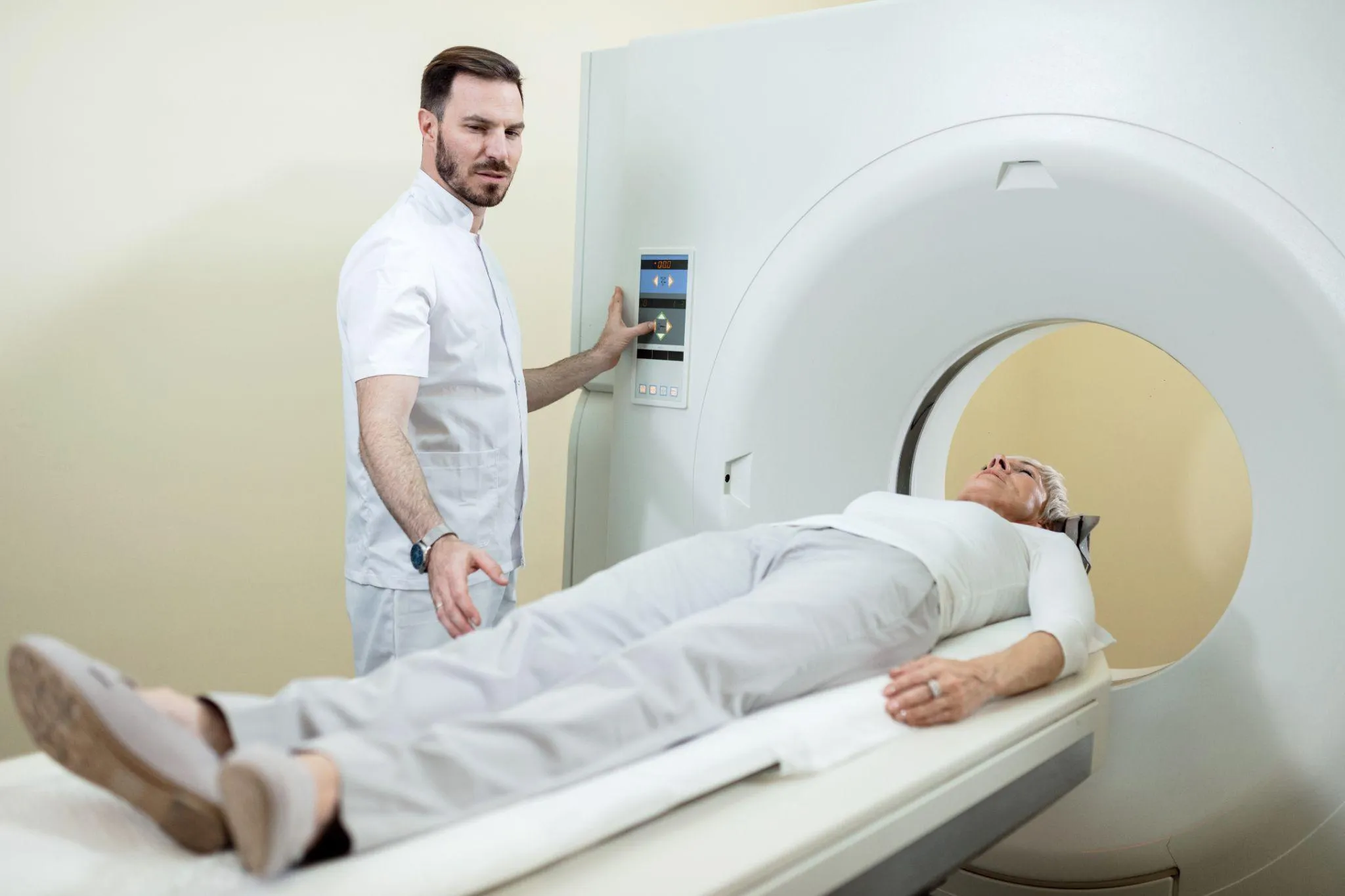
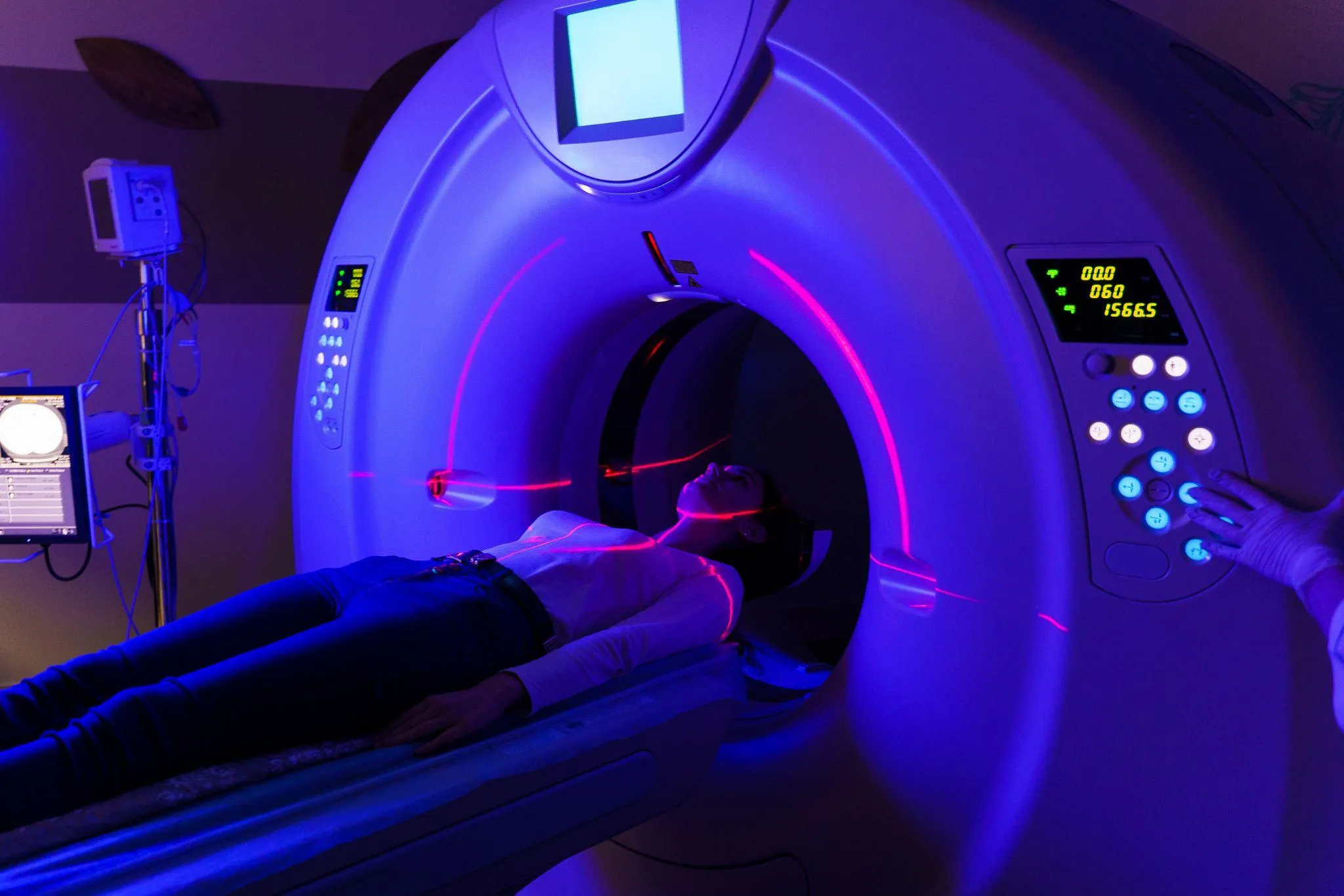
Computed Tomography (CT)
CT scans give detailed views of the body, better than X-rays. They’re great for emergency checks, like finding internal injuries. The high-resolution images from CT scans help doctors diagnose many conditions.
Magnetic Resonance Imaging (MRI)
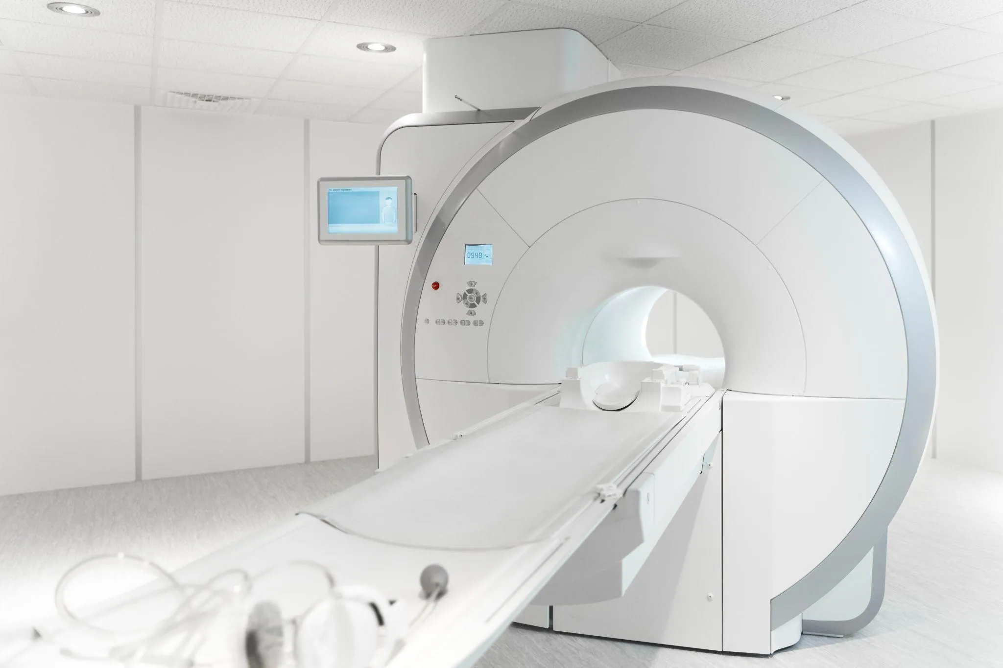
MRI uses strong magnetic fields and radio waves for detailed images. It’s best for soft tissues, like the brain and joints. MRI is very useful for many medical checks.
Ultrasound Imaging
Ultrasound uses sound waves to see inside the body. It’s often used for checking the womb and organs like the gallbladder. Ultrasound is safe and doesn’t hurt.
In summary, radiology has many imaging options. This lets doctors pick the best tool for each patient’s needs. It helps in making accurate diagnoses and treatments.
Imaging Modalities in Nuclear Medicine
Advanced imaging modalities are key in nuclear medicine. They help doctors diagnose and treat diseases well.
Single Photon Emission Computed Tomography (SPECT)
SPECT uses small amounts of radioactive tracers to create 3D images of the body. It’s great for checking the heart, bones, and some cancers.
The SPECT technology detects gamma rays from the tracer. This makes detailed images that help doctors a lot.
Positron Emission Tomography (PET)
PET is a top imaging modality in nuclear medicine. It’s very good at finding metabolic changes in the body. PET scans are used a lot in oncology, neurology, and cardiology.
The PET imaging process involves a tracer that goes to areas with high activity, like tumors. The PET scanner then shows detailed images of these areas.
Hybrid Imaging Systems (PET/CT, SPECT/CT)
Hybrid imaging systems mix the best of nuclear medicine with CT scans. This combo improves diagnosis and helps plan treatments.
PET/CT and SPECT/CT have changed nuclear medicine a lot. They give a full view of the body’s functions and structure. This leads to more accurate diagnoses and better treatments.
Anatomical vs. Functional Imaging
In medical imaging, anatomical and functional imaging have different roles. Knowing the difference helps doctors make accurate diagnoses and treatment plans.
How Radiology Visualizes Structure
Radiology mainly looks at the body’s internal structures. It uses X-rays, CT scans, and MRI to see organs, bones, and tissues. These images help doctors spot problems like fractures or tumors.
Key Features of Anatomical Imaging:
- High-resolution images of internal structures
- Ability to detect structural abnormalities
- Useful for diagnosing conditions related to anatomy
How Nuclear Medicine Visualizes Function
Nuclear medicine focuses on how the body’s organs work. It uses tiny amounts of radioactive tracers. This lets doctors see how different parts of the body function.
Key Features of Functional Imaging:
- Provides information on organ function and metabolism
- Helps in diagnosing and monitoring conditions affecting organ function
- Useful for assessing the metabolic activity of tumors
The table below shows the main differences between anatomical and functional imaging:
|
Characteristics |
Anatomical Imaging (Radiology) |
Functional Imaging (Nuclear Medicine) |
|---|---|---|
|
Primary Focus |
Structural details |
Organ function and metabolism |
|
Imaging Techniques |
X-ray, CT, MRI |
PET, SPECT |
|
Diagnostic Use |
Identifying structural abnormalities |
Assessing organ function and metabolic activity |
Both anatomical and functional imaging are key in modern medicine. They often work together to help doctors diagnose and treat patients. Understanding their roles helps healthcare providers make better decisions, leading to better patient care.
Clinical Applications of Radiology
Radiology has many uses in medicine, from imaging to procedures. It’s a key tool for doctors to find and treat many health issues.
Diagnostic Uses Across Body Systems
Radiology is key for diagnosing health problems in different parts of the body. Diagnostic imaging like X-rays, CT scans, MRI, and ultrasound help see inside the body. They help find issues like broken bones, tumors, and blood vessel problems.
In heart health, radiology checks how well the heart works and spots heart disease. It’s also important for brain health, helping find strokes and brain disorders. In cancer care, it helps track tumors and see how treatments are working.
Interventional Radiology Procedures
Interventional radiology uses images to guide small, non-surgical procedures. These can be for checking or treating problems. Examples include biopsies, angioplasties, and embolizations.
This field has changed how we treat many conditions. It’s less invasive than surgery, leading to quicker recovery, less scarring, and fewer complications.
Clinical Applications of Nuclear Medicine
Nuclear medicine is a key tool in healthcare. It lets doctors see and treat diseases at a molecular level. This makes it vital for diagnosing and treating many diseases.
Diagnostic Applications
Nuclear medicine is great for spotting problems in organs and tissues. Diagnostic applications include:
- Cancer detection and staging
- Cardiac function assessment
- Neurological disorder diagnosis
Positron Emission Tomography (PET) and Single Photon Emission Computed Tomography (SPECT) are top tools. They show how tissues work, helping catch diseases early.
Therapeutic Applications
Nuclear medicine also helps treat diseases. Therapeutic applications include using radioactive materials to target cancer cells. This is very promising.
Therapies in nuclear medicine aim at diseased cells, sparing healthy ones. This makes treatments more effective and safer.
Nuclear medicine is vital for both diagnosis and treatment. As research grows, it will play an even bigger role in healthcare. This brings hope to patients everywhere.
Radiation Safety Considerations
Keeping people safe from radiation is very important in radiology and nuclear medicine. These fields use ionizing radiation, which can harm health if not handled carefully.
Radiation Exposure in Radiology
Radiology uses imaging methods like X-rays and CT scans that involve ionizing radiation. The amount of radiation you get depends on the type of scan and the technology used. For example, a chest X-ray has a low dose, but a CT scan has a higher dose.
Factors influencing radiation exposure in radiology include:
- The type of imaging modality used
- The specific protocol followed for the imaging procedure
- The patient’s size and the area being imaged
Radiation Exposure in Nuclear Medicine
Nuclear medicine uses radiopharmaceuticals that give off radiation. The amount of radiation you get depends on the type and amount of radiopharmaceutical, and how your body processes it.
Key considerations for radiation exposure in nuclear medicine include:
- The half-life of the radiopharmaceutical
- The energy of the emitted radiation
- The biodistribution of the radiopharmaceutical in the body
Safety Protocols for Patients and Staff
To reduce radiation exposure, strict safety rules are followed in radiology and nuclear medicine. These rules help protect both patients and healthcare workers who often face radiation.
|
Safety Measure |
Description |
Benefit |
|---|---|---|
|
Personal Protective Equipment (PPE) |
Use of lead aprons, thyroid shields, and protective eyewear |
Reduces radiation exposure to staff and patients |
|
Radiation Monitoring |
Use of dosimeters to track radiation exposure |
Ensures that exposure limits are not exceeded |
|
Quality Control |
Regular maintenance and calibration of imaging equipment |
Ensures optimal performance and minimizes unnecessary exposure |
By following these safety rules and keeping up with new radiation safety info, healthcare teams can lower the risks of radiation exposure in radiology and nuclear medicine.
Technological Advancements and Future Directions
Technological innovations are changing radiological and nuclear medicine imaging. They are making diagnoses more accurate. These advancements improve image quality and reduce scan times.
Recent Innovations in Radiological Imaging
New technologies have entered radiological imaging. High-field MRI machines offer clearer images. Computed tomography (CT) technology now scans faster and uses less radiation.
Here are some recent innovations in radiological imaging and their benefits:
|
Innovation |
Benefits |
|---|---|
|
High-field MRI |
Higher resolution images, better tissue differentiation |
|
Advanced CT technology |
Faster scan times, reduced radiation exposure |
|
Artificial intelligence in image analysis |
Improved diagnostic accuracy, reduced interpretation time |
Emerging Technologies in Nuclear Medicine
Nuclear medicine is getting better thanks to new technologies. Hybrid imaging systems like PET/CT and SPECT/CT give both functional and anatomical info. New radiopharmaceuticals help target diseases more effectively.
Artificial Intelligence in Medical Imaging
Artificial intelligence (AI) is becoming key in medical imaging. AI helps analyze images and spot problems. It also aids in making diagnoses more accurate.
The future of radiological and nuclear medicine imaging is bright. Ongoing tech advancements will improve diagnostics and patient care. These technologies will shape the future of medical imaging.
Cost and Accessibility Factors
Medical imaging is changing fast. The cost and how easy it is to get these services are key. These factors affect healthcare choices a lot.
Comparative Costs of Imaging Modalities
Different imaging methods cost a lot. X-rays and ultrasounds are cheaper. But, MRI and CT scans are pricier.
|
Imaging Modality |
Average Cost Range |
|---|---|
|
X-ray |
$100-$500 |
|
Ultrasound |
$200-$1,000 |
|
CT Scan |
$500-$3,000 |
|
MRI |
$1,000-$5,000 |
|
PET Scan |
$1,500-$6,000 |
PET scans are very expensive. This is because of the cost of special drugs and the tech used.
Availability and Access to Imaging Services
Getting to imaging services is a big deal. Cities have more options, but rural areas struggle. They don’t have as many places to go.
Key factors influencing accessibility include:
- Geographical location
- Insurance coverage
- Availability of specialized centers
A healthcare expert said, “The difference in access to imaging between cities and rural areas is a big problem. We need to fix it for fair healthcare.”
To make things better, new tech and telemedicine are being used. They help bring healthcare to more people.
The Major Differences Between Radiology and Nuclear Medicine
It’s important to know how radiology and nuclear medicine differ. Both are key in healthcare for imaging the body. But they use different methods and technologies.
Technical and Methodological Distinctions
Radiology uses X-rays from outside the body to see inside. Nuclear medicine, on the other hand, uses tiny amounts of radioactive materials inside the body. These materials help show how the body works.
- Radiology uses outside radiation, while nuclear medicine uses inside tracers.
- Radiology includes X-rays, CT, MRI, and ultrasound. Nuclear medicine uses PET and SPECT scans.
- Radiology shows body structures. Nuclear medicine shows how body parts function.
Clinical Decision-Making: When to Use Each Modality
Choosing between radiology and nuclear medicine depends on the question being asked. Radiology is good for acute injuries and detailed images of tumors or vascular diseases.
- Nuclear medicine is better for looking at how body parts work, like the heart or thyroid.
When deciding, think about what each modality offers:
- Do you need to see the body’s structure or how it functions?
- How accurate does the diagnosis need to be?
- Are there any patient factors that might affect the choice?
Patient Experience Differences
The experience for patients is different between radiology and nuclear medicine. Radiology procedures like X-rays and CT scans are quick and don’t usually hurt. But they do involve some radiation.
- Nuclear medicine procedures take longer because they need time for the tracer to spread in the body.
Key differences in patient experience include:
- How long it takes to prepare and do the procedure.
- How invasive and safe the procedure is.
- What information the patient gets before, during, and after.
Knowing these differences helps doctors explain things better to patients. This way, patients can make better choices about their care.
Integration of Radiology and Nuclear Medicine in Modern Practice
In today’s medicine, radiology and nuclear medicine work together more often. This team-up helps doctors get better and more detailed info about patients. This info makes patient care better. Multidisciplinary approaches in healthcare mean teams of experts, like radiologists and nuclear medicine doctors, work together. They give a full picture of a patient’s health.
Multidisciplinary Approaches to Diagnosis
When radiology and nuclear medicine join forces, they make diagnoses more accurate. Radiology shows the body’s structure in detail. Nuclear medicine looks at how the body works. Together, they help doctors make better choices for patients.
|
Diagnostic Modality |
Primary Use |
Benefits |
|---|---|---|
|
Radiology |
Anatomical Imaging |
High-resolution images of body structures |
|
Nuclear Medicine |
Functional Imaging |
Insights into physiological processes |
|
Hybrid Imaging (e.g., PET/CT) |
Combined Anatomical and Functional Imaging |
Comprehensive diagnostic information |
Complementary Roles in Patient Care
Radiology and nuclear medicine play key roles in patient care. Radiology spots structural problems. Nuclear medicine looks at how these problems affect the body. This team effort means patients get care that fits their needs.
In cancer care, PET/CT scans are a big help. They show where tumors are and how active they are. This info is key for figuring out cancer stages, planning treatments, and checking how well treatments work.
By combining radiology and nuclear medicine, doctors can offer a more complete care plan. They use the best of both fields to improve how well they diagnose and treat patients.
Conclusion
Radiology and nuclear medicine are key in healthcare. They help doctors diagnose and treat patients. Radiology uses X-rays to see bones and dense areas. Nuclear medicine uses tiny amounts of radioactive materials to view organs and soft tissues.
To work in these fields, you need at least an Associate’s degree. Many jobs for nuclear medicine techs want a Bachelor’s degree. You can get certified by the American Registry of Radiologic Technologists (ARRT).
The future looks bright for these careers. Radiologic techs will see a 12% growth, and nuclear medicine techs a 10% growth by 2026. Knowing the differences between radiology and nuclear medicine helps healthcare professionals make better choices for imaging.
FAQ
What is nuclear imaging, and how does it differ from other medical imaging techniques?
Nuclear imaging uses tiny amounts of radioactive materials to see how organs and tissues work. It’s different from X-rays and CT scans because it shows how things function, not just their shape.
Is ultrasound considered radiology?
Yes, ultrasound is part of radiology. Radiology includes many imaging methods like X-rays, CT scans, MRI, and ultrasound. These help doctors diagnose and treat diseases.
What are the different types of radiology?
Radiology has three main types. Diagnostic radiology uses images to find diseases. Interventional radiology guides small procedures with images. Nuclear medicine uses radioactive substances to diagnose and treat diseases.
What is the major difference between MRI and other radiology equipment?
MRI uses a strong magnetic field and radio waves for images. X-rays and CT scans, on the other hand, use ionizing radiation.
How do radiopharmaceuticals work in nuclear medicine?
Radiopharmaceuticals contain tiny amounts of radioactive materials. When given to a patient, they go to specific areas. This lets nuclear medicine imaging, like PET and SPECT, see how these areas work.
What are the benefits of nuclear imaging in medicine?
Nuclear imaging shows how organs and tissues function. This helps find diseases early and treat them. It’s great for managing cancer, neurological issues, and heart disease.
What is the role of artificial intelligence in medical imaging?
Artificial intelligence helps in medical imaging. It improves analyzing images and finding problems. AI makes it easier for doctors to understand images and make diagnoses.
How do radiation safety protocols differ between radiology and nuclear medicine?
Both radiology and nuclear medicine aim to reduce radiation exposure. But nuclear medicine needs extra safety because of radioactive substances. This includes handling and storing these materials safely.
What are the clinical applications of nuclear medicine?
Nuclear medicine is used for many things. It helps diagnose and treat diseases like cancer and thyroid issues. It also checks how well treatments are working and if diseases come back.
How do radiology and nuclear medicine complement each other in patient care?
Radiology and nuclear medicine give different views of the body. They work together to understand a patient’s health better. This leads to more accurate diagnoses and better treatment plans.
References
- PRP Imaging. Differences Between Nuclear Medicine and Radiology. Retrieved from https://www.prpimaging.com.au/differences-between-nuclear-medicine-and-radiology/


