
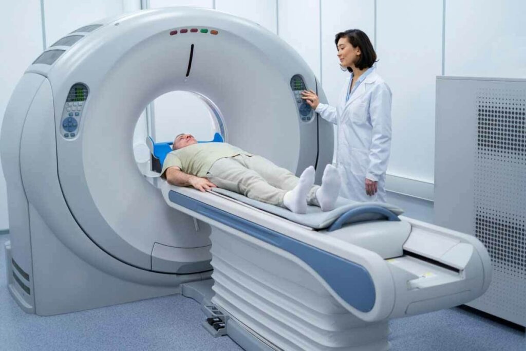
At Liv Hospital, we use advanced technology like the nuclear medicine camera, also known as a gamma camera or scintillation camera, for precise and quick diagnoses. This device detects gamma radiation emitted from special tracers in the body to produce detailed images that help in diagnosing and monitoring various conditions.
This tech helps us make detailed pictures of organs and how they work. It’s key for our non-invasive tests. This way, we can give our patients the best care possible.
Gamma cameras are key in showing the body’s inside parts and how they work. They help us get images without needing to cut open the body. This is vital for checking how organs are working and what they look like.
A gamma camera, also known as a gamma radiation camera or nuclear camera, catches gamma rays from a special tracer given to patients. These cameras are essential for seeing how the body’s parts function. They are a must-have in nuclear medicine.
Nuclear imaging started in the early 20th century. But it wasn’t until the 1950s that the first gamma camera was made by Hal Anger. Over time, big improvements have been made. These have made images clearer and helped doctors diagnose better. The history of nuclear imaging is one of constant improvement, turning it into a top-notch tool for doctors.
Gamma cameras find gamma rays from the body after a special tracer is given. They use several parts to work together. The camera heads, usually one to three, are put close to the patient for better images. Each head has a flat part that needs to be as close to the patient as possible for clear pictures.
Learning about gamma cameras helps us see their importance in nuclear medicine. They have changed how we do diagnostic imaging. Now, doctors can check how organs work and look without surgery.
Understanding a gamma camera’s parts is key to seeing its role in medical tests. We’ll look at the main elements that make up this advanced imaging tool.
The collimator is vital for guiding gamma rays into the camera. It’s made of dense materials like lead. This lets only gamma rays from a certain direction reach the detector. This makes sure the image shows the radioactive areas in the patient’s body accurately.
The scintillation crystal, often sodium iodide (NaI), turns gamma radiation into visible light. When a gamma ray hits the crystal, it creates a light flash. This light’s intensity is based on the gamma ray’s energy. This process is how images are made.
Behind the crystal are photomultiplier tubes (PMTs). They boost the weak light signals from the crystal into electrical signals. These signals help figure out where and how strong the gamma rays were. For more on how these parts work together.
Today’s gamma cameras use advanced digital systems to make images from PMT data. These systems fix issues that could mess up image quality, like scatter radiation. This leads to clear images that help doctors diagnose.
| Component | Function |
| Collimator | Directs gamma rays |
| Scintillation Crystal | Converts gamma radiation to light |
| Photomultiplier Tubes | Amplifies light signals |
| Digital Processing Systems | Reconstructs images |
Experts say, “The gamma camera’s skill in detecting and imaging gamma radiation has changed nuclear medicine. It now makes diagnoses that were once impossible” (
The gamma camera’s development has been a big step forward in nuclear medicine. It lets us see how the body works.
).
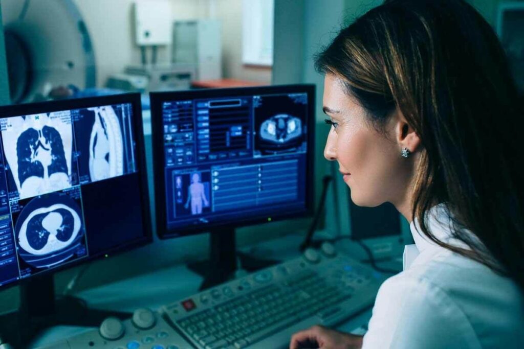
In nuclear medicine, gamma cameras are key for showing where radiopharmaceuticals are in the body. They catch the gamma rays from these substances. This lets us see how different parts of the body work.
First, a patient gets a radiopharmaceutical. This substance goes to the area we want to see. The gamma camera picks up the gamma rays from it.
The collimator, a key part of the camera, focuses these rays onto a scintillation crystal. This crystal turns the rays into light. Then, photomultiplier tubes boost this light into an electrical signal.
Creating images in gamma cameras is about catching gamma rays and where they come from. The electrical signals from the photomultiplier tubes help figure out where and how strong the rays are. This info makes an image of where the radiopharmaceutical is in the body.
Spatial resolution is about seeing two close points. Energy resolution is about telling different gamma ray energies apart. Both are key for clear, accurate images.
To work well, gamma cameras need regular checks. These include making sure the camera responds evenly, checking its spatial resolution, and verifying its energy resolution. Keeping the camera in top shape is vital for clear images and correct diagnoses.
Understanding gamma cameras shows how complex and important nuclear medicine imaging is. These cameras are essential for diagnosing and treating many health issues, like heart problems and cancers.
Scintigraphy is a key tool in nuclear medicine. It helps us see how different parts of the body work. A gamma camera picks up gamma radiation from a special tracer given to the patient.
The process starts with a tracer injected into the patient’s vein. Then, the gamma camera captures how the tracer moves. For example, scans with Technetium 99 usually last 3-4 hours.
Scintigraphy can be done in two ways: static and dynamic. Static scintigraphy takes one image at a time. It shows how organs work at that moment. Dynamic scintigraphy takes many images over time. It lets us see how organs change and work over time.
Planar imaging is a key part of scintigraphy. It uses the gamma camera to take 2D images from different angles. This method is great for checking the heart, bones, and thyroid.
Scintigraphy also lets us measure how well organs work. By tracking the tracer, doctors can get important data. This data helps with diagnosis and treatment plans.
| Scintigraphy Type | Description | Clinical Application |
| Static Scintigraphy | Single image capture | Assessing organ function at a specific time |
| Dynamic Scintigraphy | Multiple image capture over time | Observing tracer movement and organ function over time |
Advanced gamma camera imaging has changed nuclear medicine a lot. It gives more detailed and accurate info for diagnosis. This has greatly helped in diagnosing and managing many health issues.
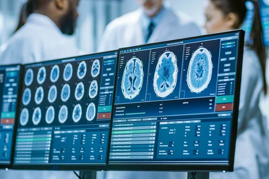
Single Photon Emission Computed Tomography (SPECT) is a top-notch imaging method. It gives three-dimensional images of the body’s inside. SPECT makes diagnosis more accurate by showing detailed cross-sections of the body.
A leading nuclear medicine expert says, “SPECT imaging is key for diagnosing and managing heart and cancer issues.”
“SPECT imaging lets doctors pinpoint radioactive tracers precisely. This helps them check organ function and spot problems accurately.”
Gated imaging is mainly for heart studies. It syncs image taking with the heart’s beat. This method gives important info on heart function and movement.
| Technique | Application | Benefits |
| Gated Imaging | Cardiac Studies | Assesses cardiac function and wall motion |
| SPECT | Oncological and Cardiac Imaging | Provides 3D tomographic images |
| Whole-Body Scanning | Oncology and Metabolic Disorders | Evaluates disease extent and metabolic activity |
Whole-body scanning is used to see how far disease has spread, mainly in cancer and metabolic disorders. It lets doctors check the body’s metabolic activity and disease spread fully.
New designs in gamma camera machines, like digital detectors and hybrid imaging, have boosted both resolution and diagnostic value. Today’s computers can quickly process complex data. This turns the radiation detected by the camera into useful info for radiologists.
Gamma camera scans have changed nuclear medicine a lot. They give us key information about many health problems. We use them to help diagnose and keep track of health issues.
“The gamma camera is a key tool in nuclear medicine,” say doctors. It lets us do scintigraphy scans. These scans show us how organs work.
Gamma camera scans are mainly used for heart imaging. They help us see how well the heart works. We can spot heart disease and check if treatments are working.
Bone scintigraphy is another big use. It shows us how bones work and helps find problems like osteoporosis. This is key for fixing bones and planning treatments.
Gamma camera scans also help with thyroid issues. They help us figure out thyroid problems. Plus, they help check how other glands work.
These scans are used for kidneys and liver too. They help find liver and bile duct problems. They’re also used for other organs, showing their wide use in medicine.
In short, gamma camera scans are a big help in medicine. They give us important info on organs. This helps doctors make better choices for patients.
Nuclear medicine has seen big changes in gamma camera tech. These changes have made images clearer and helped doctors make better diagnoses. New designs in gamma cameras have led to better diagnostic tools.
Digital detector systems are a big step forward. They replaced old analog detectors, bringing better sensitivity and clarity. Digital detectors can handle more information and give more precise readings of how radiopharmaceuticals are taken up.
Hybrid imaging has changed nuclear medicine a lot. It combines SPECT’s functional info with CT or MRI’s detailed images. SPECT/CT systems, for example, give both types of info at once. This makes diagnoses more accurate and treatment plans more precise.
There’s a push towards making gamma cameras smaller and more portable. These smaller systems are flexible and can be used in many places, like operating rooms and ICUs. Portable gamma cameras allow for live imaging during surgeries. This makes procedures like sentinel lymph node biopsies more accurate.
Software updates have also been key in improving gamma camera tech. New algorithms and techniques have made images better. Iterative reconstruction algorithms, for instance, cut down on noise and improve detail. This leads to more accurate diagnoses.
In summary, new tech in gamma cameras has greatly improved nuclear medicine. It has led to better care for patients. As tech keeps getting better, we can look forward to even more progress in this field.
Gamma camera imaging has changed nuclear medicine a lot. It gives us functional info that goes with anatomical images. We use gamma cameras to check many body functions, not just structure.
Gamma camera imaging gives us functional information about the body’s work. This is different from the detailed anatomy pictures from CT or MRI. This info is key for diagnosing and treating many health issues.
Gamma cameras are compared to other imaging methods. We look at the info they give, radiation exposure, and cost. Gamma cameras give special insights that help with diagnosis, making them very useful.
One big thing about gamma camera imaging is the ionizing radiation it uses. Even though the doses are safe, we must think about the risks and benefits for each patient.
Gamma camera imaging is cost-effective when used right. It gives us important info that helps decide treatment. This might mean we don’t need more tests.
In short, gamma camera imaging is a big plus in nuclear medicine. It gives us useful info and is affordable. But, it also has downsides like radiation exposure. Knowing these helps us use gamma cameras better in medical care.
We’ve looked into how gamma cameras work in nuclear medicine. They play a key role in helping doctors diagnose diseases. These cameras help us see how our bodies work at a molecular level.
The pictures from gamma cameras are vital for diagnosing many health issues. Even though a gamma camera itself might seem simple, its images are very important in healthcare today.
Scintigraphy is the correct term for using gamma cameras to see radioactivity in the body. This method helps doctors understand where certain substances are in the body. It’s key for diagnosing and treating diseases.
Gamma cameras are essential in nuclear medicine. They help doctors create personalized treatment plans. Their ability to show how the body functions is a big plus in healthcare today.
A gamma camera, also known as a nuclear medicine camera, detects gamma radiation. It uses a radiopharmaceutical to create images of organs and their functions.
A gamma camera detects gamma rays from a radiopharmaceutical. These rays are turned into light by a scintillation crystal. Then, photomultiplier tubes amplify this light, and digital systems turn it into an image.
The main parts of a nuclear medicine camera are a collimator, scintillation crystal, photomultiplier tubes, and digital systems. Together, they detect gamma radiation and create detailed images.
The collimator directs gamma rays to the scintillation crystal. This helps the gamma camera pinpoint where the gamma radiation comes from, making precise images.
Scintigraphy is a diagnostic technique in nuclear medicine. It uses a gamma camera to see how organs and tissues work. This is done by detecting gamma radiation from a radiopharmaceutical.
SPECT stands for Single Photon Emission Computed Tomography. It’s a three-dimensional imaging technique. It gives detailed information about where a radiopharmaceutical is in the body.
Gamma camera imaging shows how organs and tissues work. This helps diagnose diseases early. It’s often used with other imaging methods for a better understanding of the body.
Gamma camera imaging has some limits. These include radiation exposure and limited detail. Also, there can be artifacts that affect image interpretation.
Gamma camera technology has made big strides. Advances in digital detectors, hybrid imaging, and software have improved it. These changes have enhanced diagnostic abilities and patient care.
The correct term is scintigraphy.
Static scintigraphy takes one image of a radiopharmaceutical’s distribution. Dynamic scintigraphy takes multiple images over time. This shows how an organ or tissue functions and changes.
Gated imaging is used in cardiac studies. It synchronizes image acquisition with the heart’s cycle. This helps assess cardiac function and wall motion.