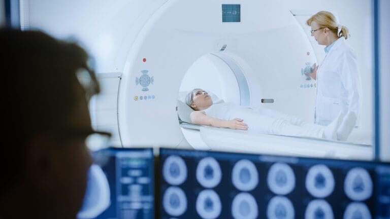
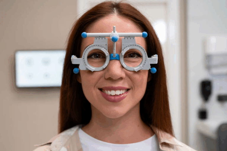
Learn how an ophthalmologist detect a brain tumor: yes, it’s possible through key signs like optic disc swelling and vision changes found during a routine eye exam.
Ophthalmologists do more than just check your eyes. They are often the first to find problems like brain tumors.
At an eye exam, an ophthalmologist might see signs of a brain tumor. These signs include swelling of the optic disc or unusual pupil reactions to light.
Key Takeaways
- Ophthalmologists can detect signs of brain tumors during routine eye exams.
- Certain eye conditions can be indicative of a brain tumor.
- Early detection is key for effective treatment.
- Ophthalmologists play a vital role in referring patients for further diagnosis.
- Understanding the link between eye health and brain tumors is essential.
The Connection Between Eyes and Brain Tumors

The eyes and brain are closely linked through the visual system. This system can be impacted by brain tumors. It’s key to grasp how brain tumors can show up through eye symptoms.
The visual pathway starts with the eyes and goes through the optic nerves and brain parts. Tumors in the brain can press or harm these areas. This leads to different eye symptoms.
How the visual system connects to the brain
The visual system is a complex network starting with the eyes and reaching the brain. The optic nerves from each eye cross over at the optic chiasm. This allows the brain to mix visual info from both eyes.
This detailed connection means brain tumors can impact vision in many ways. It depends on the tumor’s location and size.
Why eye symptoms often appear with brain tumors
Eye symptoms are common in brain tumor patients. Tumors can press or damage the optic nerves or other visual pathway parts. This can cause blurred vision, double vision, or loss of side vision.
| Symptom | Description | Possible Cause |
| Blurred Vision | Loss of sharpness in vision | Optic nerve compression |
| Double Vision | Seeing two images of one object | Cranial nerve palsy |
| Peripheral Vision Loss | Loss of side vision | Tumor pressing on optic nerves |
It’s vital to understand the link between eyes and brain tumors for early detection and treatment. Recognizing eye symptoms linked to brain tumors helps doctors start the right tests and treatments.
Role of Ophthalmologists in Brain Tumor Detection
Ophthalmologists are key in finding brain tumors through eye checks. They know how to spot signs that might mean a brain tumor is present.
Specialized Training of Ophthalmologists
Ophthalmologists learn a lot to spot eye problems that could mean something bigger, like a brain tumor. They study the eye’s structure and how it works. They also learn about diseases and how to find them.
When Ophthalmologists Suspect Neurological Issues
An eye check might show signs of brain problems, like odd pupil reactions or visual field defects. Seeing these signs means they might look deeper into a brain tumor. They look for eye diseases but also for signs of bigger brain issues.
Limitations of Ophthalmological Examinations
Even though ophthalmologists can spot signs of brain tumors, their tests can’t say for sure if there’s a tumor. More tests, like MRI scans, are needed to be sure. But, the ophthalmologist’s findings help decide what tests to do next.
Key Eye Exam Tumor Signs Ophthalmologists Look For
Certain visual changes and eye abnormalities can signal a brain tumor. Ophthalmologists are trained to spot these signs. During a detailed eye exam, they look for key signs to see if more tests are needed.
Visual Changes That May Indicate Tumors
Visual disturbances can be an early sign of a brain tumor. These changes include:
- Blurred vision or double vision
- Loss of peripheral vision
- Difficulty seeing colors or changes in color perception
- Flashes of light or floaters
These visual changes happen when a tumor presses on the optic nerve or other parts of the visual pathway. Ophthalmologists can tell if these symptoms are just vision problems or something more serious like a brain tumor.
Physical Eye Abnormalities Associated with Brain Tumors
Physical eye changes can also point to brain tumors. These include:
- Swelling of the optic disc (papilledema)
- Protrusion of the eyeball (proptosis)
- Drooping eyelid (ptosis)
- Abnormal eye movements
These signs come from the tumor’s effect on nerves controlling eye movement and the eye’s surroundings.
Subtle Early Warning Signs
Some early signs of brain tumors are subtle and don’t always seem serious. These include:
- Mild headaches that persist or worsen over time
- Nausea or vomiting, often in the morning
- Seizures or convulsions
- Changes in personality or cognitive function
While these symptoms can have many causes, seeing them with visual or physical eye changes means a detailed check is needed. Ophthalmologists are key in spotting these signs and sending patients for more tests when needed.
Papilledema: A Critical Warning Sign
Papilledema is when the optic disc swells. It’s a sign of high pressure in the brain, often from tumors. This swelling happens when the skull’s pressure goes up. It’s a big warning that needs more checking.
What is papilledema and how it’s detected
An eye doctor checks for swelling in the optic disc to find papilledema. They use special tools to look at the retina and optic disc. Finding papilledema is key to finding the cause, like tumors. Papilledema is a big warning sign, It means you need a full check of your brain.
Why increased intracranial pressure causes optic disc swelling
High pressure in the skull makes the optic disc swell. This happens because the pressure pushes on the optic nerve. Knowing this helps doctors find the cause of swelling.
Differentiating papilledema from other optic disc conditions
It’s important to tell papilledema apart from other optic disc problems. Conditions like optic neuritis or pseudopapilledema look similar. Doctors use tests like OCT to make sure it’s papilledema.
Learning about papilledema helps us see why eye exams are key. They can spot brain tumors early.
Visual Field Defects and Their Significance
Understanding visual field defects is key to finding brain tumors. These defects show where the tumor is. Visual field tests help find out how much vision is lost and where.
Types of Visual Field Loss Associated with Tumors
Brain tumors can lead to different vision problems. These include losing half or a quarter of your vision. They can also make your field of vision smaller.
A pituitary tumor can block your vision in the outer parts of your field. Tumors in one side of the brain can affect the same side of your vision in both eyes.
How Visual Field Testing Works
Visual field testing is a safe way to check your vision. You look at a point while lights are shown in different places. This helps make a map of your vision.
Today’s tests use advanced tech like automated perimetry. This makes the results very accurate. It’s important for finding and tracking vision problems from brain tumors.
What Specific Patterns Tell Doctors About Tumor Location
The way your vision is lost can tell doctors a lot. For example, a tumor on the optic nerve might cause a blind spot in the middle. Tumors near the optic chiasm can lead to losing half of your vision in both eyes.
Doctors look at these patterns to guess where the tumor is. They might then use MRI scans to confirm their thoughts.
Bitemporal Hemianopia: A Sign of Pituitary Tumors

Bitemporal hemianopia is a key sign of pituitary tumors. It affects the visual field, causing the loss of the outer half of vision in both eyes.
Understanding Bitemporal Hemianopia
This condition happens when the optic chiasm gets compressed. The optic chiasm is where the optic nerves cross. This compression stops visual signals from getting through, causing vision loss.
Pituitary tumors are often linked to this condition. These tumors, usually benign, grow on the pituitary gland. As they grow, they can press on the optic chiasm, causing vision problems.
How Pituitary Tumors Affect the Optic Chiasm
Pituitary tumors can grow too big for their space and press on the optic chiasm. This pressure is what causes the visual field defects seen in bitemporal hemianopia.
| Tumor Size | Effect on Optic Chiasm | Visual Field Defect |
| Microadenoma (<10 mm) | Minimal compression | Mild or no visual field defect |
| Macroadenoma (≥10 mm) | Significant compression | Bitemporal hemianopia |
Progressive Nature of Vision Loss with Pituitary Adenomas
The way vision loss gets worse in pituitary adenoma patients can differ. Sometimes, vision loss stays the same for a long time. But other times, it can get worse fast if not treated.
It’s important to keep an eye on this and act quickly to avoid permanent vision loss. Eye doctors are key in spotting these changes and helping patients get the right care.
Cranial Nerve Palsies and Ocular Motility Issues
Understanding cranial nerve palsies is key to diagnosing and treating eye movement problems linked to brain tumors. These nerves control our eye movements. Any disruption can cause big visual issues.
Which Cranial Nerves Affect Eye Movement
The oculomotor (III), trochlear (IV), and abducens (VI) nerves control our eye movements. They work through extraocular muscles for precise eye actions. Damage to these nerves can cause cranial nerve palsies, leading to eye movement problems.
The oculomotor nerve (III) controls several muscles for eye movement. The trochlear nerve (IV) works the superior oblique muscle. The abducens nerve (VI) controls the lateral rectus muscle. Together, they help us move our eyes in many ways.
How Tumors Can Cause Double Vision and Eye Misalignment
Brain tumors can press on or damage cranial nerves. This can cause double vision and eye misalignment. When a tumor affects a nerve, it disrupts the signals to the muscles, leading to uncoordinated eye movements.
Double vision, or diplopia, happens when the eyes can’t fuse images. This is due to muscle weakness or paralysis from nerve damage. Eye misalignment, or strabismus, can also occur from cranial nerve palsies, making the eyes not align properly.
Specific Tests for Cranial Nerve Function
Testing cranial nerve function is vital for diagnosing eye movement problems. Several tests can check the nerves controlling eye movement.
- The cover-uncover test detects strabismus and checks eye alignment.
- The Hirschberg test uses a light reflex to measure strabismus angles.
- Ocular motility testing checks eye movement range and finds any issues.
These tests, along with a detailed clinical exam, help doctors diagnose cranial nerve palsies and related eye movement problems accurately.
Pupillary Abnormalities as Tumor Indicators
Pupillary abnormalities can signal serious neurological issues, like brain tumors. The way pupils react to light and their look can tell us a lot about the brain and visual pathway’s health.
Anisocoria and Other Pupil Irregularities
Anisocoria, where pupils are not the same size, is a warning sign. Other issues, like irregular shapes or poor light response, also hint at neurological problems. These can be due to tumors affecting the nerves that control the pupils.
The Pupillary Light Reflex Examination
The pupillary light reflex test is key for checking pupil function. A light is shone into the eyes to see how the pupils react. A normal response is when the pupils get smaller with light. But, if they don’t, it could mean a problem in the nerves or muscles of the iris.
Afferent Pupillary Defects and Their Significance
Afferent pupillary defects happen when nerve signals from the eye to the brain are disrupted. This can be due to optic neuritis, nerve compression, or severe retinal disease. For brain tumors, such defects suggest the tumor is pressing on the optic nerve or its pathway. Spotting these defects early is vital for timely treatment.
| Pupillary Abnormality | Possible Cause | Significance |
| Anisocoria | Tumor affecting the nerves controlling pupil size | Potential indication of neurological issue |
| Poor Light Reflex | Afferent pathway defect due to tumor or other conditions | Indicates problem with optic nerve or retina |
| Irregular Pupil Shape | Neurological damage or tumor affecting the iris or its nerves | May indicate serious underlying condition |
Optic Nerve Compression: Causes and Detection
It’s important to know about optic nerve compression to spot brain tumors early. These tumors can press on the optic nerve, causing serious vision problems.
Causes of Optic Nerve Compression
Tumors can put pressure on the optic nerve in different ways. Usually, a tumor grows near the optic nerve and presses on it. This can mess up the optic nerve’s work, leading to vision issues.
Tests for Optic Nerve Function
Ophthalmologists use several tests to check the optic nerve’s function. These include:
- Visual Acuity Tests: To measure the sharpness of vision.
- Visual Field Tests: To assess the peripheral vision and detect any blind spots.
- Pupillary Light Reflex Tests: To evaluate the response of the pupils to light, indicating optic nerve function.
| Test | Purpose | Indications of Optic Nerve Compression |
| Visual Acuity Test | Measures the sharpness of vision | Reduced visual acuity |
| Visual Field Test | Assesses peripheral vision | Blind spots or loss of peripheral vision |
| Pupillary Light Reflex Test | Evaluates pupil response to light | Abnormal pupil reaction or afferent pupillary defect |
Visual Acuity Changes with Optic Nerve Involvement
When the optic nerve is compressed, vision changes are often the first sign. People might see things less clearly or have blurry vision. How much vision changes depends on how much the nerve is compressed.
Early detection of optic nerve compression is key to saving vision. Regular eye exams can catch signs of optic nerve compression early. This leads to more investigation and timely treatment.
Advanced Diagnostic Techniques Used by Ophthalmologists
Ophthalmologists use many advanced techniques to check eye health and find signs of brain tumors. These methods have changed ophthalmology a lot. They help doctors diagnose and keep track of conditions better.
Optical Coherence Tomography (OCT)
Optical Coherence Tomography (OCT) is a test that doesn’t hurt. It uses special light to take detailed pictures inside the eye. OCT is great for looking at the retina and optic nerve for damage or disease.
Key benefits of OCT include:
- High-resolution imaging of retinal layers
- Detection of subtle changes in the optic nerve head
- Monitoring of disease progression over time
Fundus Photography and Documentation
Fundus photography is a key tool for eye exams. It takes pictures of the inside of the eye, like the retina and optic disc. This method helps keep a record of the eye’s state, useful for future comparisons.
| Feature | Description |
| High-resolution imaging | Captures detailed views of the retina and optic disc |
| Longitudinal monitoring | Enables tracking of changes over time |
| Documentation | Provides a permanent record for patient files |
Fluorescein Angiography in Special Cases
Fluorescein angiography is a special test. It uses dye to see the blood vessels in the retina and choroid. It’s mainly used when there’s a suspicion of vascular problems.
The procedure involves:
- Injection of fluorescein dye
- Rapid-sequence photography to capture the dye’s passage through the retinal vessels
- Analysis of the images to identify abnormalities
By using these advanced techniques, ophthalmologists can understand a patient’s eye health well. They can spot signs of brain tumors early.
What to Expect During a Neurological Eye Examination
The neurological eye examination is a detailed process. It checks your vision and eye health. It’s important for finding eye problems linked to the brain.
The Step-by-Step Process of the Examination
Your ophthalmologist will follow a structured process. They will check your eye health thoroughly. The steps include:
- Visual acuity testing to assess the sharpness of your vision
- Pupillary light reflex examination to check for abnormalities in pupil reaction
- Ocular motility testing to evaluate eye movements
- Visual field testing to detect any blind spots or areas of decreased vision
- Fundoscopic examination to inspect the retina and optic nerve
Questions the Doctor May Ask About Symptoms
Your ophthalmologist will ask you questions about your symptoms. They might ask:
- When did you first notice your symptoms?
- Have you experienced any changes in your vision or eye health?
- Do you have a family history of eye or neurological conditions?
Being ready to answer these questions helps your doctor make a better assessment.
Duration and Comfort Considerations
The duration of a neurological eye examination varies. It usually takes 30 to 60 minutes. Your comfort is a priority. The exam is not painful but might involve bright lights or other stimuli.
Knowing what to expect can make you feel more comfortable and prepared for your examination.
When an Ophthalmologist Will Refer for Further Testing
An ophthalmologist might suggest more tests if they see signs of a brain tumor. This choice comes after a detailed eye check-up. They look for specific symptoms or red flags that need more investigation.
Red Flags that Prompt Immediate Referral
Some symptoms and eye exam findings can lead to an urgent need for more tests. These red flags include:
- Significant changes in visual acuity or field
- Papilledema or optic disc swelling
- Cranial nerve palsies affecting eye movement
- Unusual pupillary reactions or abnormalities
These signs point to possible neurological problems, like a brain tumor. They need a closer look.
The MRI Referral Process
Magnetic Resonance Imaging (MRI) is key for finding brain tumors. If an ophthalmologist suspects a tumor, they’ll send the patient for an MRI. The MRI referral process involves:
- Preparing the patient for the MRI by explaining it
- Sharing the patient’s medical history and findings with the radiologist
- Telling the radiologist where to focus the scan
This ensures the MRI is done well and quickly.
Urgency Levels and Timeframes
The urgency of the referral depends on the symptoms’ severity and the chance of a brain tumor. Severe symptoms or big red flags need quick action, often within days. Less urgent cases follow standard testing schedules.
Knowing the urgency helps everyone involved manage the testing process better.
Differentiating Between Brain Tumors and Other Eye Conditions
Not all eye symptoms are from brain tumors. It’s key to tell them apart. Ophthalmologists look at many other conditions that can seem like brain tumors.
Common Conditions that Mimic Tumor Symptoms
Many eye problems can look like brain tumors. These include:
- Optic neuritis, which can cause vision loss and pain
- Ischemic optic neuropathy, leading to sudden vision loss
- Glaucoma, which can cause optic nerve damage
- Diabetic retinopathy, affecting vision due to diabetes
These conditions can be mistaken for brain tumors because of similar symptoms.
How Doctors Rule Out Other Causes
Ophthalmologists use tests to figure out if it’s a brain tumor or something else. They use:
| Diagnostic Test | Purpose |
| Visual Field Testing | Assesses peripheral vision and detects defects |
| Optical Coherence Tomography (OCT) | Provides detailed images of the retina and optic nerve |
| Fundus Photography | Documents the retina and optic disc for abnormalities |
Doctors combine these tests with what the patient says and a physical check. This helps them find the right cause of symptoms.
When Symptoms Warrant Immediate Attention
Some symptoms need quick medical help. They might mean serious issues like brain tumors or other emergencies. These include:
- Sudden vision loss
- Severe eye pain
- Double vision or loss of peripheral vision
The Importance of Regular Eye Exams for Tumor Detection
Regular eye exams are key to finding tumors before they show symptoms. They help keep your vision sharp and spot serious health problems, like brain tumors. Early detection can greatly improve treatment results, making eye exams essential for health.
Recommended Frequency of Eye Examinations
How often you need an eye exam depends on your age, health history, and risk factors. Adults usually need an eye check every two years. But, if you have a family history of eye issues or certain health problems, you might need more frequent visits.
Guidelines suggest that adults without risk factors should see an eye doctor once between 20 and 30. Then, every 2-3 years until 40, and more often after that.
Risk Factors that May Require More Frequent Monitoring
Some risk factors mean you need to see an eye doctor more often. These include a family history of eye diseases, diabetes, high blood pressure, and past eye injuries or surgeries. Talk to your eye care provider about how often you need to go.
- A family history of glaucoma or other eye diseases
- Diabetes or other metabolic disorders
- History of eye injuries or surgeries
- Certain neurological conditions
How Routine Exams Can Catch Asymptomatic Tumors
Eye exams can find tumors before you notice any symptoms. An ophthalmologist can spot signs like papilledema, visual field defects, or cranial nerve palsies that might mean a tumor. Early detection through regular eye exams can be lifesaving.
“Regular eye exams are key to catching eye diseases and other health issues early, when they’re easier to treat.” – American Academy of Ophthalmology
By knowing how important regular eye exams are and following the recommended schedule, you can boost your chances of catching and treating brain tumors early.
Conclusion: Early Detection and Next Steps After Diagnosis
Early detection of brain tumors is key for better treatment and outcomes. Regular eye exams help spot signs like papilledema and visual field defects. Ophthalmologists use tools like Optical Coherence Tomography (OCT) to find these signs.
After finding a brain tumor, patients need to know what comes next. They might have more tests, like MRI scans, to learn about the tumor. A team of doctors will work together to create a treatment plan just for them.
Getting medical help quickly is very important. Using eye tumor detection methods and finding tumors early can greatly improve a patient’s life.
FAQ
Can an eye exam detect a brain tumor?
Yes, an eye exam can spot signs of a brain tumor. These include papilledema, visual field defects, and cranial nerve palsies.
What are the common eye symptoms of a brain tumor?
Eye symptoms include blurred vision and double vision. You might also lose peripheral vision or feel eye pain. Some people see less in the outer half of their vision.
How do ophthalmologists detect eye tumor signs during an exam?
Ophthalmologists use several methods. They test your vision and check your visual fields. They also look at the optic disc and retina with tools like OCT and fundus photography.
What is papilledema, and how is it related to brain tumors?
Papilledema is when the optic disc swells due to high pressure in the brain. It’s a sign of a brain tumor. Ophthalmologists can see it during an eye exam.
Can pituitary tumors cause vision problems?
Yes, pituitary tumors can lead to vision issues. They can press on the optic nerves, causing vision loss. This is because they’re near the optic chiasm.
How do cranial nerve palsies affect eye movement?
Cranial nerve palsies can cause double vision and eye misalignment. They affect the nerves that control eye movement.
What is the role of MRI in diagnosing brain tumors?
MRI is key in finding and understanding brain tumors. It shows the tumor’s size, location, and type. Ophthalmologists might suggest an MRI if they suspect a tumor.
How often should I have an eye exam to detect potentially brain tumors?
Eye exam frequency varies based on risk factors and age. Adults should get a full eye exam every two to four years. Those at higher risk might need more checks.
Can routine eye exams detect asymptomatic brain tumors?
Yes, eye exams can find brain tumors before symptoms show. Regular eye checks are very important.
What are the red flags that prompt an immediate referral to a specialist?
Red flags include sudden vision loss and severe eye pain. Double vision and changes in the optic disc or retina also require immediate action.
How do doctors differentiate between brain tumors and other eye conditions?
Doctors use exams, medical history, and tests to tell brain tumors from other conditions. This helps avoid misdiagnosis.


