


Discover the importance of Bone Health and its impact on your lifestyle. Learn the Bone Health Basics and how to protect your skeletal system for a lifetime.
Overview And Definition
What are Bone Health Basics?
Bone Health is the foundation of a functional, active body. Our bones provide structural support, protect vital organs, and store essential minerals like calcium. While we often think of bones as static structures, they are actually living tissues that are constantly being broken down and rebuilt in a process called remodeling. Bone Health Basics involve understanding that peak bone mass is typically reached in your late twenties. After this point, the body may begin to lose more bone than it creates, making early intervention and lifelong maintenance essential for mobility and strength.
The Connection Between Bone and Muscle Health
You cannot discuss the skeleton without also addressing Muscle Health. The two systems work in a mechanical partnership; muscles pull on bones to create movement, and this physical tension actually signals the bones to become denser and stronger. When muscles are weak or inactive, the bones receive fewer signals to maintain their density, which can lead to increased fragility. At Liv Hospital, we emphasize a holistic approach that treats the musculoskeletal system as a single unit, ensuring that both your framework and the power behind it remain in optimal condition.
Understanding Osteoporosis and Bone Loss
The most significant threat to long-term skeletal integrity is Osteoporosis. This is a systemic condition characterized by decreased bone mass and the deterioration of bone tissue, which leads to an increased risk of fractures—particularly in the hip, spine, and wrist. Often called a “silent disease,” it can progress for years without symptoms until a sudden fall or strain causes a bone to break. Understanding the risks associated with bone thinning is a critical part of our clinical evaluation process, allowing us to help patients take preventive action before an injury occurs.
Injury and Causes
What Causes Bone Injury and Fragility?
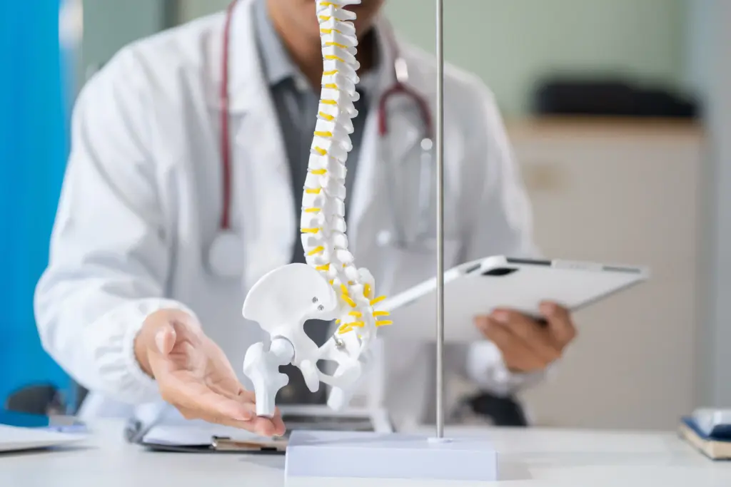
The “injury” to the skeletal system often happens at a microscopic level long before a visible break occurs. The primary cause of bone deterioration is an imbalance in the remodeling process. Factors such as hormonal changes, sedentary behavior, and poor nutrition can cause the body to reabsorb bone tissue faster than it can replace it. This internal erosion weakens the architectural “struts” inside the bone, making them susceptible to fractures even from minor impacts.
Risk Factors for Osteoporosis
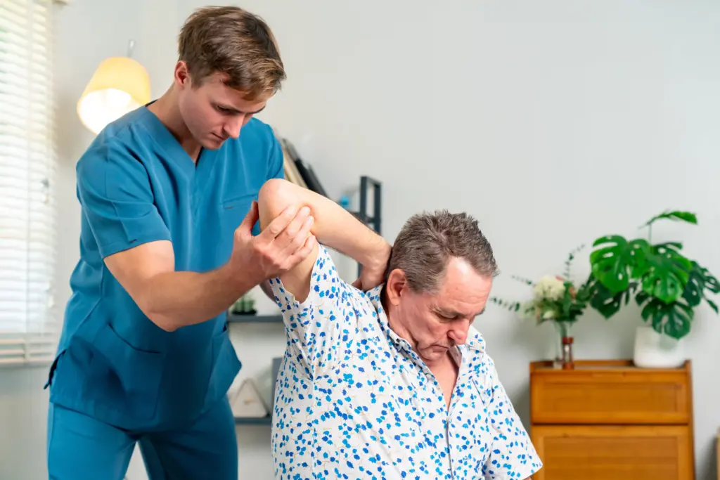
Several factors can accelerate the development of Osteoporosis. While genetics play a role, lifestyle choices are significant drivers of bone loss. Smoking and excessive alcohol consumption are toxic to bone-building cells. Furthermore, a lack of mechanical loading—meaning not enough weight-bearing activity—tells the body that the bones do not need to be strong, leading to a natural “downsizing” of bone density. At Liv Hospital, we evaluate these lifestyle factors to create a personalized risk profile for every patient.
Impact of Nutritional Deficiencies
A lack of specific “raw materials” is a leading cause of poor Bone Health. Without a steady supply of minerals and vitamins, the body is forced to “mine” the bones to maintain vital functions like heart rhythm and muscle contraction. Chronic deficiencies in calcium and Vitamin D are the most common nutritional causes of skeletal weakening. When the body is in a constant state of mineral debt, the structural integrity of the skeleton is the first thing to suffer, leading to porous, fragile bones.
Diagnosis and Imaging
How do Doctors Evaluate Bone Health?
The diagnosis of bone strength begins with a clinical assessment of risk factors and a review of any history of height loss or unexplained pain. Because you cannot “feel” your bones thinning, we rely on specialized diagnostic tools to see inside the skeletal structure. At Liv Hospital, we use these metrics to determine your “T-score,” which compares your bone density to that of a healthy young adult to assess your fracture risk.
The Role of DEXA Scanning
The gold standard for diagnosing Osteoporosis is the Dual-Energy X-ray Absorptiometry (DEXA) scan. This is a quick, non-invasive imaging test that measures the mineral content in specific areas of the skeleton, usually the hip and the lower spine. It uses a very low dose of radiation to provide a precise calculation of your bone density. This data is essential for determining if you need medical intervention or if lifestyle changes alone are sufficient to protect your health.
Advanced Imaging and Laboratory Tests
In addition to DEXA, we may use quantitative CT scans to look at the 3D structure of the bone, which provides more detail in complex cases. Laboratory work is also a vital part of the evaluation. Blood tests can measure levels of calcium, Vitamin D, and specific markers of bone turnover (how fast your bone is being broken down). These tests help us understand the “why” behind bone loss, allowing for a more targeted and effective treatment plan.

Treatment and Recovery
Clinical Strategies for Bone Restoration
Once a diagnosis of low bone density is made, the goal is to stop further loss and, if possible, increase bone strength. Treatment is multifaceted, combining medical therapy with nutritional support. For patients with high fracture risk, doctors may prescribe medications that either slow down the cells that break down bone or stimulate the cells that build new bone. These treatments are most effective when started early and monitored consistently by a clinical team.
The Role of Bone Health Supplements
Nutritional support is a non-negotiable part of treatment. Most patients require targeted bone health supplements to ensure the body has the building blocks it needs for repair. While we always advocate for a “food first” approach, the reality is that many adults struggle to absorb enough Vitamin D and minerals through diet alone. At Liv Hospital, we provide specific dosing guidelines for calcium supplements for bone health, ensuring you get enough to protect your skeleton without exceeding safe limits.
Managing Recovery from Fractures
If a patient has already suffered a fracture due to Osteoporosis, the focus shifts to recovery and the prevention of future injuries. Recovery involves stabilizing the break—often through orthopedic surgery—and then immediately addressing the underlying bone weakness. We work to optimize the healing environment through superior nutrition and the “re-introduction” of safe movement to ensure the bone heals as strongly as possible.
Rehabilitation Tips
Tips to Keep Your Bones Healthy
The long-term maintenance of your skeleton is a daily practice. Rehabilitation and prevention are centered on mechanical loading and superior nutrition. By following these tips to keep your bones healthy, you can significantly reduce your risk of future injury and maintain your independence as you age.
Nutritional Rehabilitation: Food for Healthy Bones
Your diet is your best defense. Focus on food for healthy bones by incorporating:
- Dairy and Fortified Alternatives: High in calcium and often Vitamin D.
- Leafy Greens: Kale, collards, and broccoli provide calcium and Vitamin K.
- Fatty Fish: Salmon and sardines are rare natural sources of Vitamin D.
Protein: Essential for maintaining the collagen matrix that holds the bone together.
Physical Rehabilitation: Weight-Bearing and Resistance
To support Muscle Health and stimulate bone growth, you must engage in weight-bearing exercises. This includes walking, dancing, or climbing stairs—any activity where you are working against gravity. Resistance training, such as using weights or elastic bands, is equally important as it strengthens the muscles that protect your joints and pull on your bones to keep them dense.
Why Choose Liv Hospital?
At Liv Hospital, we provide a comprehensive center for skeletal and Muscle Health. Our multidisciplinary team combines the expertise of endocrinologists, orthopedists, and nutritionists to provide a 360-degree approach to your care. From our advanced DEXA imaging technology to our personalized nutritional and exercise programs, we are dedicated to keeping you strong and mobile. We encourage you to reach out and call Liv Hospital to schedule a bone health assessment and take a proactive step toward protecting your future.
Injury and Causes
Injury and Causes
Understanding Bone Health: From Traumatic Fractures to Metabolic Loss
Bone injuries and degradation occur when the structural integrity of the skeleton is overwhelmed by external forces or internal biological failure. While the skeleton is remarkably resilient, it has physical limits. Understanding the mechanism of injury is the first step in determining the appropriate treatment and predicting the recovery timeline.
Injuries range from acute trauma, such as a clean break from a fall, to chronic conditions where the bone slowly loses density until it becomes fragile. The causes are often multifactorial, involving a combination of environmental hazards, genetic predisposition, and lifestyle choices that weaken the bone matrix over time.
Modern medicine categorizes bone issues into traumatic injuries and metabolic disorders. Traumatic injuries are mechanical failures, while metabolic disorders are systemic failures of the remodeling process. Both require distinct approaches to care and rehabilitation.
- Acute mechanical overload leading to fractures
- Repetitive stress causing microdamage
- Metabolic imbalances leading to demineralization
- Hormonal shifts accelerating bone loss
- Genetic mutations affecting collagen structure
Acute Traumatic Fractures
Traumatic fractures happen when a sudden force exerted on the bone exceeds its strength. This can occur from falls, vehicular accidents, or high impact sports collisions. The nature of the force determines the type of fracture.
A direct blow typically causes a transverse fracture, where the break is horizontal across the bone shaft. Twisting motions, common in sports, often result in spiral fractures. These injuries are immediately painful and usually result in a loss of function in the affected limb.
Compression fractures are common in the spine, particularly when vertical force is applied. In high energy trauma, the bone may shatter into multiple pieces, known as a comminuted fracture. These complex injuries often require surgical intervention to restore alignment.
- Transverse fractures from direct impact
- Spiral fractures from torsional forces
- Comminuted fractures involving multiple fragments
- Compression fractures in vertebral bodies
- Open fractures where bone penetrates skin
Stress Fractures and Repetitive Load
Stress fractures are overuse injuries that develop over time. They occur when muscles become fatigued and are unable to absorb shock. The overload is then transferred to the bone, causing tiny cracks to form in the cortex.
These injuries are prevalent among runners, military recruits, and athletes who rapidly increase their activity intensity. Unlike acute fractures, the pain from a stress fracture starts gradually and worsens with activity. It typically subsides with rest but returns upon exertion.
If left untreated, these microfractures can coalesce into a full cortical break. Early recognition is vital to prevent a simple stress reaction from becoming a debilitating fracture that requires immobilization or surgery.
- Microcracks forming in the cortical bone
- Muscle fatigue transferring shock to the skeleton
- Gradual onset of localized pain
- Worsening symptoms with weight bearing activity
- Risk of progression to complete fracture
The Pathogenesis of Osteoporosis
Osteoporosis is a metabolic disease characterized by low bone mass and deterioration of bone tissue. It leads to increased bone fragility and risk of fracture. This condition is often silent, with no symptoms appearing until a fracture occurs.
The biological cause is an imbalance in the remodeling cycle. Osteoclasts remove bone faster than osteoblasts can replace it. This results in the thinning of the cortical shell and a loss of connectivity in the internal trabecular meshwork.
Postmenopausal women are at the highest risk due to the rapid drop in estrogen, a hormone that protects bone density. However, men and younger individuals with specific risk factors can also develop the disease.
- Imbalance between resorption and formation
- Thinning of the cortical bone shell
- Disconnectivity of trabecular architecture
- Silent progression without early symptoms
- Estrogen deficiency as a primary driver
Metabolic Bone Disorders
Beyond osteoporosis, other metabolic conditions can compromise skeletal health. Osteomalacia is the softening of the bones due to defective mineralization, often caused by severe Vitamin D deficiency. This makes bones pliable and prone to bowing.
Hyperparathyroidism occurs when the parathyroid glands produce too much hormone, signaling the body to strip calcium from the bones aggressively. This leads to cystic lesions in the bone and generalized weakness.
Paget’s disease of bone disrupts the normal recycling process, leading to bones that are fragile and misshapen. The new bone is generated too quickly and is disorganized, lacking the structural integrity of normal healthy bone.
- Defective mineralization in osteomalacia
- Calcium stripping in hyperparathyroidism
- Disorganized remodeling in Paget’s disease
- Vitamin D deficiency implications
- Renal osteodystrophy in kidney patients

Lifestyle and Environmental Risk Factors
Sedentary behavior is a major cause of bone degradation. Bones adhere to Wolff’s Law, which states that bone adapts to the loads under which it is placed. Without the stimulus of gravity and muscle contraction, bone density diminishes.
Smoking is toxic to osteoblasts and restricts blood flow to the skeletal tissues. It also interferes with calcium absorption and hormone regulation. Smokers have a significantly higher risk of fracture and slower healing times.
excessive alcohol consumption inhibits bone formation and increases the risk of falls. Nutritional deficits, particularly in protein, calcium, and magnesium, starve the bone of the raw materials needed for maintenance.
- Lack of mechanical loading stimulus
- Toxicity of nicotine to bone cells
- Alcohol inhibition of osteoblast function
- Nutritional deficiencies in key minerals
- Low body mass index reducing protection
Medication Induced Bone Loss
Certain medications necessary for treating other conditions can have detrimental effects on the skeleton. Glucocorticoids, used for inflammation and autoimmune diseases, are a leading cause of secondary osteoporosis. They inhibit bone formation and increase resorption.
Androgen deprivation therapy for prostate cancer and aromatase inhibitors for breast cancer reduce sex hormones, accelerating bone loss. Antiepileptic drugs and proton pump inhibitors (for acid reflux) can interfere with calcium absorption and metabolism.
Patients on these long term medications require careful monitoring of their bone density. Clinicians often prescribe countermeasures to mitigate the skeletal side effects of these necessary drugs.
- Glucocorticoid induced osteoporosis
- Hormone suppression therapy effects
- Calcium malabsorption from gastric medications
- Antiepileptic drug interference
- Chemotherapy impact on cell turnover
Diagnosis and Imaging
The Clinical Process Of Skeletal Evaluation
The journey toward optimal Bone Health begins with a meticulously structured diagnostic phase. Because bone loss is invisible to the naked eye, a physical exam alone is not enough to assess your risk. When you visit a specialist at Liv Hospital, the process starts with a detailed medical history and a clinical risk assessment. The goal of this evaluation is to provide objective evidence of your current bone density and to identify any metabolic issues that could be driving bone loss. This baseline data is essential for tracking the effectiveness of any future treatments.
Clinical Assessment and History
The diagnostic journey starts with a conversation. Physicians evaluate risk factors such as age, gender, family history, and lifestyle habits. A history of “fragility fractures”—breaks resulting from a fall from standing height—is a major red flag for compromised bone health.
Physical examination may reveal signs of vertebral collapse, such as height loss or an increasing curvature of the upper spine (kyphosis). Localized tenderness, swelling, or deformity in a limb indicates acute trauma.
Clinicians also use risk calculation tools like FRAX. This algorithm integrates various clinical risk factors to estimate the ten year probability of a hip fracture or major osteoporotic fracture, helping to guide treatment decisions.
- Evaluation of historical fracture events
- Measurement of height loss over time
- Assessment of spinal curvature
- Palpation for localized tenderness
- Utilization of FRAX risk calculator
Dual Energy X-ray Absorptiometry (DXA)
DXA is the gold standard for measuring bone mineral density (BMD). It uses two distinct X ray beams with different energy levels. By subtracting the soft tissue absorption, the machine calculates the density of the bone in the spine and hip.
The results are reported as a T score. This score compares the patient’s bone density to that of a healthy 30 year old adult. A T score of 2.5 or lower indicates osteoporosis, while a score between 1.0 and 2.5 indicates osteopenia (low bone mass).
DXA scans are painless, non invasive, and involve very low radiation exposure. They are essential for monitoring the effectiveness of osteoporosis treatments and tracking disease progression over time.
- Gold standard for density quantification
- Comparison against healthy young adult norms
- T score derivation for diagnosis
- Low radiation dose procedure
- Monitoring of therapeutic efficacy
Conventional Radiography (X-rays)
Standard X rays remain the first line imaging modality for suspected fractures. They provide a quick and clear view of the bone structure, allowing doctors to identify breaks, dislocations, and alignment issues.
While excellent for fractures, X rays are not sensitive enough to diagnose early bone loss. A significant amount of bone mineral—often up to 30 percent—must be lost before it becomes visible on a standard X ray.
Radiographs are also useful for monitoring the healing process. Serial X rays taken over weeks or months show the formation of the callus (new bone) and the remodeling of the fracture site.
- Rapid visualization of acute fractures
- Assessment of joint alignment
- Monitoring of fracture healing progress
- Identification of significant deformities
- Limited sensitivity for early density loss
Computed Tomography (CT)
CT scans use a series of X rays taken from different angles to create cross sectional images of the bone. This technology provides detailed 3D views that are crucial for evaluating complex fractures, especially those involving joints like the knee, wrist, or pelvis.
CT is invaluable for surgical planning. It allows surgeons to see the exact position of bone fragments and determine the best approach for fixation. It can also be used to measure bone density (QCT) in the spine, although this is less common than DXA.
For patients with metal implants, specialized CT protocols reduce artifacts, allowing doctors to assess the bone around the hardware for signs of loosening or infection.
- 3D visualization of complex anatomy
- Surgical planning and mapping
- Assessment of intra articular fractures
- Evaluation of bone healing in non unions
- Visualization of bone around implants
Magnetic Resonance Imaging (MRI)
MRI uses powerful magnets and radio waves to create detailed images. Unlike X rays and CTs, MRI does not use ionizing radiation. It is exceptional for visualizing the soft tissues inside and around the bone, including the marrow, ligaments, and tendons.
MRI is the test of choice for diagnosing stress fractures before they become visible on X rays. It detects bone edema (swelling within the marrow), which is the earliest sign of stress reaction.
It is also used to evaluate infection (osteomyelitis) and tumors. The high contrast resolution allows clinicians to distinguish between diseased tissue and healthy bone with great accuracy.
- Visualization of bone marrow edema
- Early detection of stress fractures
- Assessment of surrounding soft tissues
- Diagnosis of infection and tumors
- No ionizing radiation exposure
Nuclear Bone Scans
A nuclear bone scan involves the injection of a small amount of radioactive material into the bloodstream. This tracer accumulates in areas of high bone turnover or repair activity. A special camera then detects the radiation emitted from the skeleton.
This test is highly sensitive for detecting abnormalities in the entire skeleton at once. It is often used to screen for metastatic cancer spread to the bone, infection, or occult fractures that are difficult to localize.
“Hot spots” on the scan indicate areas of increased metabolic activity. While sensitive, bone scans are not specific, meaning further imaging is often needed to determine exactly what is causing the activity.
- Whole body skeletal assessment
- Detection of metabolic “hot spots”
- Screening for metastatic disease
- Identification of occult trauma
- Evaluation of unexplained bone pain
Laboratory and Blood Analysis
Blood tests are critical for identifying the metabolic causes of bone issues. Clinicians measure calcium, phosphorus, and alkaline phosphatase levels to assess general bone turnover.
Vitamin D levels are routinely checked, as deficiency is a common and correctable cause of poor bone health. Parathyroid hormone (PTH) levels are measured to rule out hyperparathyroidism.
Markers of bone turnover, such as CTX (resorption) and P1NP (formation), can be measured in the blood or urine. These markers respond quickly to treatment, allowing doctors to gauge if a therapy is working long before changes appear on a DXA scan.
- Measurement of mineral levels (calcium/phosphate)
- Assessment of Vitamin D status
- Evaluation of parathyroid function
- Tracking of bone turnover markers
- Screening for secondary causes (thyroid/kidney)
Treatment and Recovery
Clinical Management Goals For Bone Recovery
The treatment of bone conditions encompasses a spectrum of interventions ranging from conservative management to complex surgical reconstruction. The primary goals are to restore anatomical alignment, ensure stability for healing, and return the patient to full function. For chronic conditions like osteoporosis, the goal shifts to halting density loss and preventing future fractures.
Modern orthopedics prioritizes early mobilization. Prolonged immobilization can lead to muscle atrophy and joint stiffness. Therefore, treatments are designed to stabilize the injury sufficiently to allow for movement as soon as it is safe. This approach accelerates the biological healing process and improves long term outcomes.
Recovery is a biological process that cannot be rushed, but it can be optimized. Nutrition, medication adherence, and physical therapy all play pivotal roles in how well the bone heals and how much function is regained.
- Restoration of anatomical alignment
- Stabilization of fracture fragments
- Pharmacological management of density
- Optimization of the biological healing environment
- Pain management and functional restoration
Non-Surgical Fracture Management
Many fractures can be treated without surgery. This approach relies on the body’s natural ability to heal when the bone fragments are held in a stable position. Casts made of fiberglass or plaster are the traditional method for immobilizing limbs.
Functional bracing is another option, particularly for fractures of the humerus or tibia. These braces allow for some joint movement while compressing the soft tissues around the fracture to maintain alignment. This promotes blood flow and reduces stiffness.
Closed reduction is a procedure where the doctor manually manipulates the bone pieces back into place without making an incision. This is usually done under sedation or local anesthesia before a cast is applied.
- Immobilization with fiberglass casts
- Use of functional braces for mobility
- Closed reduction maneuvers for alignment
- Splinting for acute swelling management
- Regular X ray monitoring for displacement
Surgical Fixation Techniques
When a fracture is unstable, displaced, or involves a joint surface, surgery is often required. Open Reduction and Internal Fixation (ORIF) involves making an incision to align the bones and securing them with metal plates and screws.
Intramedullary nailing is a technique used for long bones like the femur or tibia. A metal rod is inserted down the hollow center of the bone. This shares the load with the bone, allowing patients to walk on the leg much sooner than with casting.
External fixation involves placing pins through the skin into the bone above and below the fracture. These pins are connected to a rigid frame outside the body. This is often used in severe trauma with significant soft tissue damage where internal surgery is unsafe.
- Plates and screws for precise alignment
- Intramedullary rods for load sharing
- External fixation for complex trauma
- Percutaneous pinning for smaller bones
- Joint replacement for non reconstructible fractures
Pharmacological Interventions
For osteoporosis, medications are used to alter the remodeling cycle. Bisphosphonates are the most common class of drugs. They work by inhibiting osteoclasts, the cells that break down bone. This slows density loss and reduces fracture risk.
Anabolic agents, such as teriparatide, work differently. They stimulate osteoblasts to build new bone. These are typically reserved for patients with severe osteoporosis who are at high risk of fracture.
Monoclonal antibodies like denosumab block the signals that activate bone resorbing cells. These medications require strict adherence to dosing schedules to maintain their protective effect on the skeleton.
- Bisphosphonates to inhibit resorption
- Anabolic agents to stimulate formation
- RANK ligand inhibitors (denosumab)
- Hormone replacement therapy in select cases
- Calcium and Vitamin D supplementation
Orthobiologics and Regeneration
The field of orthobiologics utilizes biological substances to enhance healing. Bone grafts are used to fill voids or stimulate union in fractures that fail to heal. Autografts are taken from the patient’s own body, usually the pelvis, while allografts come from donors.
Synthetic bone substitutes made of calcium phosphate or hydroxyapatite provide a scaffold for new bone to grow. These materials eventually resorb and are replaced by the patient’s natural bone.
Growth factors, such as Bone Morphogenetic Proteins (BMPs), can be applied surgically to potentate bone formation. Stem cell therapies and platelet rich plasma (PRP) are also being investigated for their potential to accelerate the healing of complex injuries.
- Autografts for gold standard osteoconduction
- Allografts to avoid donor site morbidity
- Synthetic scaffolds for structural support
- Bone Morphogenetic Proteins (BMPs) application
- Cellular therapies for enhanced repair
The Biology of Healing (Callus Formation)
Bone healing occurs in distinct stages. First, a hematoma (blood clot) forms at the fracture site, providing a template for repair. Inflammation brings immune cells to clean up debris.
Next, a soft callus made of cartilage forms, bridging the gap between the bone ends. This eventually mineralizes into a hard callus of woven bone. Over months or years, this woven bone is remodeled into strong lamellar bone, restoring the original shape and strength.
Understanding these stages helps patients understand why they cannot bear weight immediately but also why they shouldn’t remain immobile forever. Stress is needed in the later stages to direct the remodeling process.
- Inflammatory phase and hematoma formation
- Soft callus generation via cartilage
- Hard callus mineralization
- Remodeling and restoration of shape
- Influence of mechanical stability on healing type

Pain Management Strategies
Pain control is essential for recovery. In the acute phase, opioids may be used sparingly for severe pain, but the focus quickly shifts to non opioid alternatives to avoid dependency.
NSAIDs (Non steroidal anti inflammatory drugs) reduce pain and swelling, although their use is sometimes timed carefully to avoid interfering with the early inflammatory phase of healing. Acetaminophen is a safe baseline analgesic.
Nerve blocks and regional anesthesia can provide prolonged relief after surgery. Cryotherapy (ice) and elevation are simple but highly effective physical modalities for managing pain and reducing the edema that contributes to discomfort.
- Multimodal analgesia to reduce opioid use
- NSAIDs for inflammation control
- Regional nerve blocks for post op relief
- Cryotherapy and elevation protocols
- Psychological coping strategies for chronic pain
Nutritional Support for Healing
Not all fractures heal smoothly. A non union occurs when the bone fails to heal after an extended period. This may require surgery to clean the site and add bone graft.
Malunion occurs when the bone heals in an incorrect position, potentially affecting joint function. Corrective osteotomies (cutting the bone) may be needed to realign the limb.
Infection is a serious risk, particularly with open fractures or surgical implants. Osteomyelitis (bone infection) requires aggressive treatment with antibiotics and surgical debridement to remove dead tissue and bacteria.
- Non union management via grafting
- Malunion correction via osteotomy
- Osteomyelitis treatment protocols
- Hardware failure or loosening
- Complex regional pain syndrome awareness
Complication Management
Healing requires energy and raw materials. Protein intake is crucial, as the bone matrix is made of collagen protein. Patients recovering from fractures often need to increase their protein consumption.
Calcium and Vitamin D are the bricks and mortar of bone repair. Without adequate levels, the soft callus cannot mineralize into hard bone. Magnesium, Vitamin K, and Zinc also play supportive roles in the enzymatic processes of bone formation.
Hydration is often overlooked but essential for transporting nutrients to the injury site and removing waste products from cellular metabolism.
- Increased protein requirements for collagen synthesis
- Adequate Calcium for mineralization
- Vitamin D for calcium absorption
- Micronutrient support (Magnesium, Zinc, Vit K)
- Hydration to facilitate nutrient transport
Rehabilitation Tips
The Importance Of Structured Rehabilitation
Rehabilitation is the bridge between medical treatment and the return to normal life. Healing the bone is only half the battle; restoring the function of the limb and the confidence of the patient is equally important. Rehabilitation protocols are designed to address the secondary effects of injury, such as muscle atrophy, joint stiffness, and altered movement patterns.
For patients with chronic conditions like osteoporosis, rehabilitation focuses on prevention. It involves training the body to move safely, improving balance to prevent falls, and strengthening the muscles that support the skeleton. This proactive approach empowers patients to maintain their independence.
Success in rehabilitation requires consistency. It is not just about what happens during a therapy session but about the integration of healthy movement patterns into daily life.
- Restoration of joint range of motion
- Rebuilding of muscular strength and endurance
- Retraining of balance and proprioception
- Modification of home environments for safety
- Gradual return to functional activities
Physical Therapy Protocols
Physical therapy usually begins with passive range of motion exercises to keep joints flexible without stressing the healing bone. As healing progresses, active range of motion is introduced, where the patient moves the limb under their own power.
Strengthening exercises start with isometric contractions (tensing the muscle without moving the joint) and progress to isotonic exercises with resistance bands or weights. The therapist tailors the load to the stage of bone healing.
Manual therapy, including soft tissue mobilization and joint glides, helps reduce scar tissue and improve the fluidity of movement. The therapist also educates the patient on pain science, helping them distinguish between “hurt” (damage) and “sore” (rehabilitation).
- Passive and active range of motion
- Isometric to isotonic progression
- Manual therapy for tissue mobility
- Pain education and management
- Individualized progression plans
Weight-Bearing Exercise
For bone health, weight bearing exercise is non negotiable. Bones respond to the stress of gravity by becoming denser. Walking, hiking, and stair climbing are excellent examples.
Resistance training is also vital. When muscles pull on bones during lifting, it stimulates the osteocytes to lay down more matrix. This is particularly important for the spine and hips, common sites of osteoporotic fractures.
For patients who cannot tolerate high impact, low impact options like elliptical training still provide some weight bearing benefit compared to swimming or cycling, which are non weight bearing.
- Gravity dependent activities for density
- Resistance training for site specific loading
- Progression from low to high impact
- Frequency and duration guidelines
- Modifications for joint protection
Fall Prevention Strategies
Preventing falls is the most effective way to prevent fractures in older adults. Rehabilitation focuses on identifying and mitigating fall risks. This includes assessing gait mechanics and correcting limps or shuffles.
Balance training is a core component. Exercises that narrow the base of support, such as standing on one leg or tandem walking, challenge the vestibular system. Tai Chi is widely recommended for its ability to improve balance and body awareness.
Home safety assessments are also crucial. Removing throw rugs, improving lighting, and installing grab bars in bathrooms can significantly reduce the risk of accidental falls at home.
- Gait analysis and correction
- Balance challenge exercises
- Tai Chi for neuromuscular control
- Home environment modification
- Medication review for dizziness risks
Ergonomics and Posture
Poor posture places uneven stress on the skeleton, leading to microdamage and pain. Rehabilitation involves retraining the patient’s alignment. This is especially important for spinal health to prevent compression fractures.
Ergonomics extends to daily activities. Patients are taught how to lift objects using their legs rather than their backs (hip hinging). They learn how to set up their workstations to avoid neck strain.
For those with osteoporosis, learning to avoid excessive spinal flexion (bending forward) and rotation (twisting) is critical, as these movements increase the load on the vertebrae and the risk of fracture.
- Spinal alignment education
- Safe lifting mechanics (hip hinge)
- Workstation setup optimization
- Avoidance of contraindicated movements
- Core stability for skeletal support

Adaptive Equipment and Bracing
During recovery, adaptive equipment allows patients to maintain independence while protecting the injury. Walkers, crutches, and canes offload weight from a healing leg. Reachers and sock aids help patients dress without bending potentially compromised spines.
Bracing may be used long term for support. A spinal orthosis can help maintain upright posture in patients with kyphosis. Ankle braces can provide stability for those with ligament laxity.
The goal is to use the least restrictive device necessary and to wean off the equipment as strength and stability return, preventing dependency.
- Mobility aids for weight offloading
- Daily living aids for independence
- Orthotics for structural alignment
- Weaning protocols to restore function
- Proper fitting and usage training
Return to Sport and Activity
managed through a “return to sport” protocol. It starts with low impact drills and progresses to sport specific movements.
For runners, this might mean a walk run program. For contact athletes, it involves checking that the bone is fully consolidated and strong enough to withstand impact.
Psychological readiness is also assessed. Fear of re injury can alter movement patterns, leading to secondary injuries. Building confidence through successful repetition of movements is key.
- Gradual loading protocols
- Sport specific skill reintegration
- Psychological readiness assessment
- Movement quality monitoring
- Interval progressions
Long-Term Monitoring
Bone health is a lifelong commitment. After the acute phase of rehab, patients must maintain their gains. This involves continuing a maintenance exercise program and staying active.
For those on osteoporosis medication, “drug holidays” might be discussed after several years of treatment to prevent rare side effects. Regular DXA scans every 1 to 2 years help track density and adjust the care plan.
Patients are encouraged to monitor their own height annually, as height loss is often the first sign of a new vertebral fracture.
- Maintenance exercise routines
- Regular bone density surveillance
- Medication review and holidays
- Annual height measurement
- Nutritional adherence checks
FREQUENTLY ASKED QUESTIONS
What is bone remodeling
Bone remodeling is the continuous biological process where old bone tissue is removed by osteoclasts and new bone tissue is formed by osteoblasts. This cycle replaces the entire skeleton roughly every 10 years in adults, ensuring structural integrity and mineral balance.
Why is collagen important for bones
Collagen is a protein that provides the soft framework for bone. It gives bones their flexibility and tensile strength, allowing them to absorb impact without breaking. Without collagen, bones would be extremely brittle, like chalk.
At what age do we stop building bone
Most people reach their peak bone mass around age 30. After this point, the process of remodeling shifts, and bone resorption begins to slightly outpace bone formation, leading to a gradual loss of density over time.
How does the skeleton regulate minerals
The skeleton acts as a storage bank for calcium and phosphorus. When blood levels of these minerals drop, the body signals osteoclasts to break down a small amount of bone to release minerals into the bloodstream for critical bodily functions.
Is bone a living tissue
Yes, bone is a living, vascular tissue containing cells, blood vessels, and nerves. It constantly repairs itself, adapts to physical stress, and interacts with other organ systems through hormonal signaling.
What is a greenstick fracture
A greenstick fracture is an incomplete fracture where the bone bends and cracks, similar to how a green branch breaks when bent. This type of injury is most common in children because their bones are softer and more flexible than adult bones.
Can you have a fracture without knowing it
Yes, vertebral compression fractures in the spine can often occur with minimal pain or are mistaken for general backaches. Stress fractures in the foot may also be dismissed as general soreness until they progress to a more serious state.
Why does smoking hurt bones
Nicotine constricts blood vessels, reducing the supply of oxygen and nutrients to the bone tissue. Furthermore, chemicals in cigarette smoke are directly toxic to osteoblasts, the cells responsible for building new bone, leading to lower density and slower healing.
Is osteoporosis genetic
Genetics play a strong role in determining your peak bone mass and the rate at which you lose bone. If your parents had osteoporosis or a history of hip fractures, you are statistically at a higher risk of developing the condition yourself.
What is a pathological fracture
A pathological fracture occurs when a bone breaks due to a minor force that would not normally cause an injury. This happens because the bone has been weakened by an underlying disease, such as cancer, infection, or severe osteoporosis.
What is a T score
A T score is a number derived from a DXA scan that compares your bone density to the optimal peak bone density of a healthy 30 year old adult of the same gender. A score of 0 means your density is equal to the norm; negative numbers indicate lower density.
Is an MRI better than a CT scan for bones
It depends on the problem. CT scans are generally better for looking at the fine details of the hard cortical bone and fracture patterns. MRI is superior for looking at the soft marrow inside the bone, identifying bruising, stress fractures, and surrounding ligament damage.
Does a bone density scan have a lot of radiation
No, a DXA scan has extremely low radiation levels. The amount of radiation exposure from a standard bone density test is less than one tenth of the radiation you receive from a standard chest X ray, and less than a day’s worth of natural background radiation.
Why do I need blood work for a broken bone
If you break a bone from a minor fall, it may indicate underlying metabolic issues. Blood work checks for Vitamin D deficiency, calcium imbalance, or thyroid problems that might have weakened your bones, predisposing you to the fracture.
Can you see osteoporosis on a regular X ray
Generally, no. By the time bone loss is visible on a standard X ray, you may have lost 30 percent or more of your bone mass. DXA scans are necessary to detect early stages of bone loss (osteopenia) before it becomes visible on X rays.
What is the difference between a cast and a splint
A splint, or half cast, is often used immediately after an injury to allow for swelling. It is rigid on one side and wrapped with a bandage. A cast is a full circumferential rigid dressing applied once the swelling has gone down to provide maximum stability for healing.
How long does it take for a bone to heal
Typical healing time varies by bone and age. Smaller bones like fingers may heal in 3 to 4 weeks, while large weight bearing bones like the tibia can take 3 to 6 months. Full remodeling of the bone structure can take a year or more.
Can I take ibuprofen for a broken bone
There is some debate about this. NSAIDs like ibuprofen reduce inflammation, which is a necessary first step in bone healing. Some surgeons advise avoiding them in the early stages of healing, while others believe short term use is safe. Always consult your specific surgeon.
What is a bone stimulator
A bone stimulator is a device that uses low level pulsed ultrasonic or electromagnetic waves to encourage bone healing. It is typically prescribed for fractures that are healing slowly or are at high risk of non union.
Why do I need physical therapy after the cast comes off
Immobilization causes muscles to weaken and joints to become stiff. Physical therapy is essential to restore range of motion, rebuild strength, and retrain your balance and proprioception so you can return to normal activities safely.
What is the best exercise for bone density
The best exercises are weight bearing and resistance activities. Walking, jogging, dancing, and lifting weights force your body to work against gravity, stimulating bone growth. Swimming and biking are great for the heart but do less for bone density.
How can I prevent falls at home
Remove tripping hazards like throw rugs and clutter. Ensure your home is well lit, especially hallways and bathrooms. Install grab bars in the shower and near the toilet. Wear sturdy, non slip shoes inside the house instead of loose slippers or socks.
Is it safe to do yoga with osteoporosis
Yes, but with modifications. Yoga improves balance and strength, which helps prevent falls. However, you should avoid poses that require deep forward bending or excessive twisting of the spine, as these can put too much pressure on the vertebrae.
How soon can I drive after a fracture
You can typically drive once you are off narcotic pain medication and have regained enough strength and reaction time to control the vehicle safely. If your right leg is in a cast or boot, you generally cannot drive. Always clear it with your doctor first.
Why is balance training important for bones
Balance training doesn’t make bones harder, but it stops you from falling. Since falls are the number one cause of broken bones in older adults, having good balance is effectively the best way to protect your skeleton from trauma.
Bone remodeling is the continuous biological process where old bone tissue is removed by osteoclasts and new bone tissue is formed by osteoblasts. This cycle replaces the entire skeleton roughly every 10 years in adults, ensuring structural integrity and mineral balance.
Collagen is a protein that provides the soft framework for bone. It gives bones their flexibility and tensile strength, allowing them to absorb impact without breaking. Without collagen, bones would be extremely brittle, like chalk.
Most people reach their peak bone mass around age 30. After this point, the process of remodeling shifts, and bone resorption begins to slightly outpace bone formation, leading to a gradual loss of density over time.
The skeleton acts as a storage bank for calcium and phosphorus. When blood levels of these minerals drop, the body signals osteoclasts to break down a small amount of bone to release minerals into the bloodstream for critical bodily functions.
Yes, bone is a living, vascular tissue containing cells, blood vessels, and nerves. It constantly repairs itself, adapts to physical stress, and interacts with other organ systems through hormonal signaling.


