
Ever had sudden eye strain or noticed an eyelid drooping with double vision? These could be signs of cranial nerve palsies. This condition affects nerves that control eye movement and facial functions. At Liv Hospital, we focus on early detection and effective treatment. Palsies of cranial nerves III, IV, and VI can be alarming. Our guide explains how they cause double vision and other critical eye issues.
Cranial nerve palsies happen when one of the twelve pairs of nerves that connect the brain to the head, face, and upper body stops working. This can cause eye and facial symptoms, affecting vision and eye health. Our team uses the latest diagnostic imaging and patient-centered care to improve vision and quality of life.
Key Takeaways
- Cranial nerve palsies affect the nerves that control eye movement and facial function.
- Early signs include eye strain, drooping eyelid, and double vision.
- Effective treatment requires early detection and thorough care.
- Liv Hospital offers advanced diagnostic imaging and patient-centered treatment.
- Restoring visual function and quality of life is our main goal.
Understanding Cranial Nerves and Their Function
It’s important to know about cranial nerves to understand their impact on our vision and eye health. These nerves control eye movements and vision. There are twelve pairs of cranial nerves, each with its own role.
The Twelve Cranial Nerves: An Overview
The twelve cranial nerves are labeled with Roman numerals (I to XII). They are named based on their functions or structures. These nerves are key for controlling sensory and motor functions of the body, including eye movements and vision.
Cranial Nerve | Function |
III (Oculomotor) | Controls most eye movements, eyelid opening, and pupil constriction |
IV (Trochlear) | Controls the superior oblique muscle, which rotates the eye |
VI (Abducens) | Controls the lateral rectus muscle, responsible for outward eye movement |
VII (Facial) | Innervates facial muscles, including those around the eyes |
How Cranial Nerves Connect to Eye Function
Five cranial nerves are key for eye function. The third cranial nerve (CN III) moves the eye and opens the eyelid. The fourth cranial nerve (CN IV) controls the superior oblique eye muscle. The sixth cranial nerve (CN VI) is for outward eye movement. The seventh cranial nerve (CN VII) controls the eyelid and facial muscles.
A leading neurologist says,
“The complex network of cranial nerves is essential for eye movements and vision. Problems with these nerves can cause eye movement disorders.”
It’s vital for these nerves to work right for good eye movements and vision. Knowing their roles helps in diagnosing and treating conditions related to cranial nerve palsies.
What Are Palsies of Cranial Nerves?
Palsies of cranial nerves happen when one or more of the twelve cranial nerves get damaged. These nerves control eye and facial movements. Damage to these nerves can cause symptoms that affect daily life.
Definition and General Characteristics
Cranial nerve palsies cause weakness or paralysis in the affected nerve. This leads to a loss of function in the muscles or organs it controls. The severity and impact vary based on the nerve and the damage.
These conditions can come from many causes like vascular issues, trauma, tumors, and infections. Symptoms can start suddenly and may be painful or not.
In some cases, symptoms may go away by themselves. But in others, medical help is needed to manage symptoms and find the cause.
Common Symptoms of Cranial Nerve Palsies
The symptoms of cranial nerve palsies depend on the nerve affected. Common signs include double vision (diplopia), droopy eyelids (ptosis), and trouble with eye movements. Facial weakness or numbness can also occur.
Other symptoms include:
- Eye misalignment
- Difficulty moving the eye in certain directions
- Pain around the eye or in the orbit
- Abnormal pupil size or reactivity
- Facial asymmetry or weakness
Knowing these symptoms is key to diagnosing and treating cranial nerve palsies. We will look at the effects of different palsies in the next sections.
The Five Key Cranial Nerves Affecting Eye Function
The human visual system needs many cranial nerves to work well. These nerves help our eyes move smoothly and stay in line. They control how our eyes move, stay in place, and show emotions.
Oculomotor Nerve (CN III)
The oculomotor nerve, or CN III, controls many eye movements and functions. It helps the extraocular muscles rotate the eye and elevates the eyelid. It also makes the pupil smaller.
Trochlear Nerve (CN IV)
The trochlear nerve, or CN IV, is the thinnest and longest cranial nerve. It helps the superior oblique muscle move the eye down and laterally. This nerve is key for vertical and torsional eye movements.
Abducens Nerve (CN VI)
The abducens nerve, or CN VI, controls the lateral rectus muscle. This muscle moves the eye outward. It’s important for eye alignment and horizontal gaze.
Facial Nerve (CN VII)
The facial nerve, or CN VII, controls facial muscles, including those around the eyes. It helps close the eyelid and is vital for blinking and tear distribution.
These cranial nerves work together for normal eye function. Problems with any nerve can cause symptoms like double vision, eyelid drooping, and eye movement issues.
Cranial Nerve | Primary Function | Effect of Dysfunction |
Oculomotor (CN III) | Controls most extraocular muscles and eyelid elevation | Ptosis, limited eye movements |
Trochlear (CN IV) | Controls superior oblique muscle for vertical and torsional movements | Vertical diplopia, difficulty looking down |
Abducens (CN VI) | Controls lateral rectus muscle for outward eye movement | Horizontal diplopia, inability to abduct the eye |
Facial (CN VII) | Controls orbicularis oculi for eyelid closure | Inability to close the eye, dry eye complications |
Third Nerve Palsy: Symptoms and Effects
When the third cranial nerve is damaged, it can cause many symptoms. These symptoms affect how we see and how our eyes look. The third cranial nerve, or oculomotor nerve, controls many eye movements and functions.
Ptosis and Eyelid Drooping
Ptosis, or eyelid drooping, is a common symptom. This happens because the oculomotor nerve controls the muscle that lifts the eyelid. If this muscle weakens, the eyelid may droop, making it look half-closed.
Eye Movement Limitations
Third nerve palsy also limits eye movement. The oculomotor nerve controls muscles that help us move our eyes. When these muscles are affected, moving the eye becomes hard. This can make the affected eye look outward and downward.
Pupillary Abnormalities
In some cases, third nerve palsy can cause pupillary abnormalities. The oculomotor nerve helps control how the pupil reacts to light. Damage to this nerve can make the pupil on the affected side dilate. This is a serious sign that needs quick attention.
Double Vision (Diplopia)
Diplopia, or double vision, is another symptom. It happens when the eye movements are not coordinated. This can make everyday tasks like reading or driving very hard.
Knowing these symptoms is key to diagnosing and treating third nerve palsy. We will look at what causes it and how to treat it next.
Fourth Nerve Palsy: Symptoms and Effects
The fourth cranial nerve plays a key role in eye movement. When it’s impaired, it can cause various symptoms. Fourth nerve palsy affects the superior oblique muscle, which helps rotate the eye. This makes it hard to move the eye, mainly when looking down or inward.
Head Tilting as a Compensatory Mechanism
Head tilting is a common symptom of fourth nerve palsy. People tilt their head to help with eye movement issues. This helps align the eyes and reduces double vision. The head tilt is usually toward the opposite shoulder of the affected eye, which helps to minimize the symptoms.
Vertical Diplopia
Vertical diplopia, or double vision, is another symptom. It happens because the affected eye can’t move in sync with the healthy eye. The double vision is typically vertical, meaning the images are stacked on top of each other. This can make walking downstairs or reading hard.
- Difficulty in moving the eye downward
- Vertical double vision
- Compensatory head tilting
Special Considerations in Pediatric Populations
In children, fourth nerve palsy can have extra effects. Their brains are developing, so they might adapt by tilting their head. It’s vital to diagnose and treat fourth nerve palsy early in children to prevent long-term vision problems and abnormal head posturing. Children need special care because their visual system is developing.
Fourth nerve palsy can really affect daily life. It makes tasks like reading, walking, or even simple eye movements hard. By knowing the symptoms and effects, we can manage and treat it better.
Sixth Nerve Palsy: Symptoms and Effects
Sixth nerve palsy, also known as abducens nerve palsy, affects eye movement. It happens when the sixth cranial nerve, which controls the lateral rectus muscle, is damaged. This muscle is key for moving the eye outward, and its problem causes big visual issues.
Inability to Abduct the Eye
One main symptom is not being able to move the eye outward. This is because the lateral rectus muscle, which the abducens nerve controls, is weak or paralyzed. This muscle is vital for eye movement outward.
Horizontal Diplopia
Another symptom is seeing double, known as horizontal diplopia. This happens because the eyes can’t align properly. The brain sees two images, causing double vision. This makes everyday tasks like driving and reading hard.
Esotropia (Eye Crossing)
Esotropia, or eye crossing, is when the eye turns inward. In sixth nerve palsy, this happens because the lateral rectus muscle is weak. The medial rectus muscle then pulls the eye inward. This can make vision worse and affect how the eye looks.
Knowing the symptoms and effects of sixth nerve palsy is key for diagnosis and treatment. We will look into these details more to fully understand this condition.
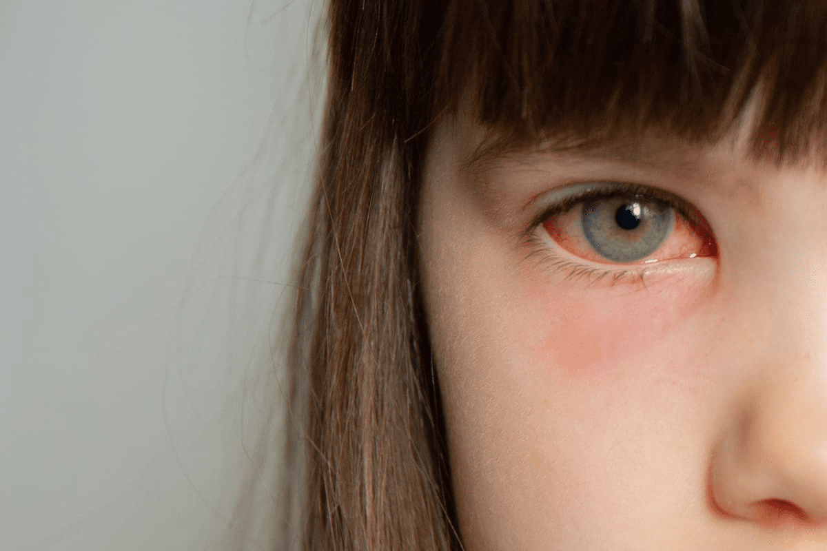
Seventh Nerve Palsy (Bell’s Palsy): Effects on the Eye
The facial nerve, or seventh cranial nerve, is key to eye health. Its palsy, known as Bell’s Palsy, impacts the muscles around the eye. This can lead to serious issues with the eye’s surface.
Inability to Close the Eye
Seventh nerve palsy makes it hard to shut the eye fully, known as lagophthalmos. This can expose the cornea, causing damage and pain.
Incomplete eyelid closure can lead to dry eye, irritation, and even corneal injury. It’s important to use lubrication and protective measures.
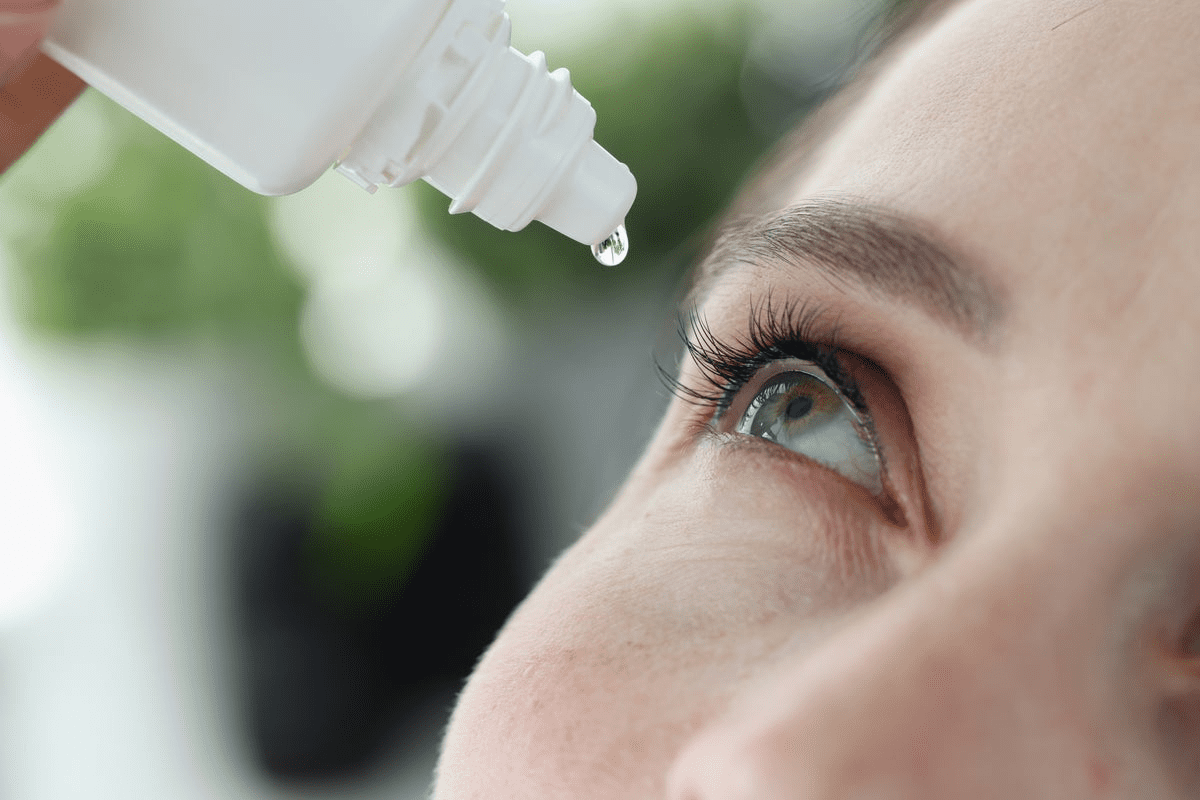
Dry Eye Complications
Dry eye is a big worry for those with seventh nerve palsy. Not being able to close the eye right disrupts tear distribution. This causes dry spots on the cornea.
We suggest using lubricating eye drops to fight dry eye symptoms and protect the eye’s surface.
Corneal Protection Concerns
Keeping the cornea safe is vital with seventh nerve palsy. The cornea is at risk from exposure and dryness. This can cause ulcers or infections if not managed well.
Using eyelid taping or patching at night can protect the cornea until the condition gets better.
Common Causes of Cranial Nerve Palsies
Cranial nerve palsies can come from many sources. These include vascular, traumatic, and inflammatory conditions. Knowing what causes them is key to treating them well.
Vascular Conditions
Vascular issues are a big reason for cranial nerve palsies. Diabetes and hypertension often play a part. They can damage the tiny blood vessels that nerves need to function.
This damage can cut off the nerves’ blood supply. This leads to palsies.
Trauma and Injury
Head or face injuries can harm cranial nerves. This damage can happen directly or through hemorrhage or edema. The severity and where the injury is affect how much damage is done.
Tumors and Space-Occupying Lesions
Tumors, whether they’re benign or cancerous, can press on or invade cranial nerves. This causes palsies. Meningiomas and schwannomas are types of tumors that can do this. Finding them early is important for treatment.
Inflammatory and Infectious Causes
Inflammation and infections can also cause cranial nerve palsies. Multiple sclerosis, sarcoidosis, and infections like Lyme disease or herpes zoster can harm nerves. Treating these quickly is important to avoid lasting damage.
In summary, cranial nerve palsies have many causes. These include vascular issues, trauma, tumors, and inflammation or infections. Knowing the causes helps in managing and treating them.
Diagnosis and Management
Diagnosing cranial nerve palsies requires a detailed approach. This includes clinical exams and specific tests. Getting the diagnosis right is key to effective treatment.
Clinical Examination Techniques
Clinical exams are vital for diagnosing cranial nerve palsies. We check visual acuity, ocular alignment, motility, pupil reaction, and eyelid function. This helps pinpoint the affected nerve and guides further tests.
A detailed clinical exam includes:
- Assessment of eye movements
- Evaluation of pupil size and reactivity
- Examination of eyelid position and function
- Testing of visual acuity
Neuroimaging Studies
Neuroimaging is essential for finding the cause of cranial nerve palsies. MRI and CT scans help spot structural issues, tumors, or vascular problems affecting the nerves.
The right neuroimaging depends on the suspected cause and the nerve involved.
Neuroimaging Modality | Use in Cranial Nerve Palsies |
MRI | Excellent for soft tissue evaluation, including cranial nerves |
CT Scan | Quick and useful for detecting acute hemorrhages or bony abnormalities |
Laboratory Tests
Laboratory tests are key for finding systemic causes of cranial nerve palsies. Tests for diabetes, infections, or inflammatory conditions are important. Blood tests help diagnose these conditions.
Common tests include:
- Blood glucose testing
- Complete blood count (CBC)
- Erythrocyte sedimentation rate (ESR)
- C-reactive protein (CRP)
Differential Diagnosis Considerations
Differential diagnosis is critical in managing cranial nerve palsies. We must consider various conditions that could mimic or cause cranial nerve palsies. This ensures we don’t miss serious conditions.
“A thorough differential diagnosis is essential to accurately diagnose and manage cranial nerve palsies.”
Treatment Approaches for Different Cranial Nerve Palsies
Treating cranial nerve palsies needs a detailed plan. It depends on the nerve and the cause. Knowing the condition well helps choose the best treatment.
Medical Management Strategies
Medical treatment is often the first step. It aims to fix the root cause, like vascular issues or inflammation. For example, managing diabetes and high blood pressure is key for some nerve problems.
Key aspects of medical management include:
- Managing underlying conditions such as diabetes and hypertension
- Using medications to control inflammation or infection
- Implementing measures to reduce the risk of further nerve damage
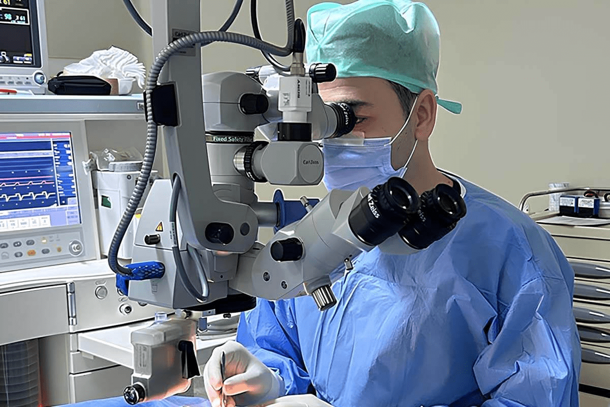
Surgical Interventions
Surgery might be needed for some nerve issues. It can relieve pressure, repair nerves, or fix other problems. Surgery is considered when nerve function is severely affected or when other treatments don’t work.
Surgical interventions may involve:
- Decompression surgery to relieve pressure on the nerve
- Nerve repair or grafting in cases of nerve damage
- Other surgical procedures to correct anatomical abnormalities
Prism Glasses and Visual Aids
Prism glasses and visual aids help with symptoms like double vision. They align images for the affected eye. This makes vision more comfortable and functional.
The benefits of prism glasses include:
- Reducing double vision and improving visual comfort
- Enhancing binocular vision and depth perception
- Providing a non-invasive solution for managing diplopia
Eye Patching and Occlusion Therapy
Eye patching or occlusion therapy helps with double vision and other vision problems. Covering the affected eye can give relief from these symptoms.
Eye patching can be useful:
- In the acute phase of cranial nerve palsy
- When prism glasses are not sufficient or feasible
- As a temporary measure until other treatments take effect
Conclusion
Cranial nerve palsies greatly affect how our eyes work. They can cause double vision, eyelids to droop, and limit eye movement. It’s key to know the causes, symptoms, and treatment options to manage them well.
We’ve looked at the five main cranial nerves that affect our eyes. We’ve also covered what causes these palsies and how to diagnose them. Treatments include medical care, surgery, and using visual aids.
Many people with cranial nerve palsies see improvement, thanks to treatable causes and managing risk factors. Getting professional help is vital for the best results. Knowing about cranial nerve palsies helps people deal with their eye issues better.
In summary, dealing with cranial nerve palsies is complex and needs thorough care. We stress the need for quick medical help and ongoing support to handle these issues well.
FAQ
What is cranial nerve palsy?
Cranial nerve palsy is when one or more cranial nerves don’t work right. This can cause problems with the eyes, face, and other areas.
What are the common symptoms of cranial nerve palsies?
Symptoms include double vision, eyelids that droop, and trouble moving the eyes. Facial weakness can also happen, depending on the nerve.
What causes cranial nerve palsies?
Causes include vascular issues, trauma, and tumors. Inflammatory and infectious diseases can also cause it. A detailed diagnosis is needed.
How are cranial nerve palsies diagnosed?
Doctors use clinical exams, neuroimaging, and lab tests. These help find the cause and how much damage there is.
What treatment options are available for cranial nerve palsies?
Treatments vary. They can include medicine, surgery, prism glasses, and eye patching. Each case is different, so treatments are tailored.
Can cranial nerve palsies be treated effectively?
Yes, with the right diagnosis and treatment, many people see big improvements. Their quality of life can get much better.
What is the role of the oculomotor nerve (CN III) in eye function?
The oculomotor nerve controls most eye movements. It also helps the pupil get smaller and the eyelid open. It’s very important for eye function.
How does third nerve palsy affect the eye?
Third nerve palsy can cause eyelids to droop and limit eye movement. It can also affect the pupil and cause double vision. It greatly impacts how well the eye works.
What are the effects of sixth nerve palsy on eye movement?
Sixth nerve palsy stops the eye from moving outward. This leads to double vision and eye misalignment. It affects how the eyes move side to side.
How does seventh nerve palsy (Bell’s Palsy) affect the eye?
Seventh nerve palsy makes it hard to close the eye. This can cause dry eyes and problems with protecting the cornea.
Are there any specific considerations for children with cranial nerve palsies?
Yes, kids need special care, like with fourth nerve palsy. It can affect their vision and how they develop.
What is the importance of seeking professional care for cranial nerve palsies?
Getting professional help is key. It ensures the right diagnosis and treatment. Early action can greatly improve symptoms and quality of life.
References
National Center for Biotechnology Information. Cranial Nerve Palsies: Impact on Eye Movement and Vision. Retrieved from https://pmc.ncbi.nlm.nih.gov/articles/PMC12087672/
