Last Updated on November 27, 2025 by Bilal Hasdemir
Panoramic x ray radiation is a key concern for patients undergoing dental imaging. At Liv Hospital, we educate patients about the risks of medical imaging, including radiation exposure from tools like panoramic x ray radiation and CT scans. Our advanced technology ensures safety and high-quality care. The average American is exposed to about 3 mSv of radiation from natural sources each year. A single chest x-ray delivers about 0.1 mSv, equivalent to 10 days of natural exposure, while panoramic x ray radiation typically involves a lower dose. For more details on radiation doses from CT scans and other imaging methods, check out this resource.

Key Takeaways
- Panoramic x-rays involve relatively low exposure xray compared to CT scans.
- The average effective dose for panoramic dental x-rays ranges from 4 to 30 microsieverts.
- A chest x-ray exposes patients to about 0.1 mSv of radiation.
- CT scans deliver significantly more radiation than x-rays.
- Low-Dose Computed Tomography (LDCT) reduces radiation exposure by around 37%.
Understanding Radiation Measurement Units in Medical Imaging
To understand the safety of medical imaging, we need to know the units for measuring radiation. These units help us see how much radiation we get from different imaging tests. This knowledge is key for both doctors and patients.
The Science Behind X-Ray Exposure Units: Sieverts, Grays, and Microsieverts
The energy from radiation that our bodies absorb is called an absorbed dose. It’s measured in a unit called the gray (Gy). The dose that takes into account how different types of radiation affect us is called the sievert (Sv).
For x-rays, we often talk about the dose in microsieverts (μSv). This is one-millionth of a sievert. For example, a chest x-ray might give you about 10 μSv.
Let’s compare this to background radiation. The average person gets about 2.4 millisieverts (mSv) of background radiation each year. Knowing these units helps us see how medical imaging compares to everyday radiation.
Interpreting X-Ray Radiation Dose Charts for Patient Safety
X-ray radiation dose charts are vital for keeping patients safe. They show the radiation doses for different x-ray tests. These charts help doctors talk about the risks and benefits of imaging tests with their patients.

For instance, a panoramic dental x-ray might expose you to 4-30 μSv. A chest x-ray is about 10 μSv. By looking at these numbers, patients and doctors can decide if an x-ray is really needed.
| X-Ray Procedure | Typical Radiation Dose |
| Panoramic Dental X-Ray | 4-30 μSv |
| Chest X-Ray | 10 μSv |
| CT Abdomen | 5-20 mSv |
In summary, knowing about sieverts, grays, and microsieverts is key. It helps us understand x-ray radiation dose charts and keep patients safe in medical imaging.
Panoramic X-Ray Radiation and Dental Imaging Safety
Panoramic x-rays are common in dental offices. It’s important to know how much radiation they use. We look at how safe panoramic x-rays and other dental images are, focusing on their radiation levels.
Low Exposure Profile: 4-30 Microsieverts in Dental X-Rays
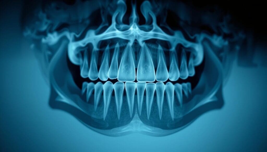
The dose from panoramic dental x-rays is about 4 to 30 microsieverts (μSv). This depends on the machine and settings used. This amount is quite low compared to other medical scans.
To understand better, let’s compare radiation doses from different dental scans.
| Dental Imaging Technique | Average Radiation Dose (μSv) |
| Panoramic X-Ray | 4-30 |
| Intraoral X-Ray | 1-10 |
| CBCT Scan | 30-1000 |

Equivalent Natural Radiation: How Dental X-Rays Compare to Daily Exposure
The dose from one panoramic x-ray is like a few days of natural background radiation. Natural background radiation is the ionizing radiation we all get from the environment. It’s measured in places far from any radiation sources.
This comparison shows that panoramic x-rays use a small amount of radiation. It’s similar to what we get from natural sources every day.
In summary, panoramic x-rays and other dental scans use a small amount of radiation. Knowing the dose and comparing it to natural background radiation helps ease worries about radiation safety in dental imaging.
Chest X-Rays vs. CT Scans: Understanding the Radiation Difference
Medical imaging techniques like chest x-rays and CT scans have different radiation profiles. We will explore these differences in detail. It’s important to understand these differences to assess the risks and benefits of each diagnostic tool.
CXR Radiation Levels: The 0.1 Millisievert Benchmark
Chest x-rays (CXR) are used to check lung and heart conditions. A chest x-ray exposes a patient to about 0.1 millisieverts (mSv) of radiation. This dose is low and is often used as a baseline for comparing other diagnostic imaging procedures.
CT Abdomen Radiation Dose: 5-20 Millisieverts Per Scan
CT scans of the abdomen use much higher doses of radiation. A CT scan of the abdomen and pelvis can expose a patient to around 10 mSv of radiation. The range is usually between 5 to 20 mSv per scan.
The difference in radiation exposure between chest x-rays and CT scans is significant. It shows the importance of choosing the right diagnostic tool for each patient. While CT scans offer detailed images, they also come with a higher radiation risk.
Healthcare providers must balance the benefits of CT scans with the risks of radiation exposure. This knowledge helps in educating patients about the risks and benefits of various diagnostic procedures.
Putting Medical Imaging Radiation in Perspective
Many patients worry about medical imaging radiation. But how does it compare to the radiation we get from nature? Understanding this helps us see the risks of medical imaging.
Comparing Medical Imaging to Natural Background Radiation
The average American gets about 3 mSv of radiation from nature each year. A chest X-ray, for example, exposes a patient to about 0.1 mSv. This is like 10 days of natural background radiation.
Diagnostic imaging, like CT scans, might use more radiation, up to 5-20 mSv per scan. But, these doses are similar to what some people get from nature over the years. It’s important to think about the benefits and risks of these tests.
Modern Protocols for Minimizing Radiation Exposure in Diagnostic Imaging
At Liv Hospital, we follow the latest international protocols to lower radiation exposure. We use the newest technology to reduce doses without losing image quality. We also follow the “as low as reasonably achievable” (ALARA) principle.
Key strategies for minimizing radiation exposure include:
- Using alternative imaging techniques that don’t involve radiation, such as ultrasound or MRI, when possible.
- Optimizing scanning protocols to reduce dose while maintaining diagnostic image quality.
- Implementing dose management systems to track and minimize radiation exposure over time.
- Ensuring that all personnel are trained in the latest dose reduction techniques.
By using these modern protocols, we aim to provide top-notch diagnostic imaging while keeping radiation exposure low. It’s a balance between getting the needed information and keeping patients safe.
Conclusion: Balancing Diagnostic Benefits with Radiation Awareness
Medical imaging is complex, and knowing about radiation is key to keeping patients safe. While there’s a risk of radiation from imaging, the benefits often make it worth it. We need to find a balance between getting accurate diagnoses and the risks of radiation.
We’ve talked about how different imaging tests use different amounts of radiation. From dental X-rays to CT scans, each has its own level. Knowing this helps doctors use less radiation while keeping tests effective.
Being aware of radiation is important for making smart choices in medical imaging. Patients and doctors can work together to get the most from tests while using less radiation. This approach is vital for quality care in medical imaging.
FAQ
What are the standard units used to measure radiation exposure in medical imaging?
Sieverts (Sv), grays (Gy), and microsieverts (μSv) are the standard units. They measure how much radiation the body absorbs during imaging.
How much radiation is typically associated with a chest x-ray?
Chest x-rays usually expose patients to about 0.1 millisieverts (mSv) of radiation.
What is the radiation dose associated with a CT scan of the abdomen?
CT scans of the abdomen can expose patients to 5 to 20 millisieverts (mSv) per scan.
How does the radiation exposure from dental x-rays compare to natural background radiation?
Dental x-rays expose patients to low levels of radiation, about 4-30 microsieverts (μSv). This is similar to the natural background radiation we get every day.
What is the difference in radiation exposure between a chest x-ray and a CT scan?
CT scans have much higher radiation doses than chest x-rays. While a chest x-ray is about 0.1 mSv, a CT scan of the abdomen can be 5-20 mSv.
How can radiation exposure be minimized in diagnostic imaging?
Modern protocols, like those at Liv Hospital, aim to reduce radiation. They use the lowest doses needed for clear images.
How do x-ray radiation dose charts help in patient safety?
X-ray dose charts help standardize and compare radiation exposure. They guide healthcare providers to reduce radiation risks.
What is the importance of understanding radiation measurement units in medical imaging?
Knowing radiation units is key for healthcare providers. It helps them accurately assess and reduce radiation exposure. This ensures patients get the benefits of imaging without risks.
References
- Radiation Exposure of Medical Imaging. (n.d.). In StatPearls. https://www.ncbi.nlm.nih.gov/books/NBK565909/


