We are on the cusp of a revolution in medical imaging, thanks to PET Fluorodeoxyglucose (18F-FDG) scans. At Liv Hospital, we focus on evidence-based, patient-centered care. This ensures every scan is precise and trustworthy.
18F-FDG is a glucose analog used in PET imaging. It helps assess tissue metabolism. The National Center for Biotechnology Information says it’s used for diagnosing and monitoring conditions like cancer and heart disease.
Understanding Fludeoxyglucose F 18 helps us see its importance in medical imaging. It builds up in tissues that use a lot of glucose, like tumors. This makes it possible to diagnose and monitor treatments accurately.

18F-FDG is a key tool in PET imaging for diagnosing diseases. It’s a radioactive glucose analog. Let’s explore its definition, chemical structure, and history.
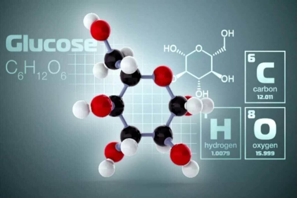
Fluorodeoxyglucose (FDG) is a glucose molecule with fluorine-18 added. This makes it radioactive and useful for PET scans. Its chemical structure is close to glucose but has fluorine-18 instead of a hydroxyl group.
This change lets 18F-FDG be taken up by cells like glucose. But, it gets stuck inside cells after being phosphorylated. This makes it detectable.
The chemical formula for 18F-FDG is C6H11FO5. It’s designed to enter cells through glucose transporters. Inside, it’s phosphorylated by hexokinase, trapping it in cells.
The journey of 18F-FDG began in the 1970s. Researchers saw its value in studying cell metabolism. But, making it was tough because fluorine-18 has a short half-life.
The first human PET scan with 18F-FDG happened in the late 1970s. It marked a new chapter in nuclear medicine. Now, 18F-FDG is a mainstay in diagnosing diseases in oncology, neurology, and cardiology.
Today, 18F-FDG is vital in PET imaging. Ongoing research aims to enhance its use and create new radiotracers.

It’s important to know how 18F-FDG works as a glucose analog. This is key to understanding its use in medical imaging. 18F-FDG, or Fluorodeoxyglucose, acts like glucose in cells.
We use 18F-FDG in PET scans to see how glucose is used in different tissues. This is very useful for finding cancer and studying the brain.
Normal cells use glucose to make energy. 18F-FDG gets into cells like glucose does. It’s then changed into 18F-FDG-6-phosphate by hexokinase.
This 18F-FDG-6-phosphate can’t be broken down further. So, it stays in the cell. This lets us check how much glucose a cell is using.
“The metabolic trapping of 18F-FDG provides a snapshot of glucose metabolism, enabling the evaluation of tissue viability and function.”
The way 18F-FDG gets trapped in cells is what makes it useful for PET scans. Once it’s changed into 18F-FDG-6-phosphate, it can’t be used by cells anymore.
This means 18F-FDG builds up in cells that use a lot of glucose. This helps us find tumors and other active areas in the body.
| Cell Type | Glucose Uptake | 18F-FDG Accumulation |
| Normal Cells | Moderate | Low to Moderate |
| Cancer Cells | High | High |
| Inflammatory Cells | Variable | Variable |
Knowing how 18F-FDG works helps us see its value in finding and treating diseases. It’s very useful for cancer diagnosis.
PET scanning uses special materials called radiopharmaceuticals or radiotracers. For fluorodeoxyglucose PET (FDG), the material is F-18 fluorodeoxyglucose. It’s a glucose-like substance that cells in the body take up.
The positron emission process is key to PET imaging. When F-18 fluorodeoxyglucose decays, it releases positrons. These are the opposite of electrons.
These positrons then collide with electrons, causing an annihilation event. This event releases energy as gamma rays.
A study on the National Center for Biotechnology Information website explains this process. It shows how detecting these gamma rays is vital for creating PET images.
PET scanners catch the gamma rays from the annihilation event. The data from these detections helps create detailed images of the body’s metabolic activity.
The steps to make these images are:
Understanding PET fluorodeoxyglucose imaging shows its importance in medicine. It helps us see how the body works. This knowledge is key for diagnosing and treating many diseases.
18F-FDG PET/CT scans are key in managing cancer. They help in detecting, diagnosing, staging, and grading tumors. They also check how well treatments work.
18F-FDG PET/CT scans are great for finding and diagnosing cancers. They work because cancer cells use more glucose than normal cells. This makes them show up well on scans.
Key benefits of using 18F-FDG in cancer detection include:
Knowing the stage and grade of a tumor is vital. It helps doctors plan treatment and predict outcomes. 18F-FDG PET/CT scans help see how far cancer has spread and how active it is.
| Tumor Type | Role of 18F-FDG PET/CT |
| Lymphoma | Staging and assessing treatment response |
| Lung Cancer | Staging and evaluating metastasis |
| Breast Cancer | Assessing recurrence and metastasis |
18F-FDG PET/CT scans also check if a tumor is responding to treatment. They look at how the tumor’s activity changes. This helps doctors see if the treatment is working.
We use 18F-FDG PET/CT scans to care for cancer patients fully. From diagnosis to treatment and follow-up, they are essential. This imaging is a key part of modern cancer care.
The use of Fludeoxyglucose F 18 in neurology has changed how we diagnose and treat many conditions. Fludeoxyglucose F 18 PET imaging helps us see how the brain uses energy. This is key to understanding many brain disorders.
18F-FDG PET scans are very helpful in studying dementia and other brain diseases. They show us how the brain uses glucose, which helps us spot certain types of dementia like Alzheimer’s. These scans also help us see how treatments like donepezil work.
They let doctors see the brain’s changes, helping them tell different types of dementia apart. This makes it easier to find the right treatment for each patient.
Fludeoxyglucose F 18 PET imaging is key in finding where seizures start in people with epilepsy. It shows us which parts of the brain are active during seizures. This info is super important for planning surgery and finding the best treatment.
Thanks to 18F-FDG PET, doctors can now treat epilepsy more effectively. This leads to better outcomes for patients.
18F-FDG PET scans are also important for checking brain tumors. They show how active the tumor is, which helps doctors figure out how serious it is. This is hard to do with regular scans.
With fluor 18 fdg PET imaging, doctors can make treatment plans that are just right for each patient. This improves their care and chances of recovery.
F-FDG PET imaging is key in checking heart health. It helps see if heart muscle is alive and spots heart inflammation. This tool is vital in cardiology, helping doctors make better treatment plans and better patient care.
Checking if heart muscle is alive is key for treating heart disease. F-FDG PET scans find hibernating heart muscle, which is alive but not working well. This helps doctors choose the right treatments, like surgery or stents.
The steps are:
This info is key for making a treatment plan that helps the patient recover better.
F-FDG PET scans are also great for finding and managing heart inflammation. They show where inflammation is in the heart. This is helpful when other tests don’t give clear answers.
The main benefits are:
Using F-FDG PET imaging helps doctors get better at diagnosing and treating heart problems.
In summary, F-FDG PET scans are a big step forward in heart disease diagnosis and treatment. They give detailed views of heart muscle and inflammation. This helps doctors make better choices and care for their patients better.
To get the best results from F 18 FDG PET scans, following specific clinical protocols is key. These protocols cover important steps like patient preparation, how much and when to give the drug, and the timing and details of the scan.
Getting ready for a F 18 FDG PET scan is very important. Patients usually need to fast for a few hours before the scan to make normal tissues take up less glucose. This helps show the difference between normal and cancerous tissues better. We also tell patients to avoid hard exercise and drink plenty of water before the scan.
For diabetic patients, controlling blood sugar is even more critical. High blood sugar can affect how 18F-FDG is taken up, which might make the images not as good. So, we ask diabetic patients to check their blood sugar levels before the scan and adjust their meds or insulin as their doctor suggests.
How much 18F-FDG is given and how it’s given are very important. Patients usually get between 5 to 15 mCi (185 to 555 MBq) of 18F-FDG, based on their weight and the PET/CT system used. The drug is given through an IV, and the patient rests for about 60 minutes after.
The imaging for F 18 FDG PET scans includes both PET and CT scans. The PET scan shows how tissues are metabolically active, while the CT scan shows their anatomy. The scan is usually done about 60 minutes after the 18F-FDG is given, so the drug can build up in the tissues.
We also think about how the patient is positioned, how long the scan takes, and what area it covers. Modern PET/CT systems help us get clearer, more accurate images.
The use of 18F-FDG PET/CT has changed how doctors manage patients. It gives both functional and anatomical details. This helps doctors make better decisions and improve treatment plans.
Research shows 18F-FDG PET/CT changes patient care in up to 27% of cases. It’s great at staging diseases, checking how treatments work, and finding cancer again. For example, a study in the Journal of Nuclear Medicine found it changed treatment plans for 23% of lymphoma patients.
This imaging isn’t just for cancer. It’s also useful in neurology and cardiology. In epilepsy, it helps find where seizures start, helping doctors plan surgery.
Many case studies show how 18F-FDG PET/CT helps doctors. A patient with ovarian cancer thought to have come back was found to have widespread disease. This changed their treatment to chemotherapy instead of surgery.
Another example is a lung cancer patient. The PET/CT scan showed cancer in the mediastinum, changing their treatment to focus on comfort instead of trying to cure.
These stories show how 18F-FDG PET/CT changes patient care. It helps doctors make better choices, leading to better outcomes for patients.
FDG-PET has changed medical imaging a lot. But, it’s not perfect. Knowing its limits helps us use it better and understand its results.
FDG-PET often shows false positives, mainly in inflammatory conditions. For example, FDG-PET can show high false positives in lymph nodes. This can confuse doctors into thinking benign conditions are cancer.
Infections, inflammation, and granulomatous diseases can also cause false positives. To avoid this, doctors need to look at the patient’s history and other tests. Using PET/CT or PET/MRI can help by giving more detailed images.
FDG-PET works differently for different cancers. Some cancers, like small or low-activity tumors, might not show up. For example, some prostate cancers and neuroendocrine tumors are hard to find with FDG-PET.
It’s important to know how different cancers take up FDG. This helps choose the right imaging and understand the results.
There are technical issues with FDG-PET imaging. Things like how the patient prepares, when the scan is done, and how the images are made can affect results. If the patient eats glucose before the scan, it can mess up the images.
Reading FDG-PET images also needs skilled people. It’s complex and requires knowledge in nuclear medicine and the patient’s situation.
By facing these challenges, we can make FDG-PET imaging better. This will help doctors make more accurate diagnoses and improve patient care.
Fluorodeoxyglucose (FDG) PET technology has seen big changes. These changes are making medical imaging better and helping patients more. New PET imaging ways are improving how we diagnose and treat diseases.
PET and Magnetic Resonance Imaging (MRI) together is a big step forward. This mix gives us detailed images of the body’s soft tissues. It’s great for oncological and neurological applications, giving us a deeper look at diseases.
This combo is also good for reducing radiation and getting more info in one go. For example, in neuroimaging, it helps spot and track neurodegenerative diseases early.
Digital PET systems are another big leap. They are more sensitive and clear than old PET scanners. This means we can spot and understand lesions better, even in hard-to-reach spots.
These systems also scan faster and might use less f-18 fluorodeoxyglucose. This makes patients more comfortable and cuts down on radiation. Soon, digital PET will be common in nuclear medicine.
Artificial intelligence (AI) is changing how we look at PET images. AI can automatically measure PET values and find patterns we might miss. This makes diagnosing diseases more accurate and quicker.
AI in PET imaging helps doctors make better decisions faster. As AI gets better, we’ll see even more ways it can help with fluoro deoxy glucose PET scans.
In short, new FDG PET tech, like PET/MRI, digital PET, and AI, is making PET scans better. These updates are making PET scans a key tool in fighting diseases. They promise to keep improving how we diagnose and treat illnesses.
The future of 18F-FDG in medical imaging looks bright. We’re seeing new uses and better tech. 18F-FDG is key for diagnosing and treating many health issues.
New uses for 18F-FDG are being found, beyond just cancer. It’s now used in neurology to study brain diseases. Studies show it helps spot Alzheimer’s early, so we can act fast.
In heart care, 18F-FDG PET scans check heart health and find heart inflammation. This is vital for knowing who needs heart surgery. Knowing this helps doctors make better choices for patients.
New tracers and imaging methods are being developed fast. Scientists are mixing 18F-FDG with other tracers for better info. PET/MRI tech is a big step, giving both function and anatomy in one scan.
AI and machine learning are being used in 18F-FDG PET scans. They help analyze images better and tailor treatments. AI in PET imaging is very promising.
As research goes on, we’ll see more 18F-FDG PET imaging breakthroughs. From better tracers to new tech, 18F-FDG will become even more vital in medicine.
We’ve looked at how pet fluorodeoxyglucose (18F-FDG) is used in medical imaging. It’s key in finding and managing many diseases. 18F-FDG PET/CT is a big help in treating cancer, brain issues, and heart problems.
The way FDG moves through the body helps doctors understand diseases better. A study in Frontiers in Nuclear Medicine shows 18F-FDG PET/CT is good at spotting cancer. But, it’s not perfect and can miss some cases.
As medical imaging gets better, 18F-FDG will play an even bigger role. It will help find new ways to diagnose and treat diseases. So, 18F-FDG will keep being a key part of modern medicine.
18F-FDG, or fluorodeoxyglucose F 18, is a special kind of sugar used in PET scans. It helps doctors see how tissues in the body work. This is key for finding and tracking diseases like cancer, brain issues, and heart problems.
18F-FDG acts like regular sugar to cells. Cells take it in and hold onto it. This lets doctors see how cells use sugar, helping spot healthy and sick cells.
In cancer care, 18F-FDG helps find tumors, figure out how big they are, and check if treatments are working. It’s very useful for cancers like lymphoma, lung cancer, and breast cancer.
Neurologists use 18F-FDG to find and manage brain diseases, like dementia. It also helps find where seizures start and check for brain tumors. It shows how the brain works and metabolizes.
In heart care, 18F-FDG helps see if heart muscle is alive and diagnose heart inflammation. It spots areas of heart muscle that can recover and finds heart inflammation.
For F 18 FDG PET scans, patients must fast and get the tracer through an IV. There are specific rules for how to prepare, give the tracer, and when to take the images.
18F-FDG PET/CT changes how doctors plan treatment. It gives detailed info on disease spread and treatment success. This leads to better care for patients.
FDG imaging can sometimes show false positives, vary in tumor uptake, and face technical and reading challenges. These need careful thought when looking at images.
New tech in FDG PET includes PET/MRI, digital systems, and AI. These advancements improve image quality, accuracy, and make things more efficient.
The future for 18F-FDG includes new uses, research, and innovations. This could mean more uses, better imaging methods, and combining with other imaging.
18F-FDG is used to check how cells use sugar. It gets taken in by cells and stays there, showing how sugar is used in different tissues.
18F-FDG is key for spotting and tracking diseases. It shows how tissues use sugar, helping doctors find and manage diseases like cancer, brain issues, and heart problems.
Subscribe to our e-newsletter to stay informed about the latest innovations in the world of health and exclusive offers!
WhatsApp us