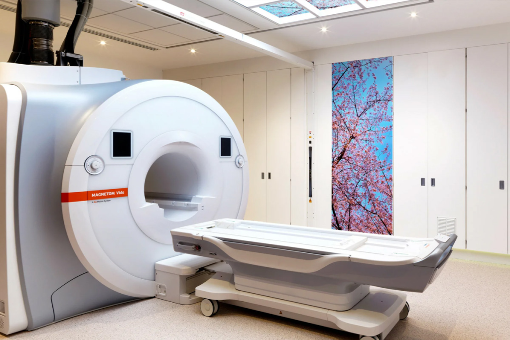
Nearly 90% of cancer deaths are due to metastasis. This is when cancer cells move from their original site to other parts of the body. It’s key to know how cancer metastasis happens. This knowledge helps in managing cancer better. PET-CT is a tool that combines PET scan cancer staging and CT scan. It’s very important for finding metastatic cancer, mainly in lung imaging.
With PET-CT, doctors can spot where cancer has spread. This helps them decide the best treatment plan.
Metastasis is when cancer cells spread to other parts of the body. It’s a complex process that changes how we treat and predict cancer outcomes.
Metastasis happens when cancer cells from the main tumor break off. They travel through the blood or lymphatic system. Then, they form new tumors in other organs.
Invasion is the first step where cancer cells invade nearby tissues. They then enter the bloodstream or lymphatic vessels, a process known as intravasation. Once in the circulatory system, cancer cells can travel to distant sites. There, they may exit the bloodstream or lymphatic vessels, a process called extravasation, and colonize new tissues.
The spread of cancer cells is not random. It involves a complex interaction between cancer cells and the host’s environment. Cancer cells must evade the immune system, adapt to new environments, and get the nutrients and oxygen they need to survive and grow.
“The metastatic process is highly inefficient, with only a small fraction of cancer cells successfully forming metastases.”
Understanding how cancer metastasizes is key to finding better treatments. Advanced imaging, like high-resolution CT scans, helps detect and track metastasis. This allows doctors to accurately stage cancer and choose the best treatment plans.
Metastasis is a key part of cancer growth. Knowing where cancer spreads is vital for treatment. Cancer cells often go to certain places in the body. This is because of blood flow and the environment of each organ.
Lymph nodes are a common place for cancer to spread. They filter lymph fluid, catching bad cells. Cancer cells can move to lymph nodes and grow, affecting cancer staging and prognosis.
Bones are often hit by cancer, like in breast, prostate, and lung cancer. Cancer cells reach bones through blood or lymph, causing damage. Common spots include the spine, pelvis, and long bones.
The liver is a common spot for cancer to spread, mainly from the gut. Its blood flow and filtering role make it a prime target. Liver metastases can greatly affect patient outcomes, needing special treatments.
Lungs can get cancer from many sources, like sarcomas, breast, and colon cancer. Their high blood flow and filtering role make them a common site. Low-dose CT scans help find lung metastases, useful for screening.
Knowing where cancer spreads is key to managing it. Imaging, like low-dose CT scans, is vital for spotting metastasis in these areas.
Different cancers spread in unique ways, affecting how we treat them. Knowing these patterns helps doctors diagnose and treat cancer better.
Breast cancer often goes to the bones, lungs, liver, and brain. The spread can depend on the type of breast cancer.
Bone metastasis is common in hormone receptor-positive breast cancer. It causes a lot of pain and discomfort.
Lung cancer spreads early, often to the brain, bones, liver, and adrenal glands. The spread pattern affects treatment choices.
Screening with low-dose CT scans can catch lung cancer early. This improves treatment outcomes.
Colorectal cancer usually spreads to the liver, lungs, and peritoneum. Knowing where it spreads helps plan treatment.
The liver is the most common place for metastasis. Removing the liver can cure some patients.
Prostate cancer often goes to the bones, like the spine, pelvis, and ribs. The spread pattern affects treatment choices.
Bone metastasis in prostate cancer can cause a lot of pain and fractures.
The following table summarizes the common metastatic patterns of these cancers:
| Cancer Type | Common Metastatic Sites |
| Breast Cancer | Bones, Lungs, Liver, Brain |
| Lung Cancer | Brain, Bones, Liver, Adrenal Glands |
| Colorectal Cancer | Liver, Lungs, Peritoneum |
| Prostate Cancer | Bones (Spine, Pelvis, Ribs) |
Cancer staging is key in oncology. It gives vital info for treatment plans and how well a patient might do. It’s a detailed process that checks how far cancer has spread in the body.
Getting cancer staging right is very important. It affects how doctors care for patients and the results of treatment. Knowing the cancer’s stage helps doctors choose the best treatment. This could be surgery, chemo, radiation, or a mix of these.
The TNM system is a common way to stage cancer. It looks at three main things: the tumor’s size and spread (T), nearby lymph nodes (N), and if cancer has spread to distant parts (M).
| TNM Component | Description |
| T (Tumor) | Size and extent of the primary tumor |
| N (Node) | Involvement of nearby lymph nodes |
| M (Metastasis) | Presence of distant metastasis |
The cancer’s stage is very important for treatment choices. For cancers caught early, treatments like surgery or radiation might be used. But for more advanced cancers, treatments like chemo or targeted therapy might be needed.
PET-CT scans are very helpful in cancer staging. They show detailed images of the body’s activity. This helps find cancer spread that might not show up on other tests.
Getting cancer staging right is vital. It makes sure patients get the best care for their cancer. This improves their chances of a good outcome and better quality of life.
PET scans are now the top choice for finding out how far cancer has spread. This new way of imaging has changed how doctors diagnose cancer. It gives them detailed info on where the cancer is.
PET scans use a tiny bit of radioactive sugar to spot cancer cells. Cancer cells use more sugar than normal cells, so they show up on scans. This lets doctors see where the cancer is by looking at where the sugar is used most.
Using PET and CT scans together is a big step forward. PET scans show how cells are working, while CT scans show their shape. This combo helps doctors find and measure cancer more accurately. It also helps them plan better treatments.
PET scans, with CT, are very good at finding where cancer has spread. They are precise in spotting cancer in lymph nodes, bones, liver, and lungs. Here’s how well PET-CT scans do compared to other methods:
| Cancer Type | PET-CT Accuracy | Conventional Imaging Accuracy |
| Breast Cancer | 95% | 80% |
| Lung Cancer | 92% | 85% |
| Colorectal Cancer | 90% | 78% |
PET-CT scans are key in finding where cancer has spread. They give doctors both how cells are working and their shape. This helps doctors make better plans for treating patients.
In oncology, chest CT and high-resolution CT have changed how we find metastasis.
These methods give us detailed views of cancer spread, often to the lungs. The lungs are a common place for cancer to spread.
Standard chest CT scans are key for spotting lung metastases and seeing how far cancer has spread.
They show the whole chest area, helping doctors find tumors that X-rays can’t see.
Key Applications:
High-resolution CT (HRCT) shows more detail than regular CT scans. It’s great for looking at the lung’s inner parts.
It’s best for spotting lung diseases and studying lung nodules closely.
Benefits of HRCT:
Low-dose CT (LDCT) is a key tool for lung cancer screening, mainly for those at high risk.
It’s good at finding lung nodules early, when they’re easier to treat.
| Imaging Modality | Primary Use | Benefits |
| Standard Chest CT | Detecting lung metastases and disease spread | Comprehensive view of the thoracic cavity |
| High-Resolution CT | Detailed assessment of lung parenchyma and nodules | Enhanced detail, improved detection of small nodules |
| Low-Dose CT | Lung cancer screening in high-risk populations | Sensitive detection of early-stage lung nodules |
Chest X-rays are a key tool for finding big metastases, though they miss small ones. They’re often the first step to check if cancer has spread, like to the lungs.
Chest X-rays are cheap and easy to get, but they can’t spot small tumors well. They work best for finding big tumors or those that change the lung or chest a lot. They’re quick and use less radiation than CT scans.
But, chest X-rays have big downsides. They might miss early or small tumors, which can delay finding out you have cancer. They also don’t show how active tumors are, which is key for cancer staging.
Chest X-rays are good for first checks or regular follow-ups in some cases. They’re helpful for watching big tumors or checking for complications like fluid in the chest. When detailed scans aren’t available or not safe, X-rays are a good backup.
Compared to chest CT scans, X-rays give less detail but are useful in certain situations. The choice between them depends on the situation, how detailed you need the scan, and the patient’s health.
In the end, chest X-rays are a basic but important tool for finding and tracking metastasis. They’re part of a bigger plan that might include more detailed scans.
Thoracic MRI is key in finding out how far cancer has spread. It shows detailed images of soft tissues. This helps doctors diagnose and stage cancer accurately.
Thoracic MRI has advantages over other imaging methods. It can see soft tissues clearly without using harmful radiation. This is great for patients who need many scans or can’t have other types of scans.
It’s also good at showing the mediastinum and hilar regions. These areas are important for checking lymph nodes and tumor spread.
The specific applications of thoracic MRI in metastasis imaging are wide-ranging. It helps see how big primary tumors are and finds metastases in the lungs, liver, and lymph nodes. MRI is good at spotting these early, which helps plan treatments.
It’s also used to check if cancer has touched important parts like the spinal cord, big vessels, and nerves. This is key for planning surgeries and knowing how well a patient might do.
Using thoracic MRI helps doctors make better diagnoses and plans. This leads to better care and outcomes for cancer patients.
Finding interstitial lung disease in cancer patients is key to managing their care well. This disease causes inflammation and scarring in the lungs. Some cancer treatments can trigger ILD, so it’s important to keep a close eye on patients getting these treatments.
Cancer treatments like chemotherapy, radiation, and immunotherapy can cause ILD. The risk depends on the treatment type, dose, and the patient’s health. Early detection is critical to avoid permanent lung damage and adjust treatments as needed.
It’s important for doctors to understand how cancer treatment and ILD are linked. This helps them tailor imaging protocols and monitoring for high-risk patients. This approach can help lower the risk of severe ILD.
Imaging is essential in spotting ILD in cancer patients. High-resolution CT (HRCT) is the top choice for diagnosing and checking how far ILD has spread. HRCT gives clear lung images, helping doctors spot ILD patterns.
Other imaging methods might be used too. Chest X-rays can hint at ILD, but HRCT is more accurate. The right imaging method depends on the patient’s situation and the need for detailed lung checks.
New imaging technologies are changing how we find and understand cancer spread. These new tools are key for better cancer staging and treatment plans.
Molecular imaging, beyond FDG-PET, is giving us new views into cancer. Probes targeting specific molecular processes are being made. They help find metastases more accurately.
Some of these methods include:
Artificial intelligence (AI) is making image analysis better. AI can analyze complex imaging data to spot patterns humans might miss. It’s used for:
Hybrid imaging technologies combine different imaging methods. They include PET-CT and PET-MRI. These tools give both functional and anatomical details.
These advanced methods are greatly improving our ability to find and understand metastases. This leads to better care for patients.
Managing metastatic cancer requires a mix of treatments. As cancer spreads, the goal shifts from curing it to making the patient comfortable. This means controlling symptoms, improving life quality, and extending life.
Systemic therapies target cancer cells all over the body. They include:
These treatments can be used alone or together. This depends on the cancer type, stage, and the patient’s health.
Localized treatments focus on specific areas with cancer. They include:
These treatments help with symptoms like pain. They can also be used with systemic therapies.
New treatments for metastatic cancer are being researched. These include:
These new methods offer hope for better outcomes for metastatic cancer patients.
| Treatment Approach | Description | Examples |
| Systemic Therapies | Treatments that reach cancer cells throughout the body. | Chemotherapy, Targeted Therapy, Hormone Therapy, Immunotherapy |
| Localized Treatments | Treatments directed at specific areas where cancer has spread. | Surgery, Radiation Therapy, Ablation Therapy |
| Emerging Approaches | New and innovative treatments being researched. | Personalized Medicine, Combination Therapies, Clinical Trials |
Imaging and treatment advances are making a big difference in fighting cancer. PET scan cancer staging plays a key role in finding out how far cancer has spread. This helps doctors create better treatment plans for patients.
New technologies like molecular imaging and artificial intelligence are changing the game. They will make diagnosing metastasis more accurate and faster. This means better care for patients in the future.
Managing metastasis needs a team effort. This includes using treatments that target cancer cells and new ways to stop cancer from spreading. More research is needed to improve cancer care.
Advanced imaging, like PET scans, will keep being important in finding and managing metastasis. As these tools get better, we’ll see even better care for patients.
Metastasis is when cancer cells spread from where they started to other parts of the body. This happens when cancer cells break into nearby tissues, then get into the blood or lymph system. They then settle in new places.
Cancer often spreads to lymph nodes, bones, liver, and lungs. Where it goes depends on the cancer type.
PET scans, often with CT scans, help see how far cancer has spread. They spot cancer by its activity.
High-resolution CT scans show detailed lung images. They help find small cancer spots that regular CT scans might miss.
Low-dose CT scans are better for lung cancer screening. They use less radiation but can find early lung cancers or nodules.
Chest X-rays can’t find small or hidden cancer spots as well as CT or PET scans. But, they’re good for first checks or follow-ups.
Thoracic MRI is great for soft tissue images. It’s useful for seeing how far tumors have grown or finding metastases in some areas.
Interstitial lung disease is lung inflammation and scarring. It can happen with some cancer treatments, like certain drugs or radiation.
New methods include molecular imaging, artificial intelligence in images, and hybrid imaging. These aim to better find and understand metastases.
Treatments include chemotherapy, targeted therapy, and immunotherapy. There are also localized treatments like surgery or radiation. New strategies aim to control the disease and improve life quality.
Staging, like the TNM system, shows how far cancer has spread. It helps decide if treatments should be local or systemic. It also predicts how well a patient might do.
Engel, R., et al. (2024). Diagnostic accuracy and treatment benefit of PET/CT in colorectal cancer: A retrospective analysis. European Journal of Radiology, 169, 110892. https://pubmed.ncbi.nlm.nih.gov/39418774/
Butt, F., et al. (2023). Diagnostic accuracy of the latest-generation digital PET/CT scanner for detection of metastatic lymph nodes in head and neck cancer. Frontiers in Nuclear Medicine, 3, 1184448. https://www.frontiersin.org/journals/nuclear-medicine/articles/10.3389/fnume.2023.1184448/full
Osman, M. M. (2010). 18F-FDG PET/CT of patients with cancer. American Journal of Roentgenology, 195(5), 1049-1059. https://ajronline.org/doi/10.2214/AJR.09.3731
Mohammadzadeh, S., et al. (2025). Comparing diagnostic performance of PET/CT, MRI, and conventional imaging in staging head and neck squamous cell carcinoma. Oral Oncology, 134, 106234. https://www.sciencedirect.com/science/article/abs/pii/S1078817425000434
Subscribe to our e-newsletter to stay informed about the latest innovations in the world of health and exclusive offers!
WhatsApp us