Knowing the stage of cancer is key to choosing the right treatment. A PET scan is a big help in this area, as doctors often use a PET scan for cancer staging to see how far the disease has spread. According to the American Cancer Society, proper staging helps determine the extent of cancer and guides the best treatment options.
A PET scan, or Positron Emission Tomography scan, uses a special sugar solution to find cancer cells. It shows where cancer is by looking at where sugar is used most. This helps see how big the tumors are and where they are.
So, what does a PET scan show? It tells a lot about the cancer. It shows where the tumors are, how big they are, and if they have spread. This info is key to figuring out the cancer stage and planning treatment.
Key Takeaways
- A PET scan is a diagnostic tool used to detect cancer cells in the body.
- Accurate cancer staging is key for choosing the right treatment.
- A PET scan helps understand how far the disease has spread.
- The scan gives important info on tumor size, location, and spread.
- This info is vital for making a good treatment plan.
What is a PET Scan and How Does it Work?

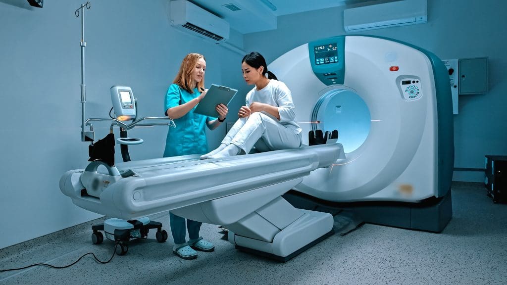
A PET scan, or Positron Emission Tomography, is a cutting-edge medical imaging method. It shows how active different parts of the body are.
To get how PET scans work, we need to know the basics. They use tiny amounts of radioactive tracers injected into the body. These tracers go to areas that are very active, like growing cancer cells.
Definition and Basic Principles
Positron Emission Tomography (PET) is a way to see inside the body. It starts with a small amount of radioactive tracer, often Fluorodeoxyglucose (FDG), being injected. This tracer is taken up by cells in the body.
The tracer then sends out positrons. These positrons meet electrons and create gamma rays. The PET scanner catches these rays to make detailed images of the body’s activity.
The Science Behind Positron Emission Tomography
PET scans rely on nuclear physics and biochemistry. When a radioactive tracer is given, it decays and sends out positrons. These positrons meet electrons and create gamma rays.
The PET scanner catches these gamma rays. It uses them to make images of how the body works. This lets -see not just what organs look like but how they function.
Types of Radioactive Tracers Used
There are many radioactive tracers for PET scans, each for different uses. The most common is FDG (Fluorodeoxyglucose). It shows how much cells use glucose.
| Tracer | Application |
| FDG (Fluorodeoxyglucose) | Cancer imaging, infection, and inflammation |
| Fluorothymidine (FLT) | Assessing cell proliferation in tumors |
| Oxygen-15 | Measuring blood flow and oxygen metabolism |
Knowing about the different tracers helps pick the right PET scan for a patient.
PET Scan for Cancer: Primary Uses in Oncology
PET scans are key in fighting cancer. They help find and treat cancer better. These scans show how tumors work, which is vital for planning treatment.
Why Oncologists Order PET Scans
Oncologists use PET scans for many reasons. They help see how far cancer has spread. They show where tumors are active and how they react to treatment.
This info is key for knowing the cancer’s stage and the best treatment. PET scans also check if treatment is working. By comparing scans, can adjust treatment plans as needed.
Types of Cancer Commonly Evaluated
PET scans check many cancers, like lymphoma and lung cancer. They’re great for cancers that grow fast or spread easily.
PET scans are vital in oncology. They help understand different cancers and plan treatments. This ensures each patient gets the right care.
FDG Uptake and Cancer Cell Metabolism
Fluorodeoxyglucose (FDG) is the main tracer in PET scans. It’s a special sugar that cancer cells love. This makes cancer cells show up bright on scans.
The more FDG a tumor takes in, the more aggressive it is. use this to decide how hard to treat the cancer. It helps them choose the best treatment plan.
Cancer Staging Fundamentals
The stage of cancer at diagnosis greatly affects treatment choices and outcomes. Staging cancer is complex. It looks at how far the tumor has spread. This is key for picking the right treatment.
The TNM Classification System Explained
The TNM system is a common way to stage cancer. It considers three main parts:
- T (Tumor): The size and spread of the main tumor.
- N (Node): If nearby lymph nodes are involved.
- M (Metastasis): If cancer has spread to distant places.
Healthcare teams use these to find the cancer’s overall stage. This helps in planning treatment.
Stages I through IV: What They Mean
Cancer stages range from I to IV. Stage I is the least severe, and Stage IV is the most advanced. Here’s a quick summary:
- Stage I: Cancer is small and hasn’t spread.
- Stage II: Cancer has grown but hasn’t spread far.
- Stage III: Cancer has reached nearby lymph nodes or tissues.
- Stage IV: Cancer has spread to distant organs or tissues.
Why Accurate Staging is Crucial for Treatment Planning
Accurate cancer staging is essential for treatment choices. For example, early-stage cancers might need surgery or local radiation. But advanced stages might require chemotherapy or targeted therapy.
Precise staging also helps predict outcomes and improves communication among healthcare teams. Knowing the cancer stage helps patients understand their diagnosis and treatment plan.
How PET Scans Contribute to Determining Cancer Stage
PET scans have changed how fight cancer. They show detailed metabolic info about tumors. This helps see how far cancer has spread, which is key for treatment.
Detecting Primary Tumor Size and Activity
PET scans are great at finding the size and activity of the main tumor. The FDG uptake level in the tumor shows how aggressive it is. Tumors with high FDG uptake need more intense treatment.
“The intensity of FDG uptake is a valuable prognostic indicator,” says a renowned oncologist. “It helps us understand the tumor’s behavior and make informed decisions about treatment.”
Identifying Regional Lymph Node Involvement
PET scans also spot if cancer has reached nearby lymph nodes. This is vital for accurate staging and treatment planning. They check the metabolic activity in lymph nodes to see if cancer has spread.
- PET scans can detect cancer spread to lymph nodes that may not be visible on other imaging tests.
- The sensitivity of PET scans in detecting lymph node involvement helps in planning surgical or radiation interventions.
Discovering Distant Metastases
PET scans are best at finding cancer in distant parts of the body. They help stage cancer right and pick the best treatment. This might include chemotherapy or targeted therapy.
As noted by the American Cancer Society, “PET scans are very good at finding metastases in organs like the liver, lungs, and bones.” This is key for knowing the cancer’s stage and guiding treatment.
What Cancer Looks Like on a PET Scan
PET scans give a special view into how cancer cells work. They show how active the cells are, unlike other scans that just show what the body looks like. This helps see how far the disease has spread.
Interpreting “Hot Spots” and Color Mapping
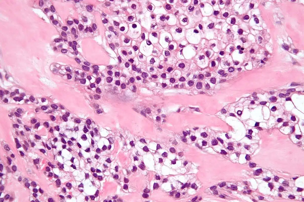
PET scan images use colors to show how active the body’s tissues are. Cancer cells, which are very active, show up as “hot spots” in red or yellow. This helps spot cancer easily.
The colors on PET scans are key to understanding the images. The most common tracer, Fluorodeoxyglucose (FDG), goes to areas that use a lot of sugar, like cancer cells. This sugar use is shown in the colors, helping see where the cancer is.
Differentiating Between Cancerous and Non-Cancerous Activity
PET scans are great at finding cancer, but they can also find other issues. This means they can sometimes show false positives. So, look at the whole picture, not just the scan, to make sure.
To tell if something is cancer, look at how much FDG is used, the pattern of uptake, and what other scans show. This helps them make sure they’re looking at cancer, not something else.
Examples of Different Cancer Types on PET Images
Each type of cancer looks different on PET scans because of how active it is. For example, fast-growing lymphomas show up brightly because they use a lot of sugar. But slower-growing tumors might be harder to see.
| Cancer Type | Typical PET Scan Appearance |
| Lymphoma | High FDG uptake, intense hot spots |
| Lung Cancer | Variable FDG uptake, often visible as a hot spot in the lung |
| Breast Cancer | Can vary; some types show high FDG uptake, while others may be less visible |
Knowing how different cancers look on PET scans is key. It helps make the right treatment plans.
The PET-CT Combination: Enhanced Diagnostic Accuracy
The mix of PET and CT technologies has greatly boosted how well we can diagnose diseases. It helps in more accurate cancer staging and tailoring treatments. This combo uses the best of both worlds to give a full view of tumors and how far they’ve spread.
Why PET and CT Scans Are Often Performed Together
PET and CT scans are often used together because they work well together. PET scans show how active a tumor is, like its glucose use. CT scans give detailed pictures of the body’s structures. Together, they give a clear picture of the tumor’s size, location, and activity.
PET-CT scans are key in fighting cancer because they spot the main tumor, check lymph nodes, and find cancer elsewhere. This info is key for staging cancer right and planning treatments.
How Anatomical and Metabolic Imaging Complement Each Other
CT scans show the body’s structure and where tumors are. PET scans show how active the tumor is, which hints at its aggressiveness. Together, they paint a full picture of the disease.
Improved Precision in Cancer Staging
The PET-CT combo makes cancer staging much more precise. It helps spot the main tumor, check lymph nodes, and find cancer elsewhere. This info is essential for making effective treatment plans.
| Benefits of PET-CT Combination | Description |
| Enhanced Diagnostic Accuracy | Combines metabolic and anatomical information for more accurate diagnosis |
| Improved Cancer Staging | Accurately identifies primary tumor, lymph node involvement, and distant metastases |
| Personalized Treatment Planning | Enables healthcare providers to develop targeted treatment strategies based on precise disease staging |
PET-CT scans are a big leap forward in cancer care. They give a detailed look at tumors and how far they’ve spread. This helps give patients care that’s just right for them.
Limitations of PET Scans in Cancer Staging
PET scans are a key tool in finding cancer. But, they have some big limits that affect how well they can stage cancer. It’s important for to know these limits to make the best choices for their patients.
Cancer Types Less Visible on PET Scans
Not every cancer shows up well on PET scans. Some cancers, like prostate cancer and lobular breast cancer, are harder to spot. This is because PET scans rely on glucose analogs like FDG, which not all cancers take up equally.
For example, neuroendocrine tumors and mucinous carcinomas often have low FDG uptake. This makes them harder to see on PET scans. might need to use other tracers or imaging methods to find and stage these cancers correctly.
Size Limitations for Tumor Detection
PET scans can only find tumors that are at least 8-10 mm in size. Tumors smaller than this might not be seen. This can lead to cancer being understaged.
- Tumors smaller than 8 mm may not be detectable.
- Small tumors near other structures can be hard to spot.
- New technology is helping to find smaller tumors.
When Additional Diagnostic Methods Are Necessary
Because of PET scan limits, often need other tests to stage cancer well. MRI and CT scans can give more information. They’re useful for tumors that PET scans can’t see well or when detailed anatomy is needed.
“Using PET with CT or MRI makes diagnosis more accurate. It helps get around some of PET’s own limits.”
In some cases, biopsy is the best way to diagnose and stage cancer. This is true when imaging results are unclear.
Knowing the limits of PET scans and using them with other tests helps stage cancer more accurately. This leads to better treatment plans for patients.
Comparing PET Scans to Other Cancer Imaging Techniques
It’s key to know how different cancer imaging techniques work. They help find and treat cancer better. Each method gives unique insights into the disease.
PET vs. CT Scans: Key Differences
PET and CT scans are used to find cancer, but they look at different things. PET scans check how cells work, helping spot cancer cells that use more energy than normal cells.
CT scans show the body’s structure, like where tumors are and how big they are. CT scans are great for seeing the body’s layout. But, they might not tell cancer cells apart from normal cells as well as PET scans do.
- PET scans are better at finding cancer because they see metabolic changes.
- CT scans are better for seeing the body’s layout, which helps with surgery planning.
PET vs. MRI: When Each is Preferred
MRI is another tool for finding cancer. MRI gives clear pictures of soft tissues, which is good for looking at tumors in the brain and spine.
Choosing between PET and MRI depends on the situation. PET scans are best for seeing how tumors work. MRI is better for soft tissue details.
| Imaging Modality | Strengths | Preferred Use |
| PET | Metabolic activity assessment | Detecting cancer spread, monitoring treatment response |
| CT | Anatomical detail | Planning surgery, assessing tumor size and location |
| MRI | Soft tissue contrast | Examining tumors in complex anatomy, such as brain and spine |
Complementary Roles of Different Imaging Modalities
Imaging methods work together, not against each other. For example, PET-CT combines metabolic info from PET with CT’s body layout. This gives a full picture of the disease.
Using PET with MRI gives both metabolic and detailed soft tissue info. This is very helpful for some cancers.
- PET-CT is great for cancer staging and checking treatment results.
- PET-MRI is best for tumors in hard-to-reach places.
Knowing what each imaging method does helps choose the right tests. This leads to better diagnoses and treatment plans.
PET Scans Throughout the Cancer Journey
The journey with cancer is complex. PET scans help guide treatment decisions. They are key in many stages, from diagnosis to follow-up. They give vital info for healthcare providers to make smart choices.
Initial Diagnosis and Staging
PET scans start by diagnosing and staging cancer. They find the main tumor and check if it’s active. They also look for cancer in lymph nodes or other organs. This info helps plan treatment.
- Accurate staging: PET scans give detailed images for accurate cancer staging. This ensures treatment plans fit the cancer well.
- Tumor characterization: PET scans show how active tumors are. This helps understand how aggressive the cancer is.
Monitoring Treatment Response
During treatment, PET scans check how cancer responds. They compare scans before, during, and after treatment. This helps adjust plans if needed.
- Early assessment: PET scans show early if treatment works. This lets make quick changes.
- Treatment modification: Based on PET scan results, treatment plans can be changed to better outcomes.
Surveillance and Detecting Recurrence
After treatment, PET scans watch for cancer return. Regular scans catch recurrence early. This makes treatment more effective.
- Early detection of recurrence
- Guiding follow-up treatment
- Providing peace of mind through regular monitoring
In summary, PET scans are key in the cancer journey. They help from the start to follow-up. Their detailed info is essential in modern cancer care.
Advances in PET Scan Technology for Cancer Detection
PET scan technology is changing how we fight cancer. It gives more accurate diagnoses and better treatment plans. New tech has made PET scans more sensitive and specific, helping find cancer early.
Evolution of PET Imaging Capabilities
PET scan tech has grown a lot over time. Better scanners, algorithms, and software have improved images and speed. Now, can spot small tumors and stage cancer more accurately.
New Tracers Beyond FDG
Fluorodeoxyglucose (FDG) is the top PET tracer, but new ones are coming. Fluorothymidine (FLT) checks cell growth, and Fluoromisonidazole (FMISO) finds tumor hypoxia. These tracers help tailor treatments to each patient.
Future Directions in Cancer Imaging
The future of PET scans in cancer imaging is bright. Research is creating smarter tracers and better scanners. Artificial Intelligence (AI) and Machine Learning (ML) will make analysis even better. These advances will be key in fighting cancer.
and Coverage for PET Scans
For those facing cancer, knowing the of PET scans and insurance coverage is key. It helps manage healthcare expenses.
The of PET scans is a big deal for patients and . Prices change based on location, scan type, and where it’s done.
Average in the United States
PET scans in the U.S. can between $1,000 and $5,000 or more. This depends on the scan’s complexity and the facility’s fees. PET-CT scans, which add CT images, are usually pricier.
Patients should talk to their and insurance to get a better estimate.
Coverage Considerations
Most insurance plans cover PET scans for cancer when needed. But, coverage can differ a lot between plans.
Before a PET scan, patients should check their insurance. This helps them know what they’ll pay out of pocket, like deductibles and copays.
When Multiple Scans Are Medically Necessary
Some patients need more than one PET scan. This might be for diagnosis, after treatment, or follow-ups.
Getting insurance for multiple scans can be tricky. Patients should work with their to explain why these scans are needed.
Understanding PET scan and insurance is vital. It helps patients get the care they need without financial stress.
Conclusion: The Value of PET Scans in Modern Cancer Care
PET scans are key in modern cancer care. They offer a detailed look at tumors, helping diagnose and treat cancer more accurately. This leads to better patient outcomes.
PET scans can spot cancer at different stages. This helps create treatment plans that work better. It makes cancer care more effective.
Using PET scans in cancer care gives patients a better understanding of their disease. As imaging technology gets better, PET scans will play an even bigger role in fighting cancer.
Healthcare teams use PET scans to give patients more precise and personal care. This leads to better results for patients. Adding PET scans to cancer care is a big step forward in treating cancer.
FAQ
Are there any advances in PET scan technology that improve cancer detection?
Yes, PET scan technology has improved a lot. New imaging abilities, tracers beyond FDG, and future directions aim to better detect cancer. These advancements aim to improve accuracy and detection.
Can PET scans be used throughout the cancer journey, from diagnosis to treatment monitoring?
Yes, PET scans are used from diagnosis to tracking treatment and watching for recurrence. They provide important info about the cancer’s status and help guide treatment.
How are PET scan images interpreted, and what do “hot spots” indicate?
PET scan images are analyzed for color and intensity. “Hot spots” mean high activity, possibly cancer. But, it’s important to tell cancer from non-cancer activity, as some conditions can also show high uptake.
What are the limitations of PET scans in cancer staging?
PET scans have some limits. They might not show all cancer types well. They can miss small tumors or have false positives or negatives. More tests might be needed to confirm findings.
How do PET scans compare to other cancer imaging techniques, such as CT and MRI?
PET scans show metabolic activity, unlike CT and MRI, which show anatomy. While CT and MRI give detailed images, PET scans highlight areas of high activity. Combining PET with CT or MRI can improve accuracy.
What is the TNM classification system, and how is it used in cancer staging?
The TNM system is a way to stage cancer. It looks at the tumor size (T), lymph node involvement (N), and distant metastases (M). It helps decide the cancer’s stage and treatment.
How does FDG uptake relate to cancer cell metabolism?
FDG uptake shows how active cancer cells are. Cancer cells use more glucose than normal cells. The FDG tracer builds up in cancer cells, making them visible on PET scans.The amount of FDG uptake shows how aggressive the cancer is. It also helps track how well treatments are working.
What types of cancer are commonly evaluated using PET scans?
PET scans are used for many cancers, like lung, breast, colon, lymphoma, and melanoma. They’re most helpful for aggressive or high-risk cancers.
How does a PET scan help determine the stage of cancer?
A PET scan helps find the cancer’s size and spread. It looks for the main tumor, checks lymph nodes, and finds distant metastases. This info is key for accurate staging and treatment planning.
What is a PET scan, and how does it work?
A PET (Positron Emission Tomography) scan is a medical test that uses a radioactive tracer. It shows how active the body’s cells are. A small amount of radioactive material, like FDG, is injected into the body. This material is then absorbed by cells.


