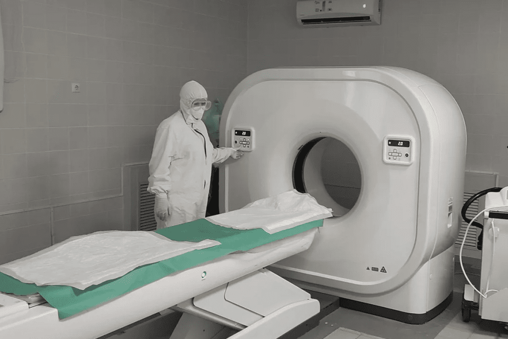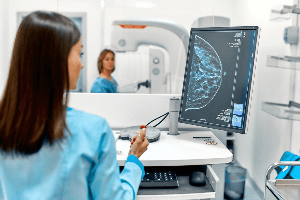
Cancer diagnosis has become more precise with new medical imaging. Early detection significantly improves treatment outcomes. It’s important to pick the right tool for diagnosis.
Scans like MRI, CT scans, and positron emission tomography (PET) scans are used to find cancer. For lung cancer, a thoracic PET scan is very good. It checks how far the disease has spread and if treatment is working.
The accuracy of a scan depends on the cancer type and where it is. Knowing the strengths of each diagnostic tool is key for managing cancer well.
Key Takeaways
- Cancer diagnosis relies heavily on accurate imaging techniques.
- PET scans are very useful for finding lung cancer.
- The choice of scan depends on the cancer type and location.
- Early diagnosis improves cancer treatment outcomes.
- Thoracic PET scans help in assessing lung cancer spread.
Understanding Cancer Detection Through Medical Imaging

Medical imaging is key in finding cancer early. It uses different methods to help doctors spot cancer early. This makes it easier to track how well treatments work.
The Role of Medical Imaging in Cancer Diagnosis
Medical imaging is vital for cancer diagnosis. It lets doctors see inside the body. Tools like CT scans, MRIs, and PET scans help find different cancers.
How Cancer Appears on Different Imaging Modalities
What is a PET scan of the lungs?Cancer looks different on each imaging tool. For example, lung nodules show up on CT scans and PET scans. The PET scan shows how active the cells are. Knowing this helps doctors make the right diagnosis.
| Imaging Modality | Cancer Detection Capability | Key Benefits |
| CT Scan | Detailed cross-sectional images | Helps in detecting small lung nodules |
| PET Scan | Metabolic activity visualization | Useful in identifying malignant lung nodules |
| MRI | Soft tissue visualization | Effective in assessing tumor extent and spread |
The Importance of Early and Accurate Detection
Finding cancer early and accurately is very important. Lung PET imaging and pulmonary PET scans are great for lung cancer. They help doctors act fast.
Using different imaging tools helps doctors get better at diagnosing. This leads to better treatment plans.
Common Types of Scans Used for Cancer Detection

Many scans help find and diagnose cancer. Each scan has its own benefits. The right scan depends on the cancer type, where it is, and how far it has spread.
X-rays and Their Limitations
X-rays are often used because they are fast, painless, and easy to find. But, they can’t see soft tissues well. They work best for bones and some lung issues.
Limitations of X-rays in Cancer Detection:
- They struggle to spot soft tissue tumors
- It’s hard to tell different tissues apart
- They use some radiation, but it’s usually small
Ultrasound Imaging for Cancer
Ultrasound uses sound waves to see inside the body. It’s great for checking organs like the liver and kidneys. It also helps guide biopsies.
Advantages of Ultrasound in Cancer Detection:
- It’s safe and doesn’t use radiation
- It shows things in real-time
- It’s cheaper than other scans
CT Scans: Detailed Cross-Sectional Imaging
CT scans show detailed pictures of the body. They mix X-rays and computer tech. They’re good for finding tumors in the lungs and liver. They also check if cancer has spread.
A study showed CT and PET scans together are better for lung cancer. Using lung pet ct scan helps find cancer better.
| Imaging Modality | Strengths | Limitations |
| X-rays | Quick, widely available | Limited soft tissue contrast |
| Ultrasound | Non-invasive, real-time imaging | Limited depth penetration |
| CT Scans | Detailed cross-sectional images | Involves radiation, contrast agents |
| MRI | Excellent soft tissue contrast | Expensive, claustrophobic |
MRI: Soft Tissue Visualization
MRI is key for soft tissue images. It’s vital for brain and spine tumors. MRI shows tumor size, location, and spread.
“MRI has revolutionized cancer diagnosis. It offers unmatched soft tissue detail. This helps doctors make accurate diagnoses.”
” An Oncologist
Different scans like X-rays, ultrasounds, CT scans, and MRI are all important for cancer detection. Knowing their strengths and weaknesses helps pick the best scan for each cancer type.
PET Scan Lungs: A Powerful Tool for Cancer Detection
PET scans are key in finding lung cancer. They show how active lung tissue is. This helps spot cancer cells, which use more energy than normal cells.
How PET Scans Work for Lung Cancer Detection
PET scans find lung tumors by tracking radiotracers. These substances go to very active areas, like cancer. This gives important info on tumor size and activity.
First, a tiny amount of radiotracer is given to the patient. It spreads through the body. Then, the PET scanner picks up the radiation, making detailed images of lung activity.
Radiotracer Uptake in Lung Tumors
Lung tumors take up more radiotracers than normal cells. This is because cancer cells are more active. This helps PET scans find cancer in the lungs.
There are many radiotracers, each suited for different cancers. The right one can make the scan more accurate.
Metabolic Activity Visualization in Lung Tissue
PET scans show how active lung tissue is. This helps tell if a lung nodule is cancerous. Cancer cells are usually more active.
This info helps doctors understand how aggressive the cancer is. It helps them choose the best treatment.
Types of Radiotracers Used for Lung Imaging
FDG (Fluorodeoxyglucose) is a common radiotracer for lung scans. It goes to cells that use a lot of sugar, like cancer cells. This makes it great for finding lung cancer.
Other radiotracers are used for different cancers or needs. The right one depends on the cancer type, its stage, and the patient’s health.
PET-CT Combination: Enhanced Diagnostic Accuracy
PET and CT imaging together are a powerful tool for finding lung cancer. They combine PET’s metabolic info with CT’s detailed images. This makes for better accuracy in diagnosis.
Integration of Functional and Anatomical Imaging
The PET-CT combo lets us get both functional and anatomical info at once. PET scans show how active the tumor is, while CT scans give us detailed images. This helps us pinpoint where the tumor is in the lung.
This combo helps doctors understand lung cancer better. It’s key for accurate staging and planning treatment.
Benefits of Combined Imaging for Lung Cancer
PET-CT has many benefits for lung cancer diagnosis. It spots small lung nodules better and tells the difference between harmless and harmful growths. It also makes staging lung cancer more accurate.
- Improved detection of small lung nodules
- Enhanced differentiation between benign and malignant findings
- More accurate lung cancer staging
Case Studies: Improved Detection Rates
Many case studies show PET-CT’s success in finding lung cancer early. Early detection means the cancer is easier to treat.
A study in a top medical journal found PET-CT found lung cancer when CT scans couldn’t. This shows PET-CT’s power in early detection.
Workflow and Protocol Optimization
To get the most from PET-CT, we need to improve our workflow and protocols. This means making sure patients are well-prepared, like following dietary rules and managing meds. It also means choosing the right radiotracer uptake time and image reconstruction techniques for the best images.
Comparing Accuracy: PET vs. Other Imaging Modalities
It’s important to compare PET scans to other imaging methods to find the best way to diagnose lung cancer. This comparison helps doctors make better choices for their patients.
Sensitivity and Specificity Rates
PET scans are very good at finding lung cancer. They often do better than CT scans. For example, a study in the Journal of Nuclear Medicine found PET scans were 95% accurate, while CT scans were 80% accurate.
“PET scans offer a unique advantage in lung cancer diagnosis due to their ability to provide both functional and anatomical information.” This helps doctors see if a lung nodule is cancerous by looking at its metabolic activity.
Detection of Small Lung Nodules
Finding small lung nodules is hard, and different methods are better at it. CT scans are great at spotting small nodules. But PET scans can tell if these nodules are likely to be cancerous by looking at their metabolic activity.
- PET scans can detect the metabolic activity of small lung nodules.
- CT scans are highly sensitive for detecting small nodules.
- Combining PET and CT scans can improve diagnostic accuracy.
Differentiating Benign vs. Malignant Findings
Telling if a lung finding is cancer or not is key. PET scans are good at this because cancerous tumors have more metabolic activity. But, PET scans can sometimes show false positives, like in cases of inflammation or infection.
“The use of PET scans has revolutionized the field of oncology, enabling clinicians to non-invasively assess the metabolic activity of tumors and make more informed treatment decisions.” “ An oncologist
Evidence-Based Comparison Studies
Many studies have looked at how well PET scans compare to other methods for finding lung cancer. These studies show PET scans are very accurate, often better than CT scans alone. For example, a study in the European Journal of Nuclear Medicine and Molecular Imaging found PET-CT was more accurate than CT for lung cancer detection.
| Imaging Modality | Sensitivity | Specificity |
| PET-CT | 95% | 90% |
| CT | 80% | 85% |
Preparing for a Lung PET Scan
To get ready for a lung PET scan, patients need to follow certain steps. This preparation is key for getting accurate results.
Pre-Scan Instructions
Before the scan, patients get detailed instructions. They need to arrive early to fill out paperwork and wear a comfortable gown.
Dietary and Medication Considerations
Patients might need to follow dietary rules. They might have to fast or avoid sugary foods and drinks. This helps the scan work better.
Some medications might need to be changed or stopped before the scan. It’s important to tell the healthcare provider about any medications being taken.
What to Expect During the Procedure
A small amount of radioactive tracer is injected into the patient’s blood during the scan. Then, the patient lies on a table that slides into a PET scanner.
The scan itself is usually painless. But, some might feel uncomfortable because they have to stay very quiet and calm for a long time.
Duration and Comfort Measures
A lung PET shttps://www.ncbi.nlm.nih.gov/books/NBK559089/can usually takes 30 to 60 minutes. Patients are told to wear comfy clothes and might get blankets to stay warm during the scan.
| Preparation Step | Description |
| Pre-Scan Instructions | Arrive early, complete paperwork, and change into a gown |
| Dietary Restrictions | Fast or avoid sugary foods and drinks |
| Medication Adjustment | Inform healthcare provider about current medications |
| During the Scan | Receive radioactive tracer injection, lie on scanning table |
| Scan Duration | Typically 30 to 60 minutes |
By following these steps, patients can make sure their lung PET scan goes well.
Interpreting PET Scan Results for Lung Cancer
Understanding PET scan results is key to diagnosing and managing lung cancer. It involves a deep look into lung metabolic imaging.
Understanding SUV Values
The Standardized Uptake Value (SUV) is a key part of PET scans. It shows how much the radiotracer is taken up by lung tissue compared to the body’s average. Higher SUV values often mean more metabolic activity, which can point to cancer. But, SUV values can change based on the radiotracer, scan timing, and patient factors.
Common Findings and Their Meanings
PET scans can show different things in the lungs, from benign changes to cancer. Increased radiotracer uptake can mean tumors, inflammation, or infection. The pattern and intensity of uptake, along with the patient’s history, help doctors tell the difference.
False Positives and False Negatives
PET scans are very good at showing metabolic activity in lung tissues. But, they’re not perfect. False positives can happen due to inflammation or other conditions that look like cancer. False negatives might be because the tumor is small or has low activity. Knowing these can help doctors get a clearer picture.
The Radiologist’s Role in Interpretation
Radiologists are very important in reading PET scan results. They use their knowledge of lung anatomy, pathology, and imaging to make accurate diagnoses. Their skill is key in telling apart benign and malignant findings, helping decide on treatment.
By correctly reading PET scan results, doctors can make better decisions about lung cancer diagnosis, staging, and treatment. This can lead to better patient outcomes.
Limitations and Considerations of PET Scans
PET scans are great for finding lung cancer, but they have some downsides. These issues can affect how useful and accessible PET scans are for patients.
Radiation Exposure Concerns
PET scans use small amounts of radioactive tracers. This means patients get exposed to some radiation. This is a big worry, mainly for younger people and those needing many scans. The chance of getting cancer from this radiation is small but real.
Doctors need to think carefully about the benefits of PET scans against the risks of radiation. They should look for other imaging options and use PET scans wisely.
Cost and Insurance Coverage
PET scans are pricier than X-rays or CT scans. Insurance coverage for PET scans can vary, leaving some patients with big bills. This cost can stop some people from getting PET scans.
| Imaging Modality | Average Cost | Insurance Coverage |
| PET Scan | $1,000 – $3,000 | Variable |
| CT Scan | $500 – $1,500 | Generally Covered |
| X-ray | $100 – $500 | Generally Covered |
Availability and Access Issues
PET scan facilities aren’t everywhere. Rural areas often have few PET scan options, forcing patients to travel far. This is tough for those who can’t travel easily or need ongoing care.
Patient-Specific Contraindications
Some patients can’t have PET scans. For example, people with diabetes need special prep because their blood sugar affects the scan. Also, those with claustrophobia might struggle with the scan’s enclosed space.
It’s key for doctors to know these limitations. This helps them decide when to use PET scans for lung cancer. By weighing the pros and cons, doctors can use PET scans better and help patients more.
PET Scans for Lung Cancer Staging and Treatment Planning
PET scans have greatly improved lung cancer diagnosis and treatment planning. They show how active tumors are, helping doctors understand how far the disease has spread.
TNM Staging Using PET Imaging
TNM staging is key in lung cancer diagnosis, and PET imaging is essential. PET scans check the activity of tumors and possible metastases. This helps doctors stage lung cancer accurately, which is vital for choosing the best treatment.
Mediastinal Lymph Node Assessment
Assessing mediastinal lymph nodes is important in lung cancer staging. PET scans are very good at spotting active lymph nodes. This helps doctors decide if a patient needs more aggressive treatment.
Impact on Treatment Decision-Making
PET scans greatly influence treatment choices for lung cancer patients. They help doctors stage the disease and find where it might spread. This allows for treatment plans that fit each patient’s needs, improving results and avoiding unnecessary treatments.
Radiation Therapy Planning Applications
PET scans are also useful in planning radiation therapy for lung cancer. They give detailed info on tumor location and activity. This helps radiation oncologists target tumors better, giving them the right dose of radiation while protecting healthy tissues.
Monitoring Treatment Response and Recurrence
PET scans are key in checking if lung cancer treatments are working. They help spot any signs of cancer coming back. This is vital for changing treatment plans and helping patients get better.
Evaluating Therapy Effectiveness
PET scans are important in lung cancer care. They show if the tumor is responding to treatment. By looking at metabolic activity, doctors can see if the treatment is effective.
Key indicators of therapy effectiveness include:
- Reduction in SUV (Standardized Uptake Value) on follow-up PET scans
- Decrease in tumor size or metabolic activity
- Absence of new areas of increased uptake
Detecting Residual Disease
After treatment, PET scans can find out if cancer cells are left behind. This info is key for deciding what to do next, like more treatment or watching closely.
Surveillance Protocols
Regular PET scans are key for lung cancer patients. They help catch cancer coming back early, so it can be treated quickly.
| Surveillance Protocol | Description | Frequency |
| Initial Follow-up | PET scan after treatment to check response | 3-6 months post-treatment |
| Regular Surveillance | Regular PET scans to watch for recurrence | Every 6-12 months |
| Symptom-Driven | PET scans when symptoms get worse | As needed |
Distinguishing Treatment Effects from Recurrence
It can be hard to tell if changes on PET scans are from treatment or cancer coming back. Radiologists use many factors, like how the uptake looks and the patient’s history, to figure it out.
Getting it right is very important. It helps avoid too much treatment or waiting too long for it. Using advanced imaging and checking with the patient’s history is key.
Emerging Technologies in Cancer Imaging
The field of cancer imaging is changing fast with new technologies. These changes help doctors diagnose cancer better, improve treatment, and change how we fight cancer.
New PET Tracers for Improved Specificity
New PET tracers are being made to find cancer cells better. These tracers help doctors avoid mistakes and get more accurate results. For example, Fluorodeoxyglucose (FDG) is common, but new ones target specific cancers like prostate cancer.
Creating these tracers is key for better PET scans, like those for lung nodules. A study found that new tracers could greatly improve lung cancer diagnosis.
Artificial Intelligence in Image Interpretation
Artificial Intelligence (AI) is being used more in cancer imaging, like PET scans. AI can look at lots of data fast and spot things humans might miss.
A study showed that AI can make diagnoses 15% more accurate.
This is very important for lung imaging, where finding nodules is critical.
Next-Generation Hybrid Imaging Systems
New hybrid imaging systems are being made. They mix different imaging types, like PET and MRI, to show more about tumors.
“Hybrid systems are a big step forward. They give us a full view of cancer, helping tailor treatments.” – A radiologist
Molecular Imaging Advances
Molecular imaging is getting better, thanks to understanding tumor molecules. This helps make new agents that target cancer markers, finding cancer early.
Combining molecular imaging with other tools will greatly help patients. As technology grows, we’ll see better ways to diagnose and treat cancer.
Conclusion: The Future of Cancer Detection Imaging
The future of cancer detection imaging looks bright. PET scans, including those for lung cancer, are getting better at finding cancer early. They help doctors plan treatments too.
When PET scans are used with CT scans, they become even more accurate. This is great for checking the lungs and other parts of the chest.
New developments in PET scans for the lungs are on the horizon. These changes will help doctors find and treat cancer better. Research is key to making these improvements happen.
As imaging technology advances, we can expect even better tools for diagnosing cancer. This will lead to better care and results for patients.
FAQ
What is a PET scan, and how is it used in lung cancer detection?
A PET scan is a test that uses a special tracer to see how active cells are in the body. It helps find lung cancer by spotting areas where cells use a lot of sugar, a sign of cancer.
How does a PET scan work for lung cancer?
A PET scan uses a tracer called FDG that goes into your blood. It sticks to cells that are very active, like cancer cells. The scanner then picks up this signal, showing where the activity is.
What are the benefits of using PET-CT scans for lung cancer detection?
PET-CT scans combine PET’s metabolic info with CT’s body maps. This gives a clearer picture of tumors, their activity, and exact location. It’s a more accurate way to diagnose.
How accurate are PET scans in detecting lung cancer?
PET scans are very good at finding lung cancer, even better when used with CT scans. But, how well they work can depend on the tumor’s size, where it is, and its type.
What are the limitations of PET scans in lung cancer detection?
PET scans have some downsides. They expose you to radiation, can be expensive, and not everyone can get one. They’re not good for people with certain health issues.
How do PET scans help in lung cancer staging and treatment planning?
PET scans are key in figuring out how far cancer has spread. They also help plan treatments, like radiation therapy, by showing how active the tumor is and where it is.
Can PET scans detect lung cancer recurrence?
Yes, PET scans can spot lung cancer coming back. They find areas where cells are using a lot of sugar, which might mean new tumor growth.
How often should PET scans be performed for lung cancer monitoring?
How often you need a PET scan depends on your treatment and health. Usually, they’re done at set times to check how well treatment is working and if cancer has come back.
What are the emerging technologies in PET scans for lung cancer detection?
New technologies in PET scans include better tracers, artificial intelligence, and advanced imaging systems. They aim to make diagnoses more accurate and help patients more.
How do PET scans compare to other imaging modalities in lung cancer detection?
PET scans have big advantages over CT scans and MRI for lung cancer. But, which one is best depends on the patient’s needs and the situation.
References
- Zhu, J., et al. (2022). Positron emission tomography imaging of lung cancer. PMC. https://pmc.ncbi.nlm.nih.gov/articles/PMC9581209/


