Last Updated on November 27, 2025 by Bilal Hasdemir

Choosing the right imaging test for cancer is key. At Liv Hospital, we offer top-notch, personalized care. PET scans and CT scans are two main tools for cancer detection. CT scans use X-rays for detailed images, while PET scans show metabolic activity with radioactive tracers. Pet scan vs ct scan for cancer: 7 key differences explained to help you choose the right scan.
Knowing the differences between PET scans and CT scans helps in making better cancer treatment choices. This article will dive into the main differences between these scans. We’ll see how they work together for a full cancer diagnosis.
Key Takeaways
- CT scans provide detailed anatomical images using X-rays.
- PET scans visualize metabolic activity with radioactive tracers.
- Both scans are painless and take about 30 minutes to complete.
- PET/CT scans combine the benefits of both imaging techniques.
- Understanding the differences between PET and CT scans can aid in informed cancer treatment decisions.
Understanding Medical Imaging in Cancer Diagnosis

Medical imaging is a key tool in fighting cancer. It helps find where tumors are, how big they are, and how active they are. PET and CT scans are now essential in cancer care.
Imaging tests are vital for diagnosing and treating cancer. They give doctors the info they need to plan treatments and check how patients are doing. As we learn more about cancer, the need for precise imaging grows.
The Role of Advanced Imaging in Modern Oncology
PET and CT scans give a lot of information about tumors. They show where tumors are, how big they are, and how active they are. This info is key for figuring out how serious the cancer is, planning treatment, and seeing if it’s working.
“The mix of PET and CT scans has changed how we diagnose cancer,” says a top oncologist. This mix of imaging types helps doctors understand tumors better. It makes diagnosis more accurate and helps decide on treatments.
Why Accurate Imaging Is Critical for Cancer Management
Accurate imaging is the basis of good cancer care. It lets doctors create specific treatment plans, watch how the disease grows, and change treatments if needed. Modern imaging has made early detection and treatment more successful.
Quick and accurate diagnosis is key to fighting cancer. Imaging technologies are central to this fight. They help doctors make better diagnoses, plan better treatments, and improve patient care.
What Is a CT Scan?

CT scans are a key tool in modern medicine. They use X-rays to make detailed images of the body. These scans are very useful in cancer diagnosis and tracking.
Basic Principles and Technology Behind CT Scanning
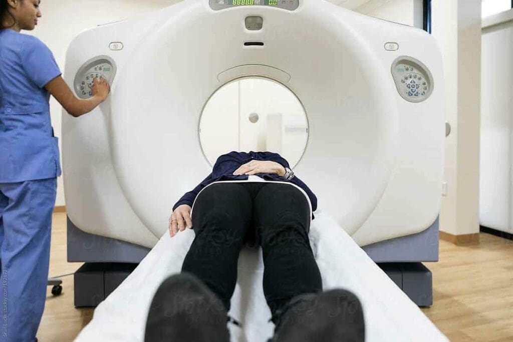
CT scanning uses X-rays to make images of the body’s cross-sections. It works by moving an X-ray source and detectors around the body. This captures data that turns into images.
The key components of a CT scanner include:
- The gantry, which houses the X-ray source and detectors
- A table that moves through the gantry
- A computer system to reconstruct images
How CT Scans Create Anatomical Images
CT scans make detailed images by showing how different body tissues absorb X-rays. This information is turned into pictures we can see.
Types of CT Scans Used in Cancer Detection
There are several CT scans for cancer detection:
- Contrast-enhanced CT: Uses a contrast agent to highlight specific areas or structures.
- High-resolution CT: Provides detailed images of small structures, useful for detecting tumors in organs like the lungs.
- Low-dose CT: Minimizes radiation exposure, making it suitable for screening and follow-up scans.
Each CT scan type has its own use in cancer care. They help from the first diagnosis to tracking how well treatments work.
What Is a PET Scan?
PET scans are advanced tools that help doctors see how the body works. They show how tissues and organs function, unlike CT scans, which focus on body structure. This makes PET scans great for checking how cells are doing.
We use PET scans to see how cells are working, which is key in finding cancer. We inject a tiny bit of a radioactive substance, like FDG, into the body. This substance lights up areas where cells are very active, like in cancer.
Fundamental Principles of PET Imaging
PET imaging works by looking at how cells use energy. Cancer cells use more energy than normal cells, so they take in more glucose. The FDG tracer goes to these cells and sends signals that the PET scanner picks up.
How Radioactive Tracers Highlight Metabolic Activity
Radioactive tracers like FDG find their way into cells based on how active they are. Cancer cells take in more of this tracer because they’re more active. This makes them show up clearly on PET scans, helping us spot cancer.
The FDG Tracer and Cancer Cell Detection
The FDG tracer is great for finding cancer because it shows where cells are using a lot of glucose. Cancer cells use more glucose than normal cells. So, FDG-PET scans are a powerful tool for finding and tracking cancer.
| Characteristics | PET Scan | CT Scan |
| Primary Use | Metabolic activity assessment | Anatomical imaging |
| Tracer Used | FDG (Fluorodeoxyglucose) | Contrast agents (optional) |
| Cancer Detection | Effective for many cancer types | Provides anatomical details |
PET Scan vs CT Scan for Cancer: Key Differences in Technology
PET scans and CT scans use different technologies to show different things for cancer diagnosis. Knowing these differences helps doctors pick the best imaging for patients.
Anatomical vs Metabolic Imaging
CT scans show detailed pictures of body structures, like tumors. On the other hand, PET scans highlight where cells are most active, like in cancer cells.
This is key because cancer cells use more energy than normal cells. So, PET scans can spot cancer cells early, even when they’re small. For example, they can find cancer in lymph nodes that look normal on a CT scan.
Radiation Exposure Comparison
Both PET and CT scans use radiation, but in different ways. CT scans use X-rays for detailed body images. The dose can vary based on the scan and body part.
PET scans use tiny amounts of radioactive tracers, like Fluorodeoxyglucose (FDG), to find active areas. They have lower radiation exposure.
Using a PET/CT scan together means getting both detailed images and metabolic info in one go. This might reduce the need for more scans and lower radiation overall.
Image Resolution and Detail Differences
CT scans are known for their high detail, showing body structures clearly. They’re great for checking tumor size and location.
PET scans don’t show as much detail as CT scanss but are better at showing tissue function. They’ve gotten better over time,, but don’t match CT scans in detail.
PET/CT scanners combine the best of both worlds. They offer detailed images and metabolic info. This combo helps doctors diagnose and plan treatment better.
Difference Between PET CT Scan and CT Scan in Cancer Detection
When it comes to finding cancer, doctors often choose between PET/CT scans and CT scans. Each has its own strengths in spotting and managing cancer. Let’s dive into what makes them different and how they help in cancer detection.
Early Detection Capabilities
PET/CT scans are great at finding cancer early. They spot changes in cells before they show up in the body. This early catch is key to starting treatment fast and helping patients get better. PET scans can spot metabolic changes in cancer cells, even before they cause big changes seen on a CT scan.
Sensitivity and Specificity Rates
PET/CT scans and CT scans have different levels of accuracy in finding cancer. PET/CT scans are very good at spotting and figuring out how far cancer has spread. They mix metabolic info from PET with body details from CT. This combo improves how well they diagnose and helps see how far cancer has spread. CT scans, while showing body details well, might miss early signs of cancer.
Limitations of Each Imaging Method
PET/CT scans give a lot of info but come with higher costs and more radiation. CT scans are faster and cheaper but don’t give the same metabolic insights as PET/CT scans. Knowing these downsides helps doctors choose the best test for each patient.
The Combined Power of PET/CT Imaging
PET/CT fusion imaging is a big step forward in cancer diagnosis. It combines PET scans’ metabolic info with CT scans’ detailed images. This gives us a clearer view of cancer.
How Fusion Imaging Works
The PET/CT scan uses one device for both PET and CT scans. It shows how active cells are and their structure. This helps pinpoint cancer’s location and size.
First, a tracer is given to highlight active cells, like cancer. The PET scan picks up this activity. The CT scan then shows the body’s layout. Together, they help spot and measure cancer.
Key benefits of PET/CT imaging include:
- Improved diagnostic accuracy
- Enhanced detection of cancer metastasis
- Better assessment of treatment response
- More precise planning for radiation therapy
Clinical Outcomes Improvement with Combined Imaging
PET and CT together have greatly improved cancer care. They give a detailed look at cancer, helping doctors plan better treatments.
Research shows PET/CT scans often change treatment plans. They help find more cancer or show how much is present. This leads to better care.
The combined power of PET/CT imaging lies in its ability to provide both functional and anatomical information, allowing for a more nuanced understanding of cancer biology.
When Is a CT Scan Preferred for Cancer Patients?
CT scans are key in cancer care, mainly in emergencies. They give quick, accurate images. This helps us make fast decisions.
Specific Cancer Types Best Visualized by CT
Some cancers are easier to see with CT scans. For example, lung, liver, and colon cancers are often checked with CT. They show tumor size and location well.
This is important for tracking changes and measuring tumor size. It also helps see if cancer has spread.
CT scans are great for lung and liver tumors. They give clear images for staging and planning treatment. This helps doctors decide the best course of action.
Emergency Situations and Rapid Assessment
In emergencies, speed is vital. CT scans are quicker than PET scans. They’re the go-to for fast assessments.
For example, in suspected internal bleeding or acute symptoms, CT scans are fast. They provide the needed info quickly.
CT scans are lifesaving in emergencies. They help doctors make quick, important decisions. Whether it’s for injury assessment or acute diagnosis, CT scans are reliable and fast.
When comparing CT versus PET scans for cancer, each has its benefits. But in urgent cases, CT scans are essential in cancer care.
When Is a PET Scan More Valuable in Oncology?
In oncology, PET scans are key for diagnosing and managing cancer. They help us understand how tumors work. This is very useful in some cases.
Cancer Types Where PET Excels
PET scans work best for lymphoma, breast cancer, and lung cancer. They can spot cancer growths that other scans miss.
- Lymphoma: PET scans are very good at finding lymphoma, helping with diagnosis and treatment checks.
- Breast Cancer: They can find aggressive breast cancer types and see if the cancer has spread.
- Lung Cancer: PET scans help figure out how far lung cancer has spread.
Staging and Treatment Response Evaluation
PET scans are key for staging and checking how well treatment works. They show whether tumors are active after treatment.
Key benefits of PET scans in staging and treatment response include:
- They help accurately stage cancer, guiding treatment choices.
- They let us quickly see if treatment is working, so we can change plans if needed.
- They find any cancer left behind, which is important for future care.
Detecting Recurrence and Metastasis
PET scans are also great for finding cancer that comes back or spreads. They can spot cancer spread that other scans can’t see.
PET scans can find cancer recurrence and spread early. This can greatly improve patient outcomes. It leads to quicker action and can help patients live longer.
We use PET scans to give the best care to cancer patients. They are very helpful in complex cases where detailed information is needed.
Clinical Applications: CT Scan or PET Scan for Cancer
Choosing between a CT scan and a PET scan for cancer patients depends on the situation. Each has its own strengths for different cancer management needs.
Initial Diagnosis Decision-Making
The choice between CT and PET scans for initial diagnosis varies by cancer type and patient symptoms. CT scans are best for finding tumors in organs like the lungs and liver. PET scans, on the other hand, show how active tumors are by looking at glucose uptake.
| Imaging Test | Strengths in Initial Diagnosis | Common Applications |
| CT Scan | Anatomical detail, quick assessment | Lung nodules, liver lesions, and emergencies |
| PET Scan | Metabolic activity assessment | Cancer staging, detecting metastasis, assessing treatment response |
Treatment Planning Considerations
Both CT and PET scans are key in treatment planning. CT scans give detailed anatomy for surgery or radiation planning. PET scans show disease extent and treatment targets. Together, they give a full picture of the cancer.
Follow-up and Surveillance Protocols
In follow-up, CT or PET scans depend on treatment response and recurrence risk. CT scans track tumor size changes. PET scans spot early metabolic activity changes, showing recurrence or treatment failure.
The choice between CT and PET scans for cancer patients should consider the patient’s condition and care needs. Knowing each modality’s strengths helps healthcare providers make the best decisions for patient care.
Patient Experience and Preparation Differences
Cancer patients often wonder about the differences in preparation and experience between PET and CT scans. These differences can greatly affect their comfort and understanding. Being informed can help reduce anxiety and make the experience smoother.
Preparing for a CT Scan
Preparing for a CT scan is simple. Patients usually need to avoid eating or drinking for a few hours beforehand. This rule can change based on the scan’s needs and the patient’s health. It’s important to tell your healthcare provider about any allergies to contrast dyes, used in CT scans.
In some cases, patients might need to drink a contrast agent or get an IV line. The scan is quick and painless, lasting just a few minutes. But patients must stay very quiet and not move during the scan to get clear images.
Preparing for a PET Scan
Preparing for a PET scan requires more steps than a CT scan. Patients must fast for several hours before the scan. They might also be told to avoid hard exercise for a day or two beforehand. The PET scan uses a radioactive tracer, like FDG, which goes to areas with high activity, like cancer cells.
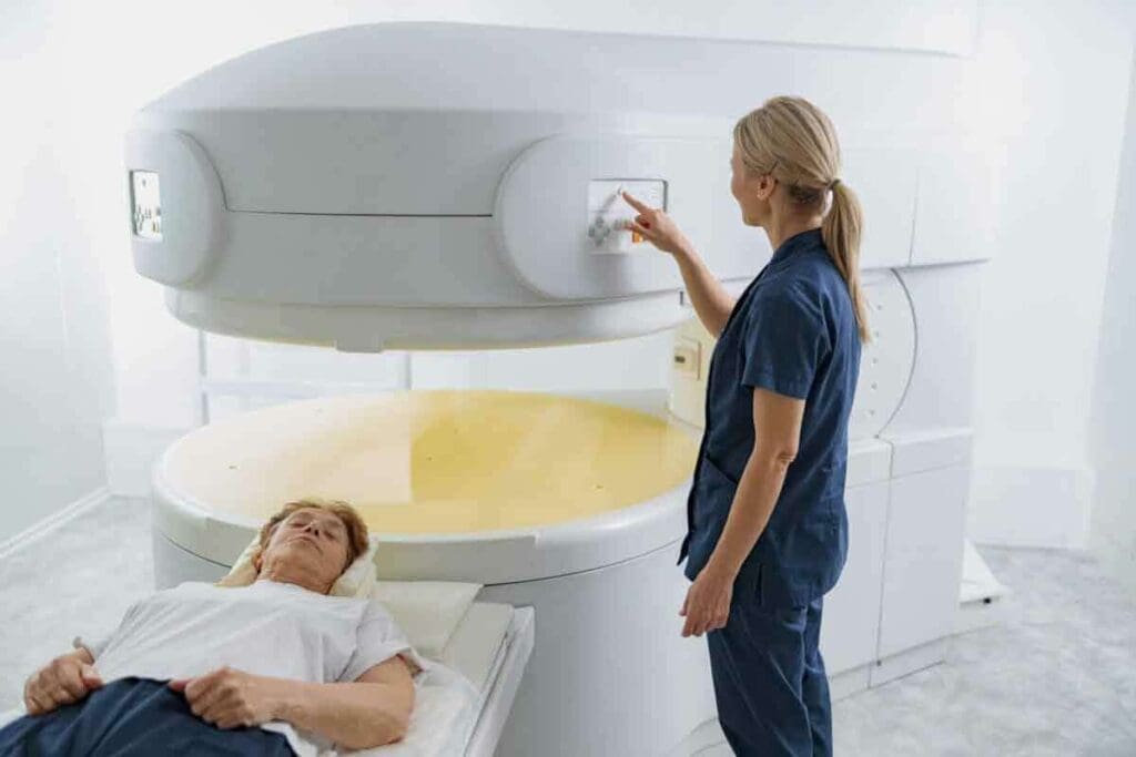
The tracer is given through a vein in the arm and circulates for about an hour before the scan. During this time, patients rest in a quiet, warm room. This helps the tracer focus on cancer cells. The actual scan involves lying on a table that slides into a scanner to detect the tracer’s activity.
What Patients Can Expect During Each Procedure
During a CT scan, patients lie on a table that moves through a doughnut-shaped machine. The scan is fast, and patients might be asked to hold their breath briefly. For a PET scan, patients lie on a table while the scanner looks for the tracer’s activity. The PET scan usually takes longer, around 30 to 60 minutes, including preparation.
“The key to a successful scan is preparation and understanding what to expect. By knowing the differences between PET and CT scans, patients can better prepare themselves for the diagnostic process.” –
Both scans are important for diagnosis, and knowing their differences can help patients feel more ready and less worried. By understanding what to expect, patients can fully cooperate with the scanning process. This ensures the best results.
Future Developments in Cancer Imaging Technology
The world of cancer imaging is about to change a lot. New technologies are making it better at finding and treating cancer. We’re seeing big improvements in how we detect, watch, and treat cancer.
Advances in CT Technology
Computed Tomography (CT) scans are key in cancer imaging. New CT tech, like dual-energy CT, helps spot cancer better. It also cuts down on radiation while keeping images clear.
These changes help doctors get better images. This is key to making the right diagnosis and treatment plan. Plus, artificial intelligence (AI) in CT scans is making image analysis faster and more accurate.
Innovations in PET Scanning
Positron Emission Tomography (PET) scans are also vital. They show how tumors work. New PET tracers are being made to better find cancer types. For example, tracers other than Fluorodeoxyglucose (FDG) are being developed.
Theranostics is another exciting area. It uses PET tracers for both diagnosis and guiding treatment. This could lead to better treatment results.
Emerging Hybrid Imaging Techniques
The future of cancer imaging is in combining different scans. PET/CT and PET/MRI scans give both metabolic and anatomical info. This makes diagnosis more accurate and is key to personalized medicine.
Research on liquid biopsies and other non-invasive tests is also growing. These could give us a deeper understanding of cancer.
As we look ahead, it’s clear that new tech in cancer imaging will help patients more. These advancements will improve how we diagnose and treat cancer, making a big difference in care.
Conclusion: Making Informed Decisions About Cancer Imaging
Knowing the difference between PET scans and CT scans is key for smart choices in cancer care. We’ve looked at how these tests work, their uses, and their benefits. This shows their special roles in fighting cancer.
When deciding between a PET scan CT scan for cancer, each has its own strengths. PET scans are great at spotting metabolic activity. CT scans give detailed views of the body’s structure. Choosing the right test helps doctors plan better treatments and improve patient results.
As we keep improving in cancer imaging, staying up-to-date is vital. This way, patients and doctors can make informed decisions for better care and survival rates.
The choice between a PET scan and a CT scan for cancer depends on the patient’s needs and care plan. Knowing the good and bad of each test helps ensure patients get the best care possible.
FAQ
What is the main difference between a PET scan and a CT scan for cancer detection?
PET scans show how cells are working in the body. CT scans give detailed pictures of body parts. They help doctors in different ways.
Which is better for cancer diagnosis, a PET scan or a CT scan?
It’s not about which one is better. They are used for different things. CT scans help find tumors. PET scans check how active cancer cells are.
What is a PET/CT scan, and how does it differ from a CT scan alone?
A PET/CT scan combines two scans into one. It shows both body structure and cell activity. This helps doctors make better treatment plans.
When is a CT scan preferred over a PET scan for cancer patients?
CT scans are better in emergencies or when quick results are needed. They’re also good for checking some cancers like lung, liver, and colon cancer.
In what situations is a PET scan more valuable than a CT scan for cancer?
PET scans are key for detailed cancer staging and checking how treatments work. They also spot cancer coming back and find where it has spread.
How do PET scans and CT scans differ in terms of radiation exposure?
Both scans use radiation, but in different ways. CT scans use X-rays, and PET scans use radioactive tracers. The amount of radiation varies with each scan.
Can PET scans and CT scans be used together for cancer diagnosis and treatment?
Yes, using PET/CT scans together gives a full picture of cancer. This helps doctors make more accurate diagnoses and tailor treatments better.
How should patients prepare differently for a PET scan versus a CT scan?
For PET scans, patients often need to fast and get a radioactive tracer. CT scans might need contrast agents and fasting, depending on the type.
Are there any emerging technologies that might change how PET scans and CT scans are used in cancer care?
Yes, new technologies in CT and PET scanning, and hybrid imaging, are coming. These could make diagnosis and treatment even better for cancer patients.
How do PET scans and CT scans contribute to the overall management of cancer?
Both scans are vital for managing cancer. They help doctors diagnose, plan treatments, and check how well treatments are working. This leads to better care for patients.
Referecnes
- Delbeke, D., & Martin, E. C. (2018). Positron emission tomography/computed tomography in the evaluation of cancer patients. Seminars in Nuclear Medicine, 48(6), 480-495. https://www.ncbi.nlm.nih.gov/pmc/articles/PMC6524530/
- Sah, B. R., Heverhagen, J. T., & Maderwald, S. (2018). Magnetic resonance imaging and computed tomography in oncology: comparison of techniques and their applications. European Journal of Radiology, 103, 114-123. https://pubmed.ncbi.nlm.nih.gov/30121618/


