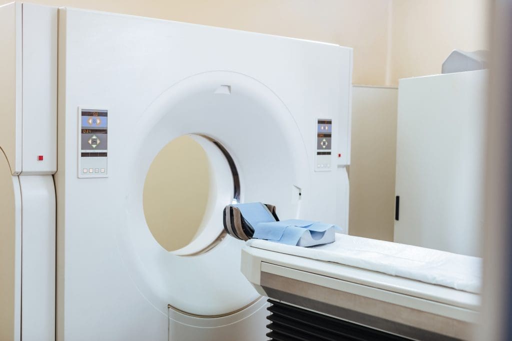
Medical imaging is key in diagnosing and treating health issues. Positron Emission Tomography (PET) and Computed Tomography (CT) scans are two main tools. They help see inside the body but in different ways.
Knowing the difference between PET scan and CT scan is important. A PET scan looks at how tissues and organs work. A CT scan shows detailed pictures of the body’s inside.

Medical imaging has grown a lot over time. It has become key in today’s healthcare. These tools help us understand the body better and make treatments more effective.
It all started with Wilhelm Conrad Röntgen’s discovery of X-rays in 1895. After that, we got CT scans, PET scans, and MRI. Each one has added something special to how we diagnose diseases.
These technologies have gotten better over time. They can show more detail and work faster. For example, PET scans show how tissues work, while CT scans give clear pictures of the body’s inside.
Medical imaging is vital in today’s healthcare. It helps find diseases early and track them. It’s used for many conditions, like cancer and heart diseases.
| Imaging Modality | Primary Use | Key Benefits |
| PET Scan | Cancer staging, neurological disorders | Functional information, high sensitivity |
| CT Scan | Trauma, structural abnormalities | High-resolution anatomical images, quick scan time |
The role of medical imaging in healthcare is huge. It has made patients’ lives better and helped in medical research and new treatments.
A PET scan is a tool in nuclear medicine. It shows how your body’s tissues and organs work. This is done through a special imaging test.
Positron Emission Tomography (PET) is a way to see how your body’s tissues and organs work. It uses a special tracer that is injected into your body. This tracer goes to areas where your body is very active, like growing cancer cells.
The PET scanner detects gamma rays emitted by the tracer.
Radioactive tracers are special substances with a bit of radioactive material. For PET scans, Fluorodeoxyglucose (FDG) is often used. It’s a glucose molecule with a radioactive tag. Cancer cells use more glucose than normal cells, so they take up more FDG.
These tracers work by showing where your body is most active. By choosing different tracers, PET scans can find many conditions, like cancer or brain disorders.
The PET scanner detects gamma rays emitted by the tracer.
These images give doctors a clear view of your body’s activity. They help in diagnosing and treating many health issues.
| Key Components | Function |
| Radioactive Tracer | Accumulates in areas of high metabolic activity |
| PET Scanner | Detects gamma rays emitted by the tracer |
| Image Reconstruction | Creates detailed, 3D images of internal structures |
The CT scan is a high-tech medical tool that uses X-rays to show detailed images of inside the body. It’s key in healthcare for its ability to give clear pictures of the body’s parts.
Computed Tomography (CT) is a way to see inside the body using X-rays and computers. It makes detailed images or ‘slices’ of body areas. Doctors use these images to find and track many health issues.
CT scans use X-rays, which are a type of radiation. These X-rays go through the body at different speeds, based on what they hit. This difference helps make clear images.
Key parts of X-ray technology in CT scans include:
To make cross-sectional images, the X-ray tube and detectors move around the body. They take data from many angles. Then, the computer turns this data into detailed images.
The benefits of cross-sectional imaging include:
| Benefit | Description |
| Detailed Visualization | Shows internal structures clearly. |
| Diagnostic Accuracy | Helps doctors diagnose health issues well. |
| Monitoring Progression | Allows tracking of disease changes. |
CT scans are a vital tool in healthcare. They give deep insights into the body’s inner workings, helping in patient care.
PET scans and CT scans are used for different things in medical imaging. They give unique insights into how the body works and what it looks like inside. Even though they help doctors diagnose, they do it in different ways.
PET scans look at how active the body’s tissues and organs are. This is great for spotting cancer, checking if treatments are working, and studying the brain. CT scans, on the other hand, show detailed pictures of the body’s inside parts. They’re good for finding injuries, diseases, and other structural problems.
PET scans help find areas in the body that are very active, like cancer. CT scans give clear images of the body’s inside, helping doctors find many health issues.
CT scans usually show more detail because they have better spatial resolution. This means they can show small things inside the body more clearly. But, PET scans are great for seeing how tissues are working, which is key for some diagnoses.
| Imaging Modality | Spatial Resolution | Primary Use |
| PET Scan | Lower | Functional Imaging |
| CT Scan | Higher | Anatomical Imaging |
Both PET scans and CT scans are quick, but they can take different amounts of time. CT scans are usually faster, taking just a few minutes. PET scans can take longer, from 30 minutes to several hours, depending on what’s being scanned.
Both scans are usually okay for patients, but they might feel some discomfort. This is because patients have to stay very quiet and sometimes feel a bit cramped. CT scans use X-rays, and PET scans use a tiny bit of radioactive material.
PET scans are key in modern medicine for many important reasons. They show how the body’s cells work, which helps doctors a lot. This makes them very useful in different medical areas.
In cancer care, PET scans are very helpful. They find cancer, see how far it has spread, and check if treatments are working. This is because they spot areas where cells are using a lot of sugar.
PET scans also check how the brain works and find brain problems. They see how brain cells are working, which helps with diseases like Alzheimer’s and epilepsy.
In heart care, PET scans look at how well the heart works. This is very important for treating heart disease.
PET scans have many uses in medicine and are getting even better. They give detailed info on how cells work, helping doctors diagnose and treat many diseases.
CT scans are used in many ways in medicine. They help check for injuries, find problems in the body, and guide treatments.
CT scans are key in emergency care. They can quickly show how bad an injury is and if there’s bleeding inside. This helps doctors and nurses make fast, smart choices.
For example, if someone has a bad head injury, a CT scan can spot bleeding in the brain. This might mean they need surgery right away.
CT scans are great at finding problems in the body. They can spot tumors, cysts, and other issues. This lets doctors know what’s wrong and how to fix it.
They’re better than regular X-rays at finding lung problems. This means doctors can catch cancer early.
CT scans are also good at helping with biopsies and other treatments. They show doctors exactly where to go, making these procedures safer and more likely to work.
For example, when doctors do a biopsy, CT scans help them get the right tissue. This makes it easier to figure out what’s wrong.
| Clinical Application | Description | Benefits |
| Trauma and Emergency | Rapid assessment of injuries | Quick decision-making, life-saving interventions |
| Detecting Structural Abnormalities | Identification of tumors, cysts, vascular issues | Accurate diagnosis, treatment planning |
| Guiding Biopsies and Interventions | Precision targeting of lesions or areas of interest | Reduced complications, improved procedural success |
The PET-CT hybrid combines PET and CT scans into one. This has changed how we diagnose diseases. It uses the best of both worlds to understand a patient’s health better.
PET-CT imaging mixes PET’s metabolic info with CT’s detailed images. It uses one device to get both PET and CT data. This makes sure the images match up perfectly.
First, a radioactive tracer is given to the patient. It goes to active areas in the body. The PET scan finds this activity. The CT scan shows the body’s structure. Together, they give a full view of the body’s inside.
The PET-CT hybrid has many benefits. It helps doctors diagnose better and manage patients better. It combines body function and structure info. This helps doctors plan treatments and check how well they work.
Key benefits include:
PET-CT is best for complex cases. This includes cancers and some brain disorders. It’s needed when detailed info is key for diagnosis or treatment.
In short, the PET-CT hybrid is a big step forward in medical imaging. It helps doctors diagnose and treat complex conditions better.
To make sure your PET scan goes well, follow certain preparation steps. A PET scan is a detailed test that needs some prep to work best.
Before your PET scan, you need to follow certain diet rules. You’ll likely be asked to not eat for 4 to 6 hours before. This helps the tracer work right in your body.
Dietary Recommendations:
Also, don’t do hard exercise for a day or two before. Wear comfy, loose clothes and no metal items like jewelry.
| Preparation Step | Guideline |
| Fasting | 4 to 6 hours before the scan |
| Avoid Sugary Foods | At least 24 hours before the scan |
| Hydration | Drink plenty of water |
On the scan day, you’ll get a radioactive tracer through an IV. You’ll wait about an hour for it to spread in your body.
While waiting, stay calm and quiet. This helps the tracer spread right. Then, you’ll lie down for the scan.
“The PET scan process is generally painless and straightforward. Most people find it a relatively comfortable experience.” – Nuclear Medicine Specialist
After the scan, you can usually go back to normal unless told not to. Drink lots of water to get rid of the tracer.
Post-Scan Care:
Your PET scan results will be checked by a specialist. Your doctor will talk about them with you later.
To get the most out of your CT scan, it’s key to know what to expect. A CT scan gives detailed images of the body. It helps doctors diagnose and treat many health issues.
Before your CT scan, you might get contrast agents. These agents make certain body parts more visible. Your healthcare provider will tell you how to use them, including any diet or drink rules.
You might need to fast for a few hours or drink a special liquid. This helps the scan see your digestive tract better. Tell your doctor about any allergies, like to iodine or contrast agents, to avoid bad reactions.
During the scan, you’ll lie on a table that moves into a big machine. The scan is quick, lasting just a few minutes. You might need to hold your breath for a bit to get clear images.
The person running the scanner will talk to you the whole time. It’s important to stay very quiet and not move to get good pictures. The scan is designed to be as comfortable and safe as possible.
After the scan, you can usually go back to your normal day unless told not to. If you had contrast agents, you might be watched for any bad reactions. Drinking lots of water helps get rid of the agents.
Your doctor will talk to you about the scan results later. These results will help decide what to do next in your treatment.
Knowing what to expect from your CT scan helps you prepare. This makes the experience smoother and the results more accurate.
PET and CT scans involve radiation, but how much and what kind varies. This raises important safety questions.
PET scans use radioactive tracers that emit positrons. The dose depends on the tracer and the patient’s health. CT scans, on the other hand, use X-rays to create detailed images. They have a higher dose than standard X-rays but are considered low.
Key differences in radiation exposure:
Medical facilities have strict safety rules to reduce radiation risks. These include:
Patients can help too by:
Some groups need extra care when it comes to radiation. These include:
Doctors consider these factors when choosing between PET or CT scans for these groups.
The world of medical imaging is about to see big changes. These changes come from new PET and CT technologies. Soon, doctors will be able to diagnose diseases more accurately and care for patients better.
New technologies in PET and CT scans aim to make images clearer and scans faster. For example, new detector materials will make PET scans better. They will help spot smaller tumors and track how well treatments work.
CT scans are also getting better. Spectral CT can tell different tissues apart. This could mean fewer tests for patients and better diagnosis of lesions.
Artificial intelligence (AI) is becoming a big part of medical imaging. AI helps look at images, find problems, and give data for doctors to make decisions.
AI could make doctors better at finding diseases. It can also help doctors do less routine work. This lets them focus on harder cases.
There’s a push to use less radiation in scans. Makers of scanners are working on low-dose CT protocols and advanced algorithms. These help keep scans good quality while using less radiation.
Future PET scans will be even better. They will be more sensitive and have higher resolution. This means doctors can find and track smaller problems. It’s a big step in fighting cancer and other diseases.
Knowing the differences between PET scans and CT scans is key. This knowledge helps us make better choices when it comes to medical imaging. We’ve looked at how these technologies work and what they’re used for.
PET scans show how cells are working. They’re great for finding cancer, brain problems, and heart issues. On the other hand, CT scans give detailed pictures of the body’s structure. They’re best for checking injuries, finding structural problems, and helping with procedures.
Understanding what each scan can do helps patients make smarter choices. When PET and CT scans are used together, like in PET-CT scans, they offer even better results. This helps doctors and patients get more accurate information.
As medical imaging keeps getting better, it’s important to stay up to date. Knowing the differences between PET scans and CT scans helps us make informed choices about our health.
A PET scan uses a radioactive tracer to see how cells work. A CT scan uses X-rays to show the body’s structure. They help doctors in different ways.
A PET scan usually takes about 30 minutes to an hour. This time can change based on the scan’s details and the body part being checked.
A PET-CT scan combines PET’s metabolic imaging with CT’s detailed anatomy. It’s mainly used for cancer diagnosis and tracking. This gives doctors a full view of the body’s health.
To prepare for a PET scan, you might need to eat less, avoid exercise, and have a full bladder. Your healthcare provider will give you specific instructions.
PET and CT scans use radiation, which has a small risk of causing cancer or genetic changes. But, the scans’ benefits usually outweigh the risks. Efforts are made to keep radiation low.
Some medical conditions or implants might not be safe for PET or CT scans. Always tell the scanning facility or healthcare provider about any conditions or implants before the scan.
The time to get PET or CT scan results varies. It usually takes a few hours or days, depending on the scan’s complexity and the facility.
PET and CT scans are usually painless. But, you might feel some discomfort because you need to stay very quiet and in one position.
Pregnancy and breastfeeding are considered when deciding on PET or CT scans. The healthcare provider will weigh the risks and benefits to guide you.
A PET scan looks at how cells work, while an MRI shows the body’s anatomy. MRI uses magnetic fields and radio waves for soft tissue images. PET scans are for functional imaging.
Subscribe to our e-newsletter to stay informed about the latest innovations in the world of health and exclusive offers!