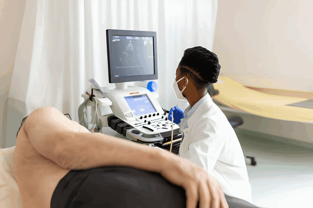
Heart failure is a big health issue worldwide, affecting over 6 million adults in the U.S. alone. Spotting symptoms early is key for good care. At Liv Hospital, we stress the need for a detailed physical exam to diagnose and manage heart failure.
Checking for heart failure involves several important steps. These include a general look, taking vital signs, and checking the heart and lungs. By doing these steps, doctors can find signs like trouble breathing, extra fluid, and odd heart sounds.
Discover the main steps in the physical examination of heart failure for early diagnosis.
Our team uses the latest methods and puts patients first, aiming for the best care in every heart failure check. Learning the physical examination helps doctors give their patients the best care.

Physical examination is key in managing heart failure. It gives doctors important clues about a patient’s health. This helps them understand how severe the heart failure is, track its progress, and adjust treatment plans.
Heart failure is a big problem in the U.S., affecting over 6 million adults. It shows how important it is to have good management strategies. Physical exams play a big part in this.
Studies show that a detailed physical exam is vital. It helps spot the effects of heart problems. This lets doctors catch heart failure signs and keep track of how it’s changing.
Finding heart failure early through physical exams can really help patients. It means doctors can start treatment sooner. This can slow down the disease and make life better for patients.
Research from places like UTSW shows the value of standard physical exams for heart failure. Using the same exam methods helps doctors make more accurate diagnoses. This leads to better care plans for patients.

Before starting a physical exam for CHF, healthcare providers must get ready. They need the right equipment and a good environment. This is key for accurate results and the best care for heart failure patients.
To do a thorough heart failure exam, providers need specific tools and a great setting. The essential tools are:
The best setting is quiet, comfy, and well-lit. This helps patients relax and makes the exam more accurate.
| Equipment | Purpose |
| Stethoscope | Auscultation of heart and lung sounds |
| Blood pressure cuff | Measurement of blood pressure |
| Pulse oximeter | Monitoring oxygen saturation |
How the patient is positioned is very important. They should be comfortable and easy to examine. Usually, they lie down with their head slightly raised. This helps check the jugular venous pressure.
Building trust and easing anxiety are vital for a good exam. When patients feel at ease, they cooperate better. Providers can do this by explaining things clearly, being kind, and staying professional.Key strategies for establishing rapport include:
By taking these steps, providers can give a detailed and effective heart failure assessment. This leads to better care for patients.
The first step in checking a patient with heart failure is a general inspection and checking vital signs. This step gives us important information about the patient’s health. It helps us decide what to do next.
We look for signs of heart trouble during the general inspection. Signs like dyspnea (trouble breathing), orthopnea (breathlessness lying down), and cyanosis (skin turning blue) are important. They show how serious the heart failure is.
We also check for peripheral edema (swelling in legs and feet) and distended neck veins. These signs help us understand how bad the heart failure is.
Checking blood pressure is key in assessing vital signs. Blood pressure can be elevated, normal, or low in heart failure. High blood pressure can make heart failure worse, while low blood pressure might mean the heart is not working well.
Heart rate, respiratory rate, and oxygen saturation are also important. A fast heart rate can mean the heart is working too hard. A high respiratory rate might show the lungs are full of fluid.
Oxygen saturation tells us how well the patient is getting oxygen. If it’s low, they might need oxygen to breathe. We check these signs to see how serious the heart failure is and what to do next.
By looking at these first findings, we can plan the best care for each patient with heart failure. This helps us give them the best treatment possible.
Evaluating jugular venous pressure is key in checking for heart failure. It tells us a lot about how well the heart is working. This helps doctors figure out what’s wrong and how to fix it.
To measure jugular venous pressure (JVP) right, the patient needs to sit at a 45-degree angle. We look for the jugular venous pulsation and measure from the sternal angle to its top. Getting it right is important because it shows the right atrium’s pressure.
High JVP means the right side of the heart is not working well. It can happen when there’s too much fluid or if the heart itself is not working right. It’s important to understand why it’s happening.
Hepatojugular reflux testing is another tool we use. It involves pressing on the liver gently for 10-15 seconds and watching the JVP. If JVP goes up and stays up, it could mean the right ventricle is not working right or is too full.
| Test | Normal Response | Abnormal Response |
| Hepatojugular Reflux | No significant change in JVP | Sustained increase in JVP |
| JVP Measurement | <8 cm H2O | ≥8 cm H2O |
By using JVP measurement and hepatojugular reflux testing together, we get a clearer picture of the heart’s health. This helps us decide the best course of action for the patient.
Checking a patient’s heart function is key in diagnosing heart failure. This step is vital for managing the condition well.
Cardiac auscultation is a key part of checking a heart failure patient. We listen with a stethoscope to hear heart sounds. This gives us important info about how the heart is working.
To do this right, we follow a specific order. We start at the apex and move to the base. This makes sure we check every part of the heart.
To check heart sounds well, we:
S3 and S4 gallops are abnormal sounds that can mean heart failure. An S3 sound happens early in diastole and shows ventricular volume overload. An S4 sound happens late in diastole and shows a stiff ventricle.
To find these sounds, we listen closely with the stethoscope’s bell. The bell picks up low-frequency sounds better.
| Gallop Type | Timing | Clinical Significance |
| S3 | Early diastole | Ventricular volume overload |
| S4 | Late diastole | Atrial contraction into a stiff ventricle |
Murmurs are sounds from blood flow turbulence. They can mean valvular disease or other heart issues. To find and describe murmurs, we look at their timing, location, intensity, and how they spread.
We listen with the stethoscope’s diaphragm for murmurs in systole and diastole. Where and how the murmur spreads can tell us what’s causing it.
By combining what we learn from auscultation, including S3 and S4 gallops and murmurs, we get a full picture of the heart. This info is key for diagnosing and treating heart failure.
Checking the lungs for congestion is key in managing heart failure. It’s important for diagnosing and treating congestive heart failure well.
Auscultation is a basic method in lung exams. We listen for unusual sounds like crackles and wheezes. These sounds can mean fluid buildup or tight airways.
Crackles often show up in heart failure patients with lung congestion. Wheezes might point to airway tightening.
Percussion is also useful for finding lung congestion. By tapping on the chest, we check for fluid or solid buildup. A dull sound might mean fluid in the lungs or around the lungs.
Pleural effusions are a common heart failure complication from too much fluid. We spot them through physical checks and clinical signs.
By using these methods in lung exams, we can find and handle lung congestion from heart failure well.
Checking abdominal and peripheral circulation is key in managing heart failure. It helps spot signs of fluid buildup and congestion, common in heart failure patients.
Liver palpation is a vital skill in heart failure assessment. We start by having the patient lie down with their knees slightly bent. We use our fingers to feel the liver, starting from the right lower quadrant and moving up.
A normal liver is not felt below the costal margin. But in heart failure, it can swell and feel tender due to fluid buildup.
Hepatomegaly means the liver is enlarged, showing right-sided heart failure. We check the liver’s size and tenderness to see how bad the congestion is.
Ascites, or fluid in the belly, is a sign of right-sided heart failure. We use percussion and the shifting dullness test to find ascites. The amount of fluid helps us grade its severity.
Peripheral edema, or swelling in the legs, is common in heart failure. We check for edema by pressing the skin on the tibia or ankle. The depth and how long it lasts help us grade its severity.
| Grade | Description |
| 1+ | Mild pitting edema |
| 2+ | Moderate pitting edema |
| 3+ | Severe pitting edema |
| 4+ | Very severe pitting edema |
By examining abdominal and peripheral circulation, we get a full picture of the patient’s health. This helps us create the best treatment plan for them.
Today, we focus on combining patient symptoms with physical checks for heart failure. This method improves how we diagnose and treat the condition.
Listening to what patients say about their symptoms is key. It adds to what we learn from physical exams. Together, we get a clearer picture of their heart health.
This way, we can make treatment plans that really fit each patient. It helps them do better.
The New York Heart Association (NYHA) and American Heart Association (AHA) systems are vital. They help us sort patients by how well they can function and their symptoms.
| NYHA Class | Description |
| I | No limitation of activities; no symptoms from ordinary activities. |
| II | Slight, mild limitation during ordinary activity; comfortable at rest or with mild exertion. |
| III | Marked limitation in activity due to symptoms, even during less-than-ordinary activity; comfortable only at rest. |
| IV | Severe limitations; experiences symptoms even while at rest, mostly bedbound. |
These systems help us decide on treatments and predict how a patient will do.
Research from the University of Texas Southwestern Medical Center (UTSW) is important. It shows the value of using the same methods for checking heart failure. This makes sure we’re always consistent and accurate.
By using the latest clinical methods, we can give heart failure patients better care. This includes listening to their symptoms and doing thorough physical exams.
To improve heart failure treatment, we need to check patients often. This helps us catch small changes early. We know managing heart failure well means more than just treating it. It also means regular check-ups to see how the disease is doing and change treatment plans if needed.
How often heart failure patients need check-ups depends on how serious their condition is and how well they’re doing. Here’s what we usually suggest:
These times help us keep a close eye on how the patient is doing. We can then make changes to their treatment plan as needed.
During check-ups, we look for small changes that might mean the heart failure is getting worse or better. We pay attention to:
| Clinical Parameter | Indicators of Worsening Condition | Indicators of Improvement |
| Weight | Unexpected weight gain | Stable or decreasing weight |
| Symptoms | Increased dyspnea, orthopnea | Reduced frequency or severity of symptoms |
| Physical Examination | Increased jugular venous pressure, peripheral edema | Decreased jugular venous pressure, reduced edema |
By watching these signs closely, we can spot early signs of getting better or worse.
We change treatment plans based on what we find during check-ups. This might mean:
We make sure treatment fits each patient’s needs. This is based on their current health and how they’ve responded to treatment before.
By regularly checking in with our heart failure patients, we can give them the best care. This improves their health and quality of life.
Mastering the physical examination is key to better heart failure care. We’ve outlined a detailed guide on how to do it. This includes checking the patient’s overall health and vital signs, and how to keep up with follow-ups.
Healthcare professionals can improve their skills in heart failure exams by following this guide. A detailed physical check is vital for spotting and managing heart failure well.
Research shows that a thorough physical exam is essential for catching heart failure early. This leads to better care and outcomes for patients. As healthcare workers, we must keep honing our skills in heart failure exams to offer top-notch care.
Physical exams are key in managing heart failure. They help doctors spot symptoms early and track how the disease is progressing. This allows them to make the right treatment changes.
The main steps in examining heart failure include checking vital signs and looking for signs of congestion. Doctors also check the abdomen and legs for circulation issues. They use modern methods and regularly update treatment plans.
To prepare for a heart failure assessment, doctors need the right tools and a good environment. They should position the patient right, build trust, and try to reduce stress.
Checking jugular venous pressure is important for diagnosing heart failure. It shows how well the heart is working. Doctors look for signs of fluid buildup and other issues.
Doctors check for pulmonary congestion by listening for sounds and tapping on the chest. They also look for fluid in the lungs and around the heart.
The NYHA and AHA systems help doctors understand how severe heart failure is. They guide treatment and help track how the disease is progressing.
Follow-up exams for heart failure patients vary based on their needs. But regular checks are key to catching small changes and adjusting treatment.
Using both patient reports and physical exams is important. It makes diagnosis more accurate and helps manage the disease better, leading to better outcomes.
Detecting small changes involves watching patient symptoms and physical signs closely. Doctors also look at lab results. This helps them make the right treatment changes.
Using research from UTSW on CHF physical assessment is key. It gives doctors evidence-based guidance. This improves diagnosis and patient care.
Government Health Resource. (2025). 7 Key Steps in the Physical Examination of. Retrieved from https://onlinelibrary.wiley.com/doi/10.1111/j.1527-5299.2007.06409.x)
Subscribe to our e-newsletter to stay informed about the latest innovations in the world of health and exclusive offers!
WhatsApp us