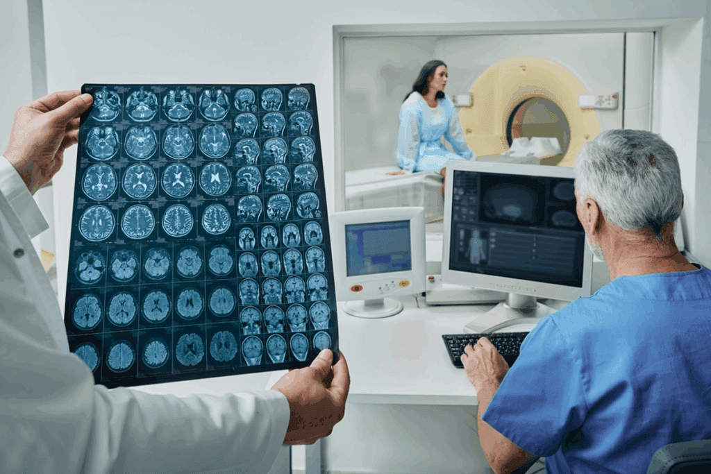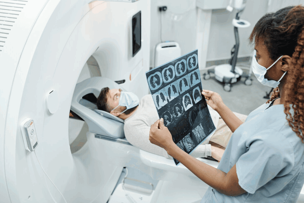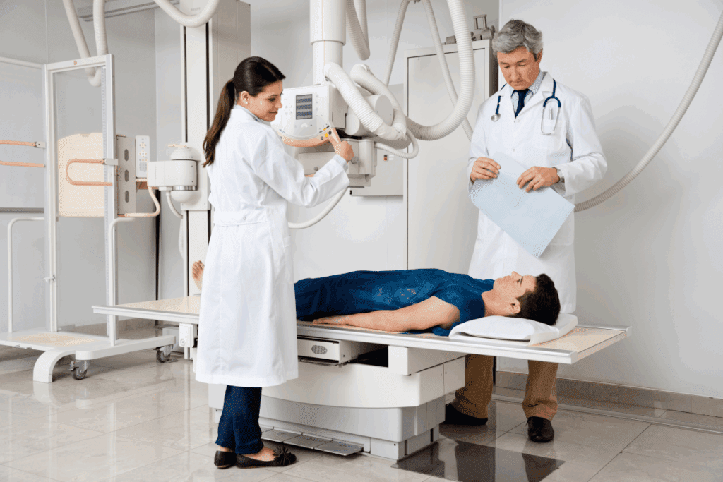
Modern healthcare relies on a key tool: medical imaging radiology. It helps us see inside the body using different methods. This way, we can find, watch, or treat diseases. Medical imaging radiology has changed how we see the human body, letting us look at organs and tissues without surgery. What is a radiology scan? Our ultimate guide for patients explains the basics of medical imaging, what to expect, and the types.
A radiology scan uses energy like X-rays, MRI, CT scans, ultrasound, and nuclear imaging. It creates images of what’s inside us. This field is vital for making accurate treatment plans.
Key Takeaways
- Medical imaging radiology is a tool for seeing inside the body.
- A radiology scan helps diagnose, monitor, or treat many health issues.
- Technologies like X-rays and MRI are used in medical imaging.
- Medical imaging radiology is key for making treatment plans.
- Non-invasive methods give us a peek into the body’s inner workings.
Understanding Radiology and Medical Imaging

Radiology plays a key role in modern healthcare. It helps doctors find problems early, like cancer and heart disease. We use tests like X-rays and MRI to see inside the body without surgery.
The Definition of Radiology in Medicine
Radiology uses imaging to diagnose and treat diseases. It’s like a window into the body, showing doctors what’s inside without surgery. Each imaging method gives a unique view of the body’s parts and how they work.
Common tools in radiology include X-rays, CT scans, and MRIs. Each has its own strengths for different needs. For example, X-rays are great for bones, while CT scans show more details inside.
The Relationship Between Radiology and Medical Imaging
Radiology and medical imaging are often used together. Medical imaging creates images of the body’s inside. Radiology goes further, interpreting these images to help diagnose and treat.
This connection is vital in today’s healthcare. Radiologists use different imaging methods to give doctors the information they need. This teamwork has greatly improved patient care for many conditions.
| Imaging Modality | Primary Use | Key Benefits |
| X-ray | Detecting fractures, lung conditions | Quick, widely available |
| CT Scan | Detailed images of internal structures | High-resolution images, useful for complex diagnoses |
| MRI | Soft tissue imaging, neurological conditions | Excellent soft tissue contrast, no radiation |
“The advancement of radiology and medical imaging has transformed the field of medicine, enabling healthcare providers to diagnose and treat conditions more effectively than ever before.”
Exploring radiology and medical imaging shows their importance. These technologies are not just for diagnosis but are key to the whole care process.
The History and Evolution of Radiology

The history of radiology is filled with innovation, starting with Wilhelm Conrad Röntgen’s discovery of X-rays in 1895. This breakthrough changed medical diagnostics and set the stage for today’s advanced imaging. Radiology has become key to modern healthcare, helping us diagnose and treat many conditions better.
From X-rays to Advanced Imaging Technologies
The journey of radiology started with X-rays, letting us see inside the body without surgery. Over time, this technology grew, leading to more advanced imaging. Computed Tomography (CT) scans, introduced in the 1970s, gave us detailed cross-sectional images of the body.
The 1980s brought Magnetic Resonance Imaging (MRI), which uses a strong magnetic field and radio waves. It’s great for seeing soft tissues, like those in the brain, spine, and joints.
Key Milestones in Diagnostic Imaging Development
Diagnostic imaging has seen many important milestones. Ultrasound technology was introduced, allowing safe imaging of pregnant women and others who couldn’t have other scans. Recently, digital imaging has made radiologic images better and more accessible, helping doctors make more accurate diagnoses.
Throughout its history, radiology has kept evolving, thanks to new technology and the need for better diagnostic tools. Today, it’s vital in healthcare, helping diagnose and monitor conditions and guide treatments. As technology keeps improving, radiology will stay at the heart of medical innovation, leading to better patient care and a deeper understanding of the human body.
What Is a Radiology Scan and How Does It Work?
A radiology scan is a high-tech way doctors find and treat health problems. It lets them see inside the body. This helps them find issues and understand how serious they are.
Scans use different technologies like X-rays, MRI, and CT scans. Each one is good for certain things. For example, X-rays are great for seeing bone fractures and lung problems. MRIs are better for soft tissues like the brain and muscles.
Basic Principles of Medical Imaging
Medical imaging through scans works by capturing images of the body’s inside. It uses energy that interacts with body tissues. For instance, X-rays use radiation to show internal structures. MRI uses a magnetic field and radio waves for soft tissues.
Choosing the right scan depends on the problem. If a bone fracture is suspected, an X-ray is often first. But for soft tissue damage, an MRI might be used.
How Images Are Created and Interpreted
Creating images through scans is complex. X-rays and CT scans use radiation. The machine sends radiation through the body, and the image is captured digitally.
MRI works differently. It uses a strong magnetic field to align body nuclei. Radio waves disturb these nuclei, and the signals are used to make detailed images.
Reading these images needs special skills. Radiologists look for problems like tumors and fractures. They then give a report to help doctors care for patients.
Digital vs. Traditional Imaging Methods
Oldly, imaging used film. Now, digital imaging is common. It’s faster, uses less radiation, and images can be easily shared.
Digital radiography lets doctors review images right away. They can also adjust brightness and contrast. This makes diagnosis more accurate and efficient.
| Imaging Modality | Key Features | Typical Uses |
| X-ray | Uses ionizing radiation, quick and widely available | Bone fractures, lung conditions |
| MRI | Uses magnetic field and radio waves, excellent for soft tissues | Soft tissue injuries, neurological conditions |
| CT Scan | Uses X-rays to create cross-sectional images, detailed | Internal injuries, cancers, vascular diseases |
Knowing the differences between imaging types is key. It shows how scans help in healthcare. The right scan helps doctors give the best care.
Common Types of Radiology Scans Explained
Radiology uses many imaging techniques. Each has its own uses and benefits. Doctors use these scans to diagnose health issues, track diseases, plan surgeries, and monitor pregnancies.
X-ray Imaging: Uses and Limitations
X-rays are a common radiological test. They’re great for finding bone fractures, lung problems, and other issues. X-rays send radiation through the body. Denser materials like bone show up white on the image.
Uses: Diagnosing bone fractures, lung conditions, and foreign objects in the body.
Limitations: Limited soft tissue contrast, possible radiation exposure.
Computed Tomography (CT) Scans
CT scans offer detailed images by combining X-rays from different angles. They’re great for looking at internal organs like the liver, pancreas, and kidneys.
| Condition | CT Scan Utility |
| Internal Injuries | Highly useful for assessing damage to internal organs. |
| Cancer Staging | Essential for determining the extent of cancer spread. |
| Vascular Diseases | Effective in visualizing blood vessels and diagnosing vascular conditions. |
Magnetic Resonance Imaging (MRI)
MRI uses a strong magnetic field and radio waves for detailed images. It’s best for soft tissues like the brain, spinal cord, and joints.
“MRI has revolutionized the field of diagnostic imaging, providing unmatched detail of soft tissues without ionizing radiation.”
Ultrasound Imaging
Ultrasound uses sound waves for body images. It’s often used during pregnancy, for gallbladder disease, and to guide procedures.
Benefits: Non-invasive, no radiation, real-time imaging.
Knowing about radiology scans helps patients and doctors make better choices for tests.
Advanced Radiology Scan Technologies
We’re seeing big changes in radiology scan tech, changing how we find and treat health issues. These new tools give us detailed views of the body. They help doctors make better choices.
Positron Emission Tomography (PET)
PET scans use a special tracer to see how the body works. A tiny bit of radioactive tracer is given, which goes to active areas like cancer cells. This helps find cancer early and see how treatments are working.
PET scans are key in fighting cancer, studying the brain, and heart health. They show where the body is working too hard or not enough. This helps doctors see if treatments are working.
Nuclear Medicine Imaging
Nuclear medicine uses tiny amounts of radioactive materials to diagnose and treat diseases. It shows how the body works, adding to what other scans like CT or MRI show.
This method is used for many health issues, like thyroid problems, bone diseases, and some cancers. It’s also great for checking the heart’s function and finding blockages.
Mammography and Breast Imaging
Mammograms use X-rays to spot breast cancer early. They make detailed pictures of the breast. This helps find tumors before they can be felt, leading to better treatment.
New mammography methods, like digital and 3D, make pictures clearer and help doctors find problems sooner. These are key in fighting breast cancer.
Fluoroscopy and Angiography
Fluoroscopy shows moving parts or tools inside the body in real-time. It helps guide procedures and find problems in the digestive and urinary systems, and blood vessels.
Angiography is a part of fluoroscopy that uses contrast to see blood vessels. It’s essential for finding and fixing blood vessel issues.
Medical Conditions Diagnosed Through Radiology
Radiology uses advanced imaging to spot serious conditions like cancer and heart disease early. It’s a key tool for doctors to find and treat many health issues. This helps plan the best treatment.
Cancer Detection and Staging
Radiology is key in finding and understanding cancer. Tools like CT scans, MRI, and PET scans find tumors and see how big they are. They also check if cancer has spread. Finding cancer early through radiology can greatly improve treatment results.
- Early Detection: Radiology spots cancer early when it’s easier to treat.
- Accurate Staging: It shows how far cancer has spread, which helps plan treatment.
- Monitoring Progress: Scans track how well cancer treatment is working.
Cardiovascular Conditions
Radiology is also key for cardiovascular conditions. Angiography and cardiac MRI give clear images of the heart and blood vessels. This helps doctors find blockages and other heart problems.
Some heart issues found through radiology include:
- Coronary artery disease
- Heart valve problems
- Aortic aneurysms
Orthopedic Injuries and Conditions
Orthopedic injuries and conditions are often diagnosed with radiology. X-rays check bone fractures, while MRI scans look at soft tissues like muscles and tendons.
Radiology helps in:
- Diagnosing fractures and assessing their severity
- Identifying soft tissue injuries
- Monitoring the healing of bones and soft tissues
Neurological Disorders
Radiology is vital for neurological disorders. MRI and CT scans show the brain and spinal cord. This helps doctors spot issues like stroke and spinal cord injuries.
Benefits of radiology for neurological disorders include:
- Accurate diagnosis of complex neurological conditions
- Guiding treatment decisions
- Monitoring disease progression
Inside the Radiology Department
In the radiology department, many processes and technologies come together to help diagnose patients. This department, also known as the imaging department, is key in modern healthcare. It offers vital diagnostic services.
Structure and Organization of Imaging Departments
The layout of a radiology department can change based on the healthcare facility’s size and type. These departments are split into areas for different imaging types, like X-ray, CT, MRI, and ultrasound. Each area has its own equipment and staff trained for that modality.
Managing a radiology department well is essential for smooth operations and quality patient care. It involves coordinating imaging services and using systems like RIS and PACS. These systems help manage workflow and store images.
The Equipment and Technology Used
Radiology departments use a variety of imaging technologies. From old X-ray machines to new MRI and CT scanners, the technology keeps getting better. It offers clearer images and more detailed diagnoses.
The choice of imaging modality depends on the patient’s condition and the clinical question. CT scans are fast and good for emergency cases. MRI is better for soft tissue and diagnosing neurological and musculoskeletal issues.
Workflow from Referral to Results
The process starts with a referral from a healthcare provider. The radiology department then schedules the exam, prepares the equipment, and gets the patient ready.
After the exam, a radiologist interprets the images and shares the findings with the referring doctor. Digital systems make this fast, allowing for quick reports. The report is usually available online, helping in making patient care decisions.
Understanding how a radiology department works helps patients and healthcare providers. It makes the imaging process better, improves patient care, and raises the quality of healthcare.
The Role of Radiologists and Imaging Specialists
In the complex world of medical imaging, radiologists are key players. They are doctors who interpret medical images, much like detectives solving mysteries. They look for clues in scans to understand what’s wrong.
Training and Expertise of Radiologists
Radiologists are doctors with a special focus on diagnostic imaging. They spend years learning to read and understand different imaging tests. This includes X-rays, CT scans, MRI, and ultrasound.
Their training lets them accurately diagnose many medical conditions. They also decide which imaging tests are best for a patient and guide procedures.
How Radiologists Interpret Scan Results
Reading scan results is a detailed task. It requires knowing a lot about human anatomy and how imaging works. Radiologists look for signs of problems like tumors or fractures.
They use their skills to tell if a condition is serious or not. They often work with other doctors to match imaging findings with symptoms and test results.
Collaboration with Other Medical Specialists
Radiologists team up with other doctors to choose tests and explain results. This teamwork is key to giving patients the best care.
In cancer, for example, they work with oncologists to plan treatment. In orthopedics, they help surgeons plan surgeries by assessing injuries.
By combining their radiology expertise with other doctors’ knowledge, radiologists greatly improve patient care and outcomes.
Preparing for Your Radiology Scan
Before your radiology scan, it’s important to know what to do. Your doctor will tell you what to expect and how to prepare. This can change based on the scan type.
General Preparation Guidelines
While your doctor will give you specific instructions, there are some general tips. You might need to arrive early to fill out paperwork or change into a gown.
Common preparations include:
- Removing any metal jewelry or objects that could interfere with the scan
- Following specific instructions regarding eating or drinking before the scan
- Informing your healthcare provider about any medical conditions or allergies you have
Specific Preparations for Different Scan Types
Each radiology scan has its own preparation needs. For example, a CT scan might ask you to drink a contrast agent. This helps show certain body areas.
| Scan Type | Preparation |
| CT Scan | May require drinking a contrast agent; possibly fasting for a few hours |
| MRI | Remove all metal objects; possibly receiving a contrast agent through an IV |
| Ultrasound | May require a full bladder for certain types of scans; wear comfortable clothing |
What to Expect During the Procedure
During the scan, you’ll lie on a table that moves into the machine. The scan is usually painless, but you might feel some discomfort from staying in one place for a long time.
We know scans can make some people anxious. Our team aims to make your experience as comfortable and stress-free as possible. If you have any worries or questions, please ask your doctor.
Safety and Radiation Considerations
Keeping patients safe during radiology scans is our top priority. We focus on managing radiation levels. Radiology scans are vital for diagnosis but involve ionizing radiation, which can pose health risks. We take these risks seriously and follow strict safety protocols to reduce exposure.
Understanding Radiation Exposure Levels
Different radiology scans expose patients to varying levels of radiation. For example, CT scans use ionizing radiation to create detailed images. The radiation from a CT scan is small, but any radiation carries some risk. We make sure the scan’s benefits outweigh these risks.
If you worry about radiation risks from scans, talk to your healthcare provider. They can offer advice tailored to your health and diagnosis needs.
Safety Protocols and Risk Minimization
We follow strict safety protocols to minimize risks. This includes using the lowest dose of radiation needed, maintaining and calibrating equipment, and following international safety guidelines.
Key safety measures include:
- Implementing dose reduction technologies
- Training staff in radiation safety practices
- Regularly reviewing and updating safety protocols
Special Considerations for Pregnant Women and Children
Pregnant women and children are more vulnerable to radiation. We take extra steps to protect them during scans. For pregnant women, we consider alternative imaging methods like ultrasound when possible.
For children, we use special protocols that account for their size and long-term effects. This includes lower doses of radiation and adapted imaging techniques.
We prioritize patient safety and follow strict radiation safety protocols. If you have concerns or questions, don’t hesitate to contact your healthcare provider.
Conclusion: The Future of Radiology and Medical Imaging
Radiology is changing fast, thanks to new tech that makes diagnosing better. Trends like Artificial Intelligence (AI), Tele-radiology, and Personalized Imaging are making big impacts.
New tech is changing radiology a lot. AI helps analyze images better. Tele-radiology lets more people get expert opinions. Personalized Imaging makes care fit each patient’s needs.
The future of radiology looks bright. We’re working hard to make patient care even better. With new tech, we’ll see more accurate diagnoses and treatments.
By going with these changes, we’re set to change radiology for the better. This will help patients more and shape healthcare’s future.
FAQ
What is radiology, and how does it relate to medical imaging?
Radiology is a branch of medicine that uses imaging technologies to diagnose and treat diseases. It includes X-ray, CT scans, MRI, and ultrasound. These tools help doctors see inside the body and find health problems.
What are the different types of radiology scans, and when are they used?
There are several types of radiology scans. X-ray is used for bone issues, MRI for soft tissue, and CT scans for many conditions. Each scan has its own use and benefits.
How do radiology scans work, and what are the basic principles of medical imaging?
Radiology scans use imaging technologies to create images of the body. They use radiation, magnetic fields, or sound waves. Radiologists then look at these images to find health problems.
What is the difference between digital and traditional imaging methods?
Digital imaging uses electronic detectors, while traditional uses film. Digital imaging is faster, has better quality, and uses less radiation.
How do I prepare for a radiology scan, and what can I expect during the procedure?
Preparing for a scan varies by type. You might need to remove metal or fast before some scans. During the scan, you’ll lie on a table. The scan’s length depends on the type and complexity.
What are the safety considerations for radiology scans, particular regarding radiation exposure?
Scans use different levels of radiation. Safety steps are taken to reduce risks. This includes using the least amount of radiation and shielding sensitive areas. Pregnant women and children get extra care.
What is the role of radiologists and imaging specialists in healthcare?
Radiologists and imaging specialists interpret scan results. They work with other doctors to make treatment plans. They help diagnose and guide treatment.
What are some advanced radiology scan technologies, and how are they used?
Advanced technologies include PET, nuclear medicine, mammography, fluoroscopy, and angiography. They help diagnose and treat many conditions, like cancer and heart disease.
How has radiology evolved over time, and what are the emerging trends in the field?
Radiology has grown a lot, starting with X-rays. New trends include artificial intelligence and personalized medicine. These advancements will improve diagnosis and care.
What is the meaning of radiology in the context of medical diagnosis and treatment?
Radiology is key in diagnosing and treating diseases. It lets doctors see inside the body. This helps them find and treat health issues.
What is the radiology department, and how is it organized?
The radiology department does scans and interprets them. It has different sections for imaging and is staffed by experts and support staff.
What is medical imaging radiology, and how does it differ from other medical specialties?
Medical imaging radiology uses imaging to diagnose and treat. It focuses on imaging and diagnosis, unlike other specialties that focus on treatment.
Reference
- Hussain, S. (2022). Modern Diagnostic Imaging Technique Applications and Risks. PMC.https://www.ncbi.nlm.nih.gov/pmc/articles/PMC9192206/


