Retina Surgery (Vitrectomy): Purpose, Procedure, and Conditions Treated
Retina surgery, also known as vitrectomy, is a specialized procedure designed to remove the vitreous gel”the clear, jelly-like substance inside the eye”or to address various issues affecting the retina. This surgery is performed to treat conditions such as retinal tears, retinal detachment, and other retinal disorders.
What is the Retina?
The retina is the sensory layer at the back of the eye responsible for enabling vision. It is located at the innermost part of the eye and is composed of billions of visual cells and nerve fibers. When images are focused onto the retina, signals are transmitted via these nerve cells to the brain, where they are interpreted as vision. The retina receives light that enters through the front of the eye”including the cornea and the intraocular lens”and converts this light into electrical signals. As the final stage of the visual process, the retina transforms these images into signals that the brain can understand.
Symptoms of Retinal Disease
The symptoms of retinal diseases can differ based on the specific type and severity of the condition. While they may vary from person to person, common symptoms generally include:
- Sudden and persistent floaters
- Decreased or blurry vision
- Vision loss or the appearance of dark spots
- Distorted or wavy vision
- Objects appearing larger or smaller than normal
- Seeing floaters or spots in front of the eyes
These symptoms can occur in a variety of retinal conditions, including retinal tears, retinal detachment, macular holes, age-related macular degeneration (AMD), and retinitis pigmentosa (night blindness).
What Are Retinal Diseases?
- Retinal Detachment
- Bleeding, edema, and detachment due to vascular diseases of the retina such as diabetes and hypertension
- Macular Hole
- Epiretinal Membrane
- Age-Related Macular Degeneration (AMD)
- Retinal Damage Due to Eye Trauma
What is Retinal Detachment?
Retinal tears lead to a condition called retinal detachment. Without treatment, detachment can result in permanent vision loss.
About 50% of patients notice flashes of light. These flashes are described as flashbulbs or lightning bolts. Black or red flying spots are observed. A dark curtain starting from the detached retina area gradually covers the entire eye, and when the central part of the eye is affected, the patient becomes unable to see anything.
At least 60% of patients experience the following symptoms during retinal tear formation:
- Flashes of light
- Sudden complaints such as seeing flies or spiders
When these complaints occur, a fundus examination is necessary. Laser treatment performed when a retinal tear occurs can prevent the development of retinal detachment. However, in some patients, retinal detachment occurs despite laser treatment.
In 40% of cases, detachment-related loss of visual field occurs without any symptoms.
The primary goal of surgical treatment is to stop the fluid passing through the tear by blocking the tear. Pumping by the pigment epithelium ensures the settling of the retina. This can be achieved in many ways.

What is Age-Related Macular Degeneration (AMD)?
It is the leading cause of vision loss in individuals over 50 in developed countries. It involves progressive damage to the macula, the central yellow spot of the light-sensitive nerve layer of the eye, along with the underlying pigment and vascular layers.
It may not present symptoms in early stages, hence regular check-ups are crucial. In later stages, symptoms such as:
- Straight lines appearing wavy or distorted
- Colors appearing less vivid than normal
- Perceiving objects as smaller than their actual size
- In advanced stages, AMD causes a significant loss of central vision, making activities like reading and recognizing faces increasingly difficult.
Although the exact causes are not fully understood, several risk factors have been identified, including:
- Age (the strongest factor)
- Family history of AMD
- Caucasian race
- Smoking
- Female gender
- High blood pressure
- High cholesterol
- Light eye color
- Sun exposure
- Myopia
- Low fish consumption
- Cardiovascular diseases
There are two main types: wet and dry. In the early stages of dry AMD, yellow deposits accumulate under the macula, and changes occur in pigment cells. In wet AMD, abnormal blood vessels grow underneath the macula, leading to leakage or bleeding in advanced stages. In dry AMD, there is a loss of cells in the retina and underlying tissues.
Although scientific studies continue for dry AMD, there is currently no proven effective treatment method. However, a combination containing Lutein, Zeaxanthin, vitamins C and E, zinc, and omega-3 has been shown to slow down the progression from moderate to advanced stages. For wet AMD, although various treatment methods have been applied in the past (laser, photodynamic therapy), the current method involves injecting anti-VEGF drugs into the eye. These drugs can be administered in various ways, with the number of applications ranging from 3 to 12 per year. While visual improvement can be achieved, the primary aim is to preserve existing vision.
AMD is not a completely blinding disease. Depending on the level of vision loss, patients can be aided with high-powered reading glasses, magnifiers, or telescopic glasses.
When Is Retina Surgery (Vitrectomy) Recommended?
Retina surgery (vitrectomy) is performed to remove the clear gel (vitreous) from the back of the eye and repair retinal damage. This procedure may be recommended in cases such as:
- Retinal tear or detachment: This occurs when the retina separates from the inner wall of the eye, which can cause sudden vision loss. Vitrectomy is performed to prevent or repair retinal detachment.
- Diabetic retinopathy: In people with diabetes, damage to the retinal blood vessels can result in vision loss or blindness. Vitrectomy may help improve vision affected by diabetic retinopathy.
- Retinal bleeding: Bleeding within the retina can cause a blood clot to form behind it, leading to vision loss. Vitrectomy can help address vision loss caused by retinal bleeding.
- Macular hole: A macular hole is a gap in the central area of the retina responsible for sharp vision. Vitrectomy can help restore or improve vision caused by a macular hole.
- Intraocular foreign body: If a foreign object enters the eye and damages the retina, vitrectomy can be used to remove the object and repair the retina.
How Is Retina Surgery (Vitrectomy) Performed?
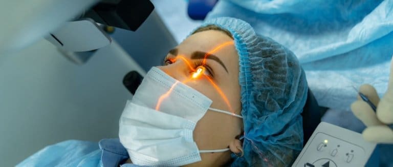
Retina surgery (vitrectomy) is an intraocular procedure typically performed under local or general anesthesia. The surgeon accesses the eye through the sclera (the white and colored part at the back of the eye). After anesthetizing the surface of the eye, the vitreous gel is carefully removed under a microscope. Depending on the condition, additional procedures may be performed to repair retinal tears or close macular holes. At the end of the surgery, the vitreous is replaced with a special gas or oil, and the eye is sealed. Recovery and outcomes vary depending on the patient’s condition and the specific procedure performed.
Preparing for Retinal (Vitrectomy) Surgery
Retinal surgery (vitrectomy) is a procedure performed to remove the transparent gel called vitreous gel from the back cavity of the eye and repair damage to the retina. Vitrectomy surgery is usually performed under local anesthesia and takes approximately 1-2 hours. Patients are typically discharged on the same day after the surgery.
Here are some precautions to take before vitrectomy surgery:
- Follow your doctor’s instructions: You may be prescribed eye drops or medications to use before surgery.
- Arrive with an empty stomach: Avoid eating or drinking anything for at least 8 hours prior to your procedure.
- Disclose all medications and supplements: Tell your doctor about any medicines or supplements you are taking, as some may impact your surgery.
- Protect your eyes after surgery: You may need to wear a bandage or protective goggles following the procedure to aid healing.
Other precautions to take before vitrectomy surgery include:
- If you have diabetes, carefully manage your blood sugar levels before surgery.
- If you take blood thinners, consult your doctor about how to manage them prior to surgery.
- Get plenty of rest after surgery to support your eyes’ healing process.
Vitrectomy is an effective treatment for many retinal diseases, but like any surgery, it carries some risks, such as infection, bleeding, and vision loss.
Caring for Your Eyes After Retinal (Vitrectomy) Surgery
Take all prescribed eye drops or medications as directed. Wear sunglasses to reduce light sensitivity and protect your eyes to lower the risk of infection. Avoid strenuous activities and, if instructed, remain in a face-down position for several days after surgery. Attend all scheduled follow-up appointments, and report any discomfort or unusual symptoms to your doctor promptly. If you smoke, quitting will support your eyes’ healing. Remember, recovery times can vary, so always follow your doctor’s instructions and care recommendations.
Retinal Surgery (Vitrectomy) Frequently Asked Questions
People frequently have questions about retinal surgery, including how vitrectomy is performed, when it is needed, how long recovery takes, and what risks and outcomes are involved. Other common inquiries cover alternative treatment options and potential complications.
Diabetes and Retinal Health
The retina is a delicate layer inside the eye that can be damaged by high blood sugar levels”a condition known as diabetic retinopathy. This occurs when elevated blood sugar affects the blood vessels in the retina, potentially leading to vision problems. Symptoms may include blurry or distorted vision, spots or floaters, flashes of light, and macular edema (central vision loss). If untreated, diabetic retinopathy can progress to severe vision loss or even permanent blindness. For this reason, diabetic patients should have regular eye exams and work to keep their blood sugar levels under control.
What Happens If the Retina is Damaged?
Damage to the retina can lead to a range of eye health problems, especially if caused by diabetes or other retinal diseases, often resulting in vision loss. Conditions such as diabetic retinopathy, macular degeneration, retinal tears or detachments, and retinitis pigmentosa may develop. These can cause symptoms like blurred vision, seeing spots or lines, changes in color perception, dark spots, or defects in the visual field.
* Liv Hospital Editorial Board has contributed to the publication of this content .
* Contents of this page is for informational purposes only. Please consult your doctor for diagnosis and treatment. The content of this page does not include information on medicinal health care at Liv Hospital .eatment. The content of this page does not include information on medicinal health care at Liv Hospital .
For more information about our academic and training initiatives, visit Liv Hospital Academy
Frequently Asked Questions
What is retina surgery (vitrectomy)?
It is a procedure to remove the vitreous gel and repair retinal problems such as tears, detachment, or bleeding.
When is vitrectomy necessary?
It is recommended for conditions like retinal detachment, macular hole, diabetic retinopathy, or retinal bleeding.
How long does retina surgery take?
The procedure usually takes 1–2 hours and is often done under local anesthesia.
Is retina surgery painful?
No, it is generally painless because local or general anesthesia is used during the operation.
How long is recovery after vitrectomy?
Recovery varies but typically takes a few weeks. Some patients may need to maintain a face-down position for several days.
What precautions should I take before surgery?
Avoid eating for 8 hours before surgery, follow your doctor’s medication instructions, and manage blood sugar if diabetic.
What should I do after surgery?
Use prescribed eye drops, wear protective eyewear, avoid strenuous activity, and attend all follow-up visits.
What are possible risks of retina surgery?
Risks include infection, bleeding, increased eye pressure, or vision changes, though complications are rare.
Can diabetic patients have retina surgery?
Yes, vitrectomy is often used to treat diabetic retinopathy and prevent further vision loss.
Where can I get retina surgery in Türkiye?
Liv Hospital in Istanbul provides advanced vitrectomy techniques and comprehensive post-surgical eye care for patients.


