
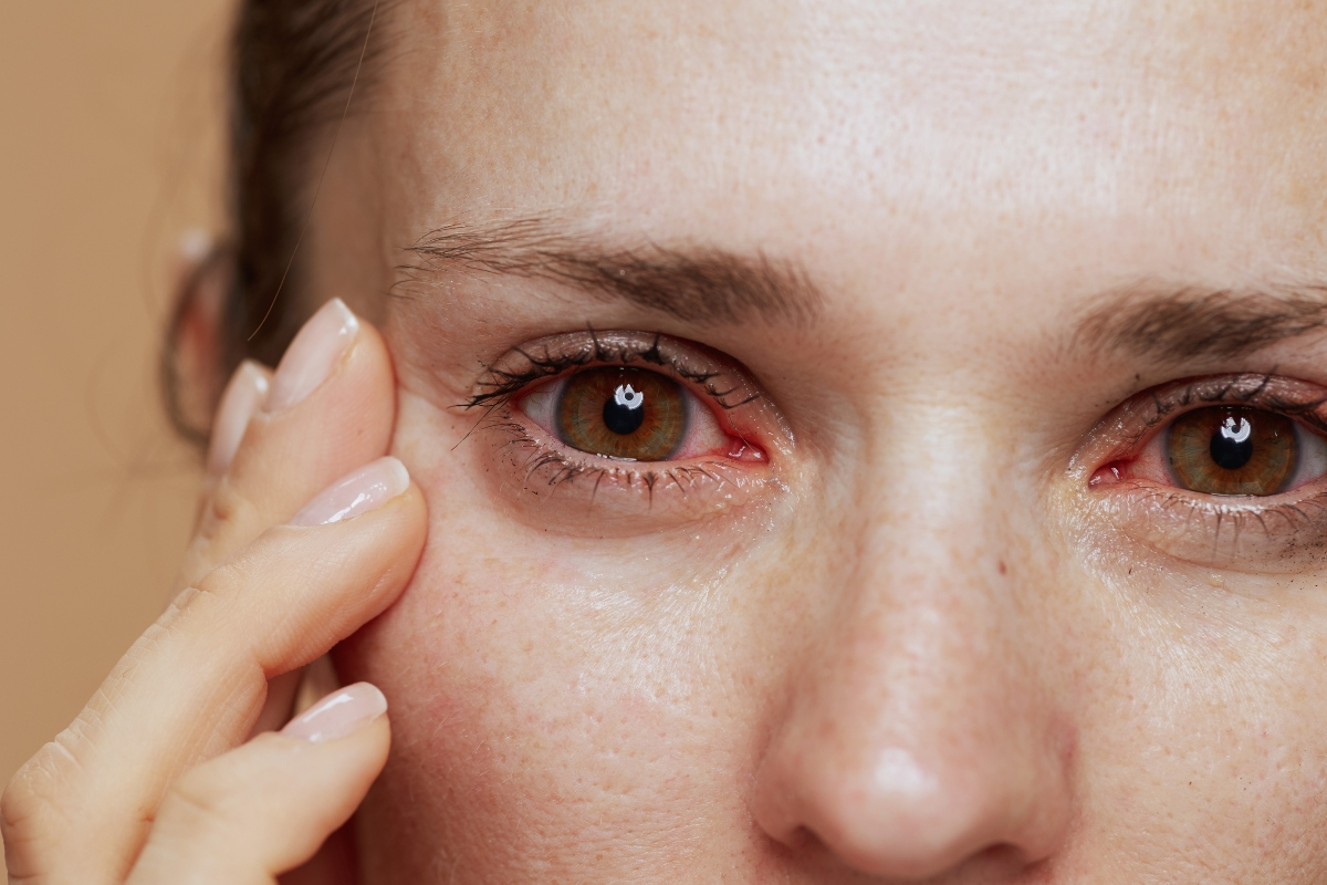
Diabetic retinopathy is a big problem for people with diabetes. It hurts their vision and changes their life. It happens when high blood sugar damages the blood vessels in the retina.severe diabetic retinopathyTop 5 Worst Autoimmune Diseases: Fatal Risks, Symptoms & Deadliest Types
We will look at how this condition gets worse, from mild diabetic retinopathy (NPDR) to severe forms. This includes proliferative disease and diabetic macular edema. Knowing these stages helps find problems early and treat them before it’s too late.
Key Takeaways
- Diabetic retinopathy is a complication of diabetes that affects the eyes.
- High blood sugar levels damage the blood vessels in the retina.
- Understanding the stages of diabetic retinopathy is key for early detection.
- Early detection and treatment can stop vision loss.
- Liv Hospital combines international expertise with patient-centered innovation.
The Global Impact of Diabetic Retinopathy

Diabetic retinopathy is a big problem worldwide, causing a lot of vision loss. It’s important to know how common it is, the cost to healthcare, and how it affects people’s lives.
Prevalence and Statistics in the United States
In the United States, about 9.6 million people have diabetic retinopathy. One in five of these cases is serious enough to threaten vision. This shows we need better screening and care.
The Centers for Disease Control and Prevention (CDC) says more people will get diabetic retinopathy. This is because more people have diabetes. We must find ways to manage this better.
Condition | Prevalence in the US | Vision-Threatening Cases |
Diabetic Retinopathy | 9.6 million | 1 in 5 |
Economic and Quality of Life Burden
Diabetic retinopathy costs a lot, both in medical expenses and lost work time. A study found the yearly cost in the US is high, with a big part due to serious cases.
A healthcare expert said, “The cost of diabetic retinopathy affects not just the person, but families and the healthcare system too.”
“The personal and economic burden of diabetic retinopathy is a critical concern, necessitating a multifaceted approach to management and prevention.”
People with diabetic retinopathy face big challenges, like losing their vision. This can make everyday tasks hard, affect their independence, and lower their happiness.
Diabetes and Your Vision: How Retinopathy Develops

Diabetic retinopathy is closely tied to blood sugar control in people with diabetes. It’s a serious problem that happens when high blood sugar damages the retina’s blood vessels.
Hyperglycemia and Vascular Damage
High blood sugar is the main cause of diabetic retinopathy. It damages the retina’s blood vessels over time. This damage can lead to nonproliferative diabetic retinopathy (NPDR), marked by microaneurysms, hemorrhages, and exudates.
The damage from high blood sugar works through several ways. It activates protein kinase C (PKC) and creates advanced glycosylation end-products (AGEs). These changes make blood vessels more leaky, cause inflammation, and lead to a lack of blood flow. All these factors help diabetic retinopathy get worse.
Timeline of Retinal Changes in Diabetic Patients
The timeline for retinal changes in diabetics varies. It depends on how long someone has diabetes, how well they control their blood sugar, and if they have other health issues like high blood pressure or high cholesterol.
Stage | Retinal Changes | Timeline |
Mild NPDR | Microaneurysms, small hemorrhages | Early in diabetes |
Moderate NPDR | More extensive hemorrhages, hard exudates | Variable, can progress from mild NPDR |
Severe NPDR | Significant ischemia, venous beading, IRMA | Can develop over several years |
Knowing how diabetic retinopathy progresses is key to preventing and treating it. By keeping blood sugar in check and watching for changes in the eyes, people with diabetes can lower their risk of losing their sight.
Risk Factors for Developing Diabetic Retinopathy
Knowing the risk factors for diabetic retinopathy is key to catching it early. This condition affects the blood vessels in the retina, leading to vision loss if not managed. It’s a complication of diabetes.
Duration of Diabetes and Glycemic Control
The longer you have diabetes, the higher your risk of diabetic retinopathy. Glycemic control is very important. Keeping blood sugar levels in check can lower your risk.
- Regular monitoring of blood glucose levels
- Adherence to medication regimens
- Lifestyle modifications such as diet and exercise
Hypertension and Hyperlipidemia
Hypertension and hyperlipidemia increase the risk of diabetic retinopathy. High blood pressure strains the retina’s blood vessels. High cholesterol can cause hard exudates.
Genetic Predisposition and Other Factors
Genetics also play a part in diabetic retinopathy. If your family has diabetes or retinopathy, you’re at higher risk. Pregnancy, kidney disease, and certain genetic markers can also raise your risk.
Healthcare providers can spot high-risk individuals. They can then take steps to prevent or delay diabetic retinopathy.
Stage 1: Mild Nonproliferative Diabetic Retinopathy
The first stage of diabetic retinopathy is mild nonproliferative diabetic retinopathy. It shows up as microaneurysms, small bulges in the blood vessels of the retina. This is the first sign of damage to the retina caused by diabetes.
Microaneurysms: The First Clinical Sign
Microaneurysms are the first signs of diabetic retinopathy. They look like small, round, red dots on the retina. They happen when the walls of the retinal blood vessels weaken because of high blood sugar levels.
Spotting microaneurysms early is key to stopping diabetic retinopathy from getting worse. It’s important to get regular eye checks to catch these signs early.
Diagnostic Criteria and Detection Methods
To diagnose mild NPDR, doctors look for microaneurysms during a detailed eye exam. They use tools like:
- Fundus photography to see changes in the retina
- Fluorescein angiography to check for leaks in blood vessels
- Optical coherence tomography (OCT) to measure the thickness of the retina
These tools help doctors find microaneurysms and other early signs of diabetic retinopathy. This makes it easier to start treatment early.
Management Strategies for Mild NPDR
Managing mild NPDR means controlling diabetes and its risks. Important steps include:
- Keeping blood sugar levels in check to slow the disease
- Controlling blood pressure and cholesterol levels
- Getting regular eye exams to monitor the condition
By following these steps, people with mild NPDR can lower their risk of the disease getting worse.
Stage 2: Moderate Nonproliferative Diabetic Retinopathy
Diabetic retinopathy gets worse and reaches a stage called moderate nonproliferative diabetic retinopathy. This stage shows more serious changes in the retina than the first stage.
Hard Exudates and Retinal Hemorrhages
In moderate NPDR, hard exudates and retinal hemorrhages are more visible. Hard exudates are fatty deposits from broken blood vessels. Retinal hemorrhages happen when these vessels burst. These signs show the disease is getting worse and can be seen during a detailed eye check.
Visual Function in Moderate NPDR
Hard exudates and retinal hemorrhages can impact how well you see. People might notice fluctuations in vision. But, losing a lot of vision is rare at this stage. It’s important to keep an eye on how well you can see.
Monitoring and Intervention Approaches
Managing moderate NPDR well means keeping a close eye on it and acting fast. We suggest:
- Regular eye exams to watch how the disease grows
- Keeping blood sugar levels in check
- Using laser treatment for retinal problems
- Injecting medicine into the eye to reduce swelling and prevent vision loss
Using these methods can help slow down diabetic retinopathy and keep your vision good.
Stage 3: Severe Nonproliferative Diabetic Retinopathy
Severe nonproliferative diabetic retinopathy (NPDR) is a serious stage of diabetic retinopathy. It shows a lot of damage to the retina. This damage increases the risk of losing vision if not treated.
The “4-2-1 Rule” for Diagnosing Severe NPDR
The “4-2-1 rule” helps doctors diagnose severe NPDR. It says severe NPDR is present if there are 4 quadrants of hemorrhages and microaneurysms, 2 quadrants of venous beading, or 1 quadrant of intraretinal microvascular abnormalities (IRMA). This rule shows how important a detailed eye check is for diabetic patients.
Clinical Feature | Description | Significance |
Hemorrhages and Microaneurysms | Presence in 4 quadrants | Indicates widespread retinal damage |
Venous Beading | Presence in 2 quadrants | Suggests significant retinal ischemia |
Intraretinal Microvascular Abnormalities (IRMA) | Presence in 1 quadrant | Indicates high risk of progression to proliferative DR |
Cotton Wool Spots and Venous Beading
Cotton wool spots and venous beading are key signs of severe NPDR. Cotton wool spots are white, fluffy lesions on the retina, showing retinal ischemia. Venous beading is when veins in the retina get irregularly dilated and constricted. These signs are not just for diagnosis but also for predicting the future of the disease.
Intraretinal Microvascular Abnormalities (IRMA)
IRMA is a critical finding in severe NPDR. It shows the dilation and twisting of retinal vessels inside the retina. These changes often happen in areas with retinal ischemia and can lead to neovascularization, a sign of proliferative diabetic retinopathy. Finding IRMA means it’s time for quick action to stop the disease from getting worse.
“The presence of IRMA and other features of severe NPDR necessitates a thorough treatment plan. This includes better blood sugar control, managing blood pressure, and possibly laser treatment or anti-VEGF therapy.”
It’s vital for doctors to know about severe nonproliferative diabetic retinopathy. By spotting the signs and using the “4-2-1 rule,” doctors can find at-risk patients. This helps start the right treatment to save their vision.
Severe Diabetic Retinopathy: Understanding Proliferative Stages
Diabetic retinopathy can get worse and turn into proliferative diabetic retinopathy. This is when new, weak blood vessels grow. These new vessels can cause serious problems like bleeding in the eye and retinal detachment.
Early Proliferative Diabetic Retinopathy
In the early stages, the retina starts making new blood vessels. These vessels are weak and can leak, causing vision issues. Early detection is key to stop things from getting worse.
High-Risk Proliferative Diabetic Retinopathy
High-risk proliferative diabetic retinopathy has new blood vessels on the disc or retina, and bleeding in the eye. This is a big risk to your vision and needs quick action. Laser treatment can help prevent more vision loss.
“The Early Treatment Diabetic Retinopathy Study (ETDRS) showed laser treatment works well in stopping vision loss in proliferative diabetic retinopathy.”
Advanced Proliferative Diabetic Retinopathy with Complications
Advanced stages can lead to serious issues like bleeding in the eye, retinal detachment, and glaucoma. These problems can cause a lot of vision loss if not treated right. Vitrectomy surgery might be needed to fix these problems and help your vision.
It’s important to understand the different stages of diabetic retinopathy to manage and treat it well. Spotting the signs early helps prevent vision loss and improves patient care.
Diabetic Macular Edema: A Vision-Threatening Complication
Diabetic macular edema is a big worry for people with diabetic retinopathy. It happens when fluid builds up in the macula, causing vision issues. We’ll look at the different types and how they affect vision and daily life.
Types of Diabetic Macular Edema
There are two main types of diabetic macular edema. Focal macular edema has fluid leaking from small blood vessels. Diffuse macular edema has fluid leaking from more blood vessels all over.
Knowing the type helps doctors choose the right treatment. It’s important for managing the condition well.
Impact on Visual Acuity and Quality of Life
Diabetic macular edema can really hurt your vision and daily life. Fluid in the macula can blur or distort vision. If not treated, it can even lead to losing sight.
It makes everyday tasks hard, like reading, driving, and seeing faces clearly. A study showed how much it affects daily life:
Daily Activity | Impact of Diabetic Macular Edema | Percentage of Patients Affected |
Reading | Difficulty reading small print | 75% |
Driving | Blurred vision at night | 60% |
Recognizing Faces | Difficulty recognizing facial expressions | 50% |
In summary, diabetic macular edema is a serious issue. It can really mess with your vision and daily life. Knowing about it helps find better ways to manage it.
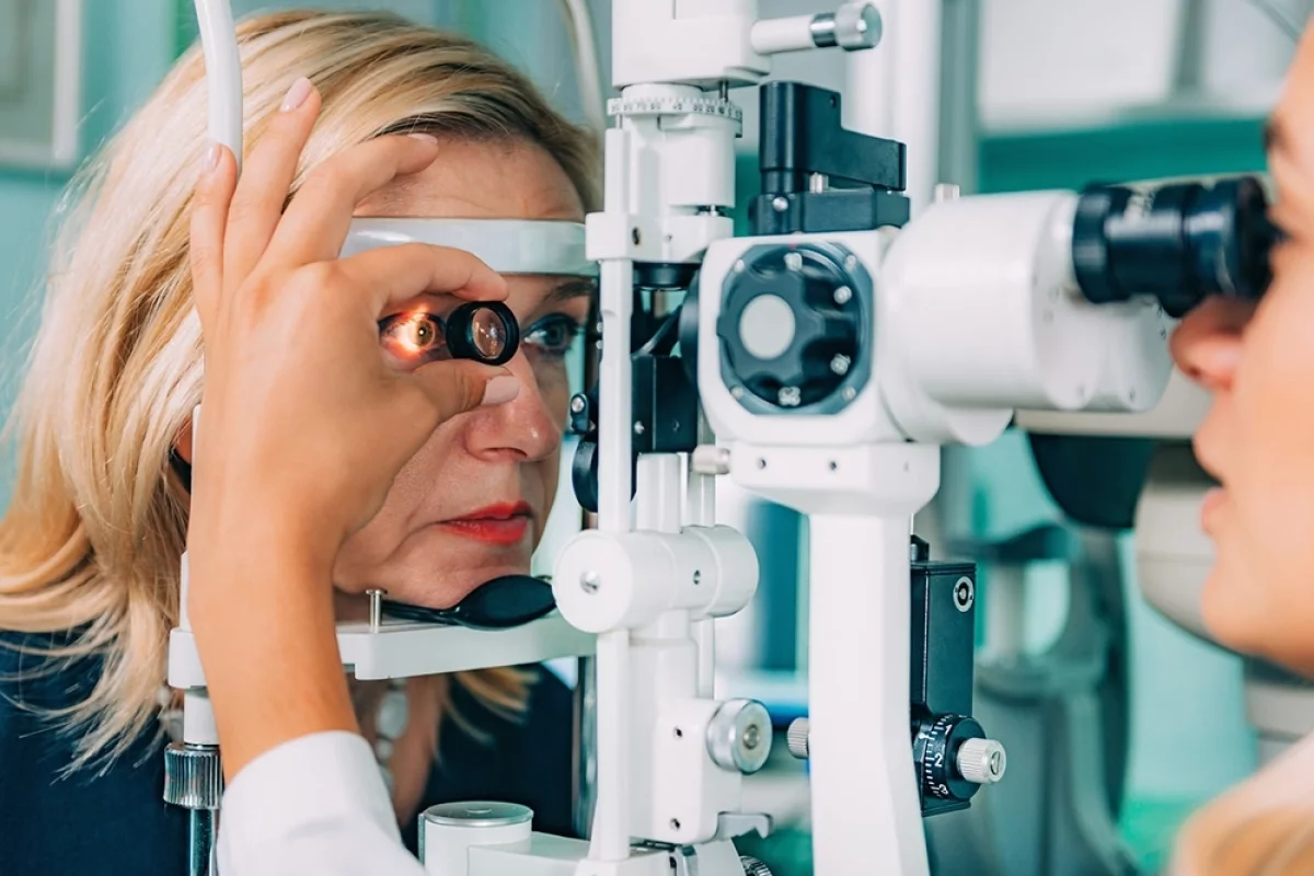
Diagnostic Technologies and Screening Protocols
Diagnosing diabetic retinopathy requires both clinical exams and advanced imaging. Finding it early is key to managing it well.
Clinical Examination Techniques
At the heart of diagnosing diabetic retinopathy are clinical exams. These include:
- Visual acuity testing to check vision sharpness
- Dilated fundus examination to see the retina
- Slit-lamp biomicroscopy for a detailed retinal look
Visual acuity testing is a basic test. It shows how diabetic retinopathy affects vision.
Advanced Imaging Modalities
Modern imaging has changed how we diagnose and treat diabetic retinopathy. These include:
- Fundus photography to record retinal changes
- Optical Coherence Tomography (OCT) for measuring retinal thickness
- Fluorescein angiography to check retinal blood vessels
Optical Coherence Tomography (OCT) is great for spotting diabetic macular edema. It gives clear images of the retina, helping measure its thickness.
Recommended Screening Intervals Based on Diabetes Type
How often to screen for diabetic retinopathy depends on diabetes type and risk factors. Here’s a table of recommended screening times:
Type of Diabetes | Recommended Screening Interval |
Type 1 Diabetes | Within 5 years of diagnosis, then annually |
Type 2 Diabetes | At diagnosis, then annually |
Gestational Diabetes | During pregnancy, as advised by the doctor |
Regular screenings and early diagnosis are vital to avoid vision loss from diabetic retinopathy. Following these guidelines is important for diabetic care.
Treatment Strategies Across the Spectrum of Disease
Managing diabetic retinopathy requires a detailed plan based on the disease’s stage and severity. It’s important to know the different treatments and when to use them.
Medical Management of Early-Stage Disease
Early diabetic retinopathy is managed by controlling risk factors like high blood sugar, high blood pressure, and high cholesterol. Glycemic control is key, as it greatly lowers the risk of retinopathy. We check hemoglobin A1c levels often and adjust treatments as needed to keep blood sugar in check.
A leading expert says tight blood sugar control is vital: “Tight glycemic control is the cornerstone of preventing diabetic retinopathy progression.” –
Medical Expert, Endocrinologist
Laser Photocoagulation: Focal vs. Panretinal
Laser treatment is a mainstay for diabetic retinopathy, mainly in later stages. There are two main types: focal and panretinal. Focal laser treatment targets specific areas to reduce fluid and improve vision. Panretinal photocoagulation (PRP) uses laser burns on the retina’s edge to stop new blood vessels from forming.
Treatment Type | Indications | Benefits |
Focal Laser | Diabetic Macular Edema | Reduces fluid accumulation, improves vision |
Panretinal Photocoagulation (PRP) | Advanced NPDR, Proliferative DR | Reduces ischemic areas, prevents neovascularization |
Intravitreal Pharmacotherapy
Intravitreal injections of anti-VEGF agents have changed diabetic retinopathy treatment, mainly for macular edema. Anti-VEGF therapy cuts down on blood vessel growth and leakage. Ranibizumab, bevacizumab, and aflibercept are common agents used.
Vitreoretinal Surgery for Advanced Complications
For severe diabetic retinopathy, surgery like vitreoretinal surgery may be needed. Vitrectomy removes the vitreous gel and blood or scar tissue. This surgery can improve vision and prevent further loss.
Early detection and treatment are key to managing diabetic retinopathy well. Knowing the treatment options helps healthcare providers tailor care for each patient, improving outcomes and quality of life.
Preventing Progression: Systemic and Ocular Approaches
We can stop diabetic retinopathy from getting worse by using a mix of systemic and ocular methods. It’s key to keep vision and quality of life for people with diabetes. This means managing blood sugar, blood pressure, and cholesterol levels, as well as eye health.
Glycemic Control and Hemoglobin A1c Targets
Keeping blood sugar in check is vital to prevent diabetic retinopathy from getting worse. Studies show that controlling blood sugar well can lower the risk of eye problems. It’s recommended to keep HbA1c levels below 7%, but targets can vary based on individual needs.
Key aspects of glycemic control include:
- Regular monitoring of blood glucose levels
- Adjusting medication, diet, and exercise as needed
- Aiming for HbA1c targets as recommended by guidelines
Blood Pressure and Lipid Management
High blood pressure and high cholesterol are big risks for diabetic retinopathy getting worse. It’s important to manage these to slow down the disease.
Effective strategies include:
- Using antihypertensive medications to control blood pressure
- Implementing lifestyle changes such as diet and exercise
- Managing cholesterol levels through statins or other lipid-lowering therapies
Emerging Preventive Therapies
New treatments are being researched to prevent or slow down diabetic retinopathy. These include:
- Anti-vascular endothelial growth factor (anti-VEGF) therapies
- Intraocular corticosteroids
- Novel oral medications targeting specific pathways involved in diabetic retinopathy
Conclusion: Living with Diabetic Retinopathy
Managing diabetic retinopathy well is key to a better life. We’ve learned that regular eye checks and quick treatment are vital. They help prevent losing vision.
Dealing with diabetic retinopathy means more than just eye care. It’s about controlling blood sugar, managing blood pressure, and keeping an eye on your retina. These steps help lower the risk of serious problems.
It takes teamwork to manage diabetic retinopathy. Doctors and patients work together to create treatment plans. This ensures the best care for each person.
People with diabetic retinopathy can keep their eyes healthy by staying informed and active. We’re here to support and guide them on their journey.
FAQ
What are the stages of diabetic retinopathy?
Diabetic retinopathy goes through several stages. These include mild nonproliferative diabetic retinopathy (NPDR), moderate NPDR, severe NPDR, and proliferative diabetic retinopathy. Diabetic macular edema can happen at any stage.
What is mild nonproliferative diabetic retinopathy?
Mild NPDR shows up with microaneurysms. These are the first signs of diabetic retinopathy. It’s the early stage of the disease.
How is diabetic retinopathy diagnosed?
Doctors use a detailed eye exam to diagnose diabetic retinopathy. This includes visual tests and a look inside the eye. They also use tools like optical coherence tomography (OCT) and fluorescein angiography.
What are the risk factors for developing diabetic retinopathy?
Several factors increase the risk of diabetic retinopathy. These include how long you’ve had diabetes, poor blood sugar control, high blood pressure, high cholesterol, and genetics. Managing these factors helps prevent diabetic retinopathy.
What is diabetic macular edema?
Diabetic macular edema is a complication of diabetic retinopathy. It happens when fluid builds up in the macula, causing vision problems. It can occur at any stage of diabetic retinopathy.
How is diabetic retinopathy treated?
Treatment for diabetic retinopathy varies. Early stages might be managed with medicine. For more advanced cases, treatments like laser therapy, injections, and surgery might be needed. The right treatment depends on the disease’s stage and severity.
Can diabetic retinopathy be prevented?
While you can’t prevent diabetic retinopathy completely, you can slow its progression. Keeping blood sugar levels in check, managing blood pressure and cholesterol, and regular eye exams are key.
What is the impact of diabetic retinopathy on quality of life?
Diabetic retinopathy can greatly affect your quality of life. Advanced stages or complications like diabetic macular edema can lead to vision loss. This can be very challenging.
How often should individuals with diabetes undergo eye screening?
The frequency of eye screenings depends on your diabetes type and if you have diabetic retinopathy. Generally, people with diabetes should get an eye exam every year. Those with diabetic retinopathy might need more frequent checks.
What is the role of glycemic control in managing diabetic retinopathy?
Keeping blood sugar levels under control is vital for preventing and managing diabetic retinopathy. Your doctor will help set a target Hemoglobin A1c level based on your health.
What are the emerging preventive therapies for diabetic retinopathy?
Researchers are exploring new ways to prevent or slow diabetic retinopathy. This includes new medicines and lifestyle changes aimed at reducing risk.
What is severe nonproliferative diabetic retinopathy?
Severe NPDR shows significant changes in the retina. These include cotton wool spots, venous beading, and intraretinal microvascular abnormalities. It means there’s a high risk of moving to the next stage.
What is proliferative diabetic retinopathy?
Proliferative diabetic retinopathy is a serious stage. It’s marked by new, fragile blood vessels in the retina. If not treated, it can cause severe vision loss.
References
National Center for Biotechnology Information. Evidence-Based Medical Guidance. Retrieved from https://www.ncbi.nlm.nih.gov/pmc/articles/PMC3322701/


