Last Updated on November 27, 2025 by Bilal Hasdemir

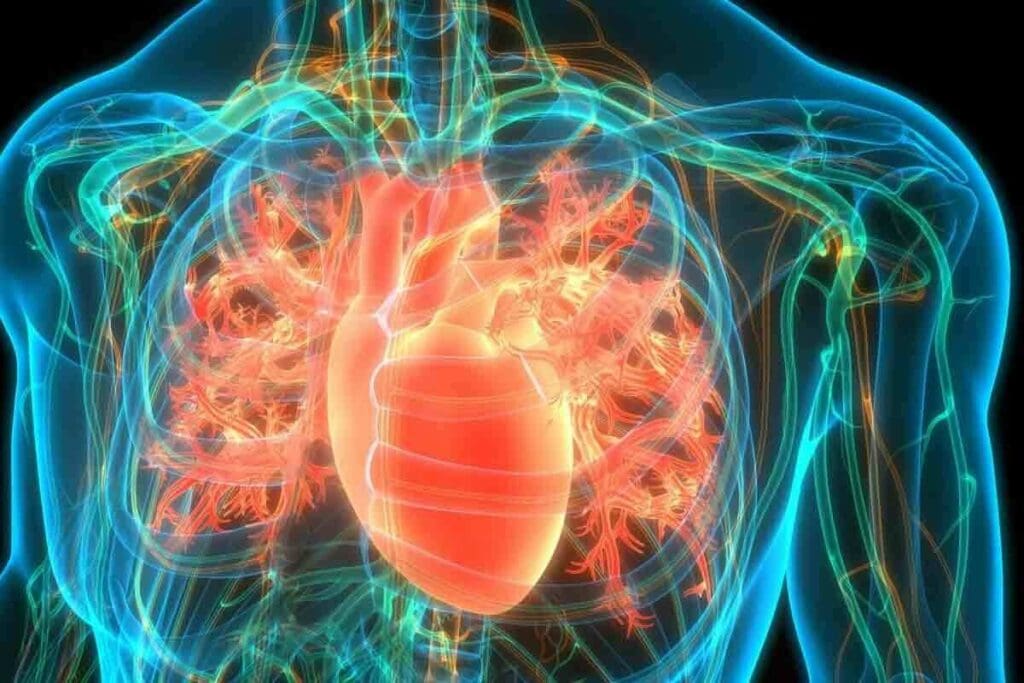
At LivHospital, we know how key accurate heart checks are. Myocardial Perfusion Imaging (MPI) is a key test. It shows how blood flows to the heart muscle, spotting blockages in coronary arteries.
Myocardial Perfusion Imaging tests, like the SPECT heart test, use safe, injectable tracers. These tracers give us insights into heart function. This info is key for spotting coronary artery disease and seeing if treatments like stents or bypass surgery worked.
We aim to give top-notch healthcare with full support for international patients. By knowing about MPI stress tests and other imaging methods, we can tailor care to meet each patient’s needs.
Key Takeaways
- Myocardial Perfusion Imaging (MPI) is a specialized test that evaluates blood flow to the heart muscle.
- SPECT heart tests use safe, injectable radioactive tracers to assess heart function.
- MPI helps diagnose coronary artery disease and detect blockages in coronary arteries.
- The test assesses the impact of blockages on heart health and the effectiveness of previous treatments.
- LivHospital is committed to delivering personalized cardiac care with advanced imaging services.
Understanding SPECT Heart Imaging: The Basics

Single Photon Emission Computed Tomography, or SPECT, is a cutting-edge imaging method in cardiology. It helps doctors check how well the heart works and spot coronary artery disease. This test uses a radioactive tracer to make detailed heart images.
What is Single Photon Emission Computed Tomography?
SPECT is a nuclear medicine test that uses a tiny bit of radioactive material. It’s great for checking heart health and finding coronary artery disease. The National Center for Biotechnology Information says SPECT is key for heart function checks.
It works by catching gamma rays from the tracer in the blood. These rays help make 3D heart images. This gives doctors a clear view of the heart’s structure and how it works.
How SPECT Heart Imaging Works
The SPECT imaging process starts with a tracer injection into the blood. This tracer goes to the heart muscle and sends out gamma rays. A special camera called a gamma camera picks up these rays.
The camera moves around the chest, taking pictures from different sides. These pictures are then mixed together to show the heart in 3D. Doctors can then see how the heart is doing and find any problems.
The Role of Radioactive Tracers
Radioactive tracers are vital in SPECT heart imaging. They are substances like technetium-99m that go to the heart muscle based on blood flow. The SPECT scanner uses these tracers to make detailed heart images.
The type of tracer used can change how clear the SPECT images are. Technetium-99m sestamibi and tetrofosmin are top choices. They help make high-quality heart images.
The Science Behind Myocardial Perfusion Imaging

Understanding myocardial perfusion imaging is key to diagnosing and managing heart disease. This imaging method checks blood flow to the heart muscle at rest and under stress. It spots areas with less blood flow, which might mean heart disease.
Blood Flow Evaluation Techniques
Checking blood flow is vital in MPI. We use special tracers that show up in the heart muscle based on blood flow. Technetium-99m sestamibi and Thallium-201 are the most used tracers. They emit gamma rays, which a camera picks up to create heart muscle images.
The process has two parts: rest and stress. During stress, the heart works harder, either through exercise or medicine. This makes blood flow increase. Images from this phase show where blood flow doesn’t rise, hinting at artery blockages.
Detecting Coronary Artery Disease
Coronary artery disease (CAD) happens when heart arteries narrow or block. MPI is great at spotting CAD by finding areas with less blood flow. The images show where the tracer doesn’t gather well, pointing to CAD.
“Myocardial perfusion imaging is a cornerstone in the diagnosis and management of coronary artery disease, providing valuable information on the extent and severity of perfusion abnormalities.”
Nuclear Cardiology Guidelines
Visualizing Heart Muscle Function
MPI also looks at heart muscle function. It checks how the heart walls move and thicken. This info is key for seeing how much damage CAD has done and for planning treatment.
| Technique | Application | Benefits |
| Rest and Stress MPI | Evaluating blood flow at rest and under stress | Identifies perfusion defects indicative of CAD |
| Radioactive Tracers | Accumulating in heart muscle proportional to blood flow | Allows for accurate assessment of myocardial perfusion |
| Gamma Camera Imaging | Detecting gamma rays emitted by tracers | Creates detailed images of heart muscle and blood flow |
Why Doctors Order MPI Stress Tests
Doctors order MPI stress tests for several reasons. They look at symptoms and risk factors. Myocardial Perfusion Imaging (MPI) is a non-invasive test. It checks blood flow to the heart muscle.
Common Symptoms That Warrant Testing
People with chest pain, shortness of breath, or fatigue might get tested. These signs show the heart might not get enough blood. MPI can spot this problem.
Risk Factors That Indicate Need for Screening
Some risk factors mean you might need a test. These include a family history of heart disease, high blood pressure, diabetes, high cholesterol, and smoking. Even if you don’t feel sick, these factors can lead to a test.
Post-Treatment Evaluation Purposes
After treatments like stenting or CABG, MPI tests check if they worked. They see if blood flow to the heart muscle is good again.
To summarize the key points:
| Category | Description | MPI Stress Test Role |
| Symptoms | Chest pain, shortness of breath, fatigue | Diagnose reduced blood flow |
| Risk Factors | Family history, high blood pressure, diabetes, high cholesterol, smoking | Screen for cardiac risk |
| Post-Treatment | After stenting or CABG | Evaluate treatment effectiveness |
The Complete SPECT Heart Testing Process
Understanding the SPECT heart testing process is important. It starts with preparation and ends with imaging. Each step is vital for a good test result.
Pre-Test Preparation Guidelines
Before a SPECT heart test, patients get specific advice. They might need to change their medications. They also have to follow certain diet rules to avoid test interference.
They should not eat caffeine or certain foods before the test. Wearing comfortable clothes is also important. If they need to exercise, they should wear the right clothes for it.
The Stress Component: Exercise vs. Pharmacological
The stress part of the test is key. It shows how the heart works when stressed. There are two ways to do this: exercise or medication.
- Exercise Stress: Patients walk on a treadmill or bike to stress their heart.
- Pharmacological Stress: For those who can’t exercise, medicine is used to mimic exercise effects.
Both methods help doctors see how well the heart works under stress. They help find any heart problems.
Rest Phase Imaging Procedures
After the stress test, patients have rest phase imaging. A radioactive tracer is injected. It helps make detailed heart images.
During this time, patients lie down while a camera takes heart images. This lets doctors compare heart function at rest and under stress. It gives them important information for diagnosis.
Interpreting Myocardial Perfusion Test Results
Understanding myocardial perfusion imaging results is key for good patient care. This imaging is vital for doctors to check heart function and spot heart disease issues.
Normal vs. Abnormal Findings
Results can be normal or abnormal. Normal findings mean the heart gets enough blood flow, both when it’s at rest and under stress. But, abnormal findings show that some heart areas might not get enough blood. This could mean heart disease or other heart problems.
It’s important to know that abnormal results don’t always mean you have heart disease. Other things can cause heart muscle issues. More tests might be needed to find out why.
Understanding Perfusion Defects
Perfusion defects are when heart muscle areas don’t get enough blood. They can be different types:
- Reversible defects: These happen when blood flow is low under stress but normal at rest. They often point to heart disease.
- Fixed defects: These show low blood flow both at rest and under stress. They might mean scar tissue from a heart attack.
- Partially reversible defects: These have both reversible and fixed traits.
What Your Doctor Looks For
Doctors look at several things when checking test results:
- The size and seriousness of perfusion defects
- Any scar tissue or past heart damage
- The heart’s overall function and how well it pumps
- Any other oddities on the images
By looking at these details, doctors can decide on the best tests, treatments, and care plans for each patient.
Benefits and Limitations of Myocardial Perfusion Imaging
Myocardial perfusion imaging (MPI) is a non-invasive way to check for heart disease. It’s very accurate and is often used to see if coronary artery disease is present. We’ll look at what makes MPI good and what might limit its use, helping us understand its role in heart care.
Diagnostic Accuracy Rates
MPI is great at finding heart disease. It shows where blood flow to the heart is low. This helps doctors catch problems early and treat them right away.
High diagnostic accuracy is key in heart care. MPI’s ability to spot heart disease makes it a top choice for doctors. It helps them make the best decisions for their patients.
Non-Invasive Nature Advantages
MPI is non-invasive, which means it doesn’t hurt. It doesn’t need to go inside the body. This makes it safer and more comfortable for patients.
“The non-invasive nature of MPI makes it an attractive diagnostic option for patients with suspected coronary artery disease.”
Being non-invasive also means MPI can be done more than once. Doctors can keep an eye on how the disease is changing or if treatments are working.
Potential Limitations and False Results
Even with its benefits, MPI has some downsides. It can sometimes give false results. This might happen if the patient moves during the scan or if there are technical problems. False results can lead to more tests or confusion, so it’s important to prepare well and interpret images carefully.
To avoid false results, it’s important to follow the right steps before the test. Also, the test should be done by skilled professionals. Despite these challenges, MPI is a valuable tool in heart care. It offers a safe and accurate way to check how well the heart is working.
Comparing SPECT Heart Tests to Other Cardiac Diagnostics
SPECT heart tests are used in cardiology, along with other tests. It’s important to know how they compare for accurate diagnosis. Healthcare providers use different tests for various heart conditions.
SPECT vs. Echocardiogram
An echocardiogram uses ultrasound to show the heart’s structure and function. It’s different from SPECT, which looks at blood flow to the heart muscle. Echocardiograms give real-time info on heart valves and sizes.
Both tests are non-invasive but serve different purposes. SPECT checks blood flow, while echocardiograms look at heart structure and function. The choice depends on the heart issue being checked.
SPECT vs. Cardiac Catheterization
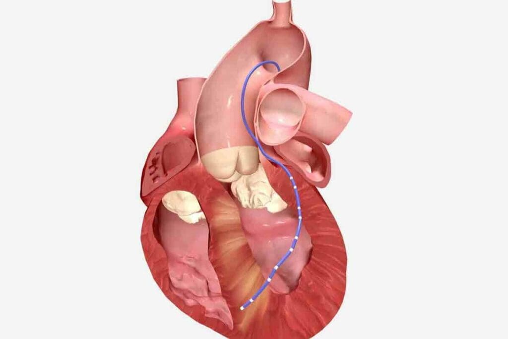
Cardiac catheterization is an invasive test that looks at the heart’s inside. It’s different from SPECT, which shows blood flow to the heart muscle. Catheterization gives detailed views of the heart’s anatomy.
SPECT heart tests are often used first to see if a patient needs catheterization. They help find coronary artery disease without the need for invasive tests.
SPECT vs. Cardiac MRI
Cardiac MRI is a non-invasive test that shows the heart’s structure and function. It looks at blood flow, viability, and scar tissue, like SPECT. But MRI gives clearer images and more info about the heart.
The choice between SPECT and MRI depends on the question being asked and the patient’s needs. Both tests have their own benefits: SPECT for blood flow, MRI for detailed heart images.
PET Scans for Cardiology: An Advanced Alternative
PET scans are a top choice for heart health checks, beating SPECT in image quality. They give a clearer view of how well the heart works and how blood flows through it.
How Cardiac PET Scans Differ from SPECT
PET scans and SPECT are both used to look at the heart. But they work in different ways and show different things. PET scans use special tracers and methods to make clearer images.
PET scans use tracers like Rubidium-82 or Nitrogen-13 ammonia. These tracers have shorter lives than those in SPECT. This means patients get less radiation when they have PET scans.
Superior Resolution Benefits
PET scans have better resolution, which helps in many ways. They can spot coronary artery disease more accurately. They also help see if heart muscle is working right, which is key for complex heart issues.
Key advantages of PET scans include:
- Higher image resolution
- Lower radiation doses
- Improved diagnostic accuracy for certain conditions
When PET is Preferred Over SPECT
PET scans are better in some situations. For example, they’re great for people who are overweight or need to check if heart muscle is working. Their high-quality images are a big plus.
| Diagnostic Criteria | SPECT | PET |
| Image Resolution | Lower | Higher |
| Radiation Exposure | Higher | Lower |
| Diagnostic Accuracy for CAD | Good | Excellent |
Cardiac PET Scan vs. Echocardiogram: Key Differences
It’s important to know the differences between cardiac PET scans and echocardiograms. These tests give different kinds of information about the heart. Each has its own benefits and limitations.
Functional vs. Structural Information
Cardiac PET scans and echocardiograms give different kinds of information. Cardiac PET scans show how the heart works, like its blood flow and metabolism. This is great for checking heart disease and how well the heart muscle is doing.
Echocardiograms, on the other hand, show the heart’s structure. They give clear pictures of the heart’s parts, like chambers and valves. Echocardiograms are good for checking heart valves and finding heart problems.
Radiation Exposure Considerations
Cardiac PET scans and echocardiograms differ in radiation exposure. PET scans use a small amount of radiation from a tracer. Echocardiograms don’t use radiation, which is safer for some patients, like pregnant women.
When choosing between these tests, we must think about radiation risks. While PET scans’ radiation is usually safe, it’s key to weigh it against the benefits of the test.
Cost and Availability Factors
The cost and availability of cardiac PET scans and echocardiograms vary. Echocardiograms are more common and cheaper than PET scans. This makes echocardiograms a good first choice for many patients.
But, PET scans are better for certain things, like checking heart muscle health and finding heart disease. The right test depends on the patient’s needs and the doctor’s goals.
In summary, both cardiac PET scans and echocardiograms are useful, but they’re used for different reasons. Knowing their differences helps doctors choose the best test for each situation.
Preparing for Your Myocardial Perfusion Stress Test
Knowing how to prepare for a myocardial perfusion stress test is key. It makes sure the test works well. This way, your doctor can make the best decisions for your health.
Medication Adjustments Before Testing
Before the test, you might need to change your medications. It’s important to listen to your doctor about any changes. Some medicines, like beta-blockers, might need to be adjusted to get the best heart rate response.
Medication Adjustment Guidelines:
| Medication Type | Adjustment Instruction |
| Beta-blockers | Stop or adjust as directed by your doctor |
| Caffeine | Avoid for 24 hours before the test |
| Nitrates | Consult your doctor for specific instructions |
Dietary Restrictions and Guidelines
There are special diet rules for your test. You might need to skip certain foods and drinks to get good results.
Dietary Guidelines:
- Avoid eating or drinking anything except water for 4-6 hours before the test
- Refrain from consuming caffeine for 24 hours before the test
- Follow any specific dietary instructions provided by your healthcare provider
What to Wear and Bring
Wear comfy clothes and shoes for the test. Don’t forget to bring important documents like insurance cards and ID.
Items to Bring:
- Comfortable clothing and shoes
- Insurance cards and identification
- A list of your current medications
- Any relevant medical records or test results
By following these tips, your test will go smoothly. This ensures the results are accurate and helpful for your doctor.
Safety and Risks of Nuclear Medicine for Heart Diagnostics
Nuclear medicine for heart diagnostics is a complex topic. It involves radiation exposure, side effects, and who should avoid these tests. Understanding both the benefits and risks is key.
Radiation Exposure Levels
Nuclear medicine tests, like SPECT and PET scans, use small amounts of radiation. The amount of radiation depends on the test, the tracer dose, and the person’s body. “The radiation exposure from these tests is generally low to moderate,” studies say.
It’s important to think about radiation exposure, more so for young patients or those needing repeated tests. A typical myocardial perfusion SPECT scan’s radiation dose is 9 to 12 millisieverts (mSv). This is similar to other common imaging tests.
Potential Side Effects and Complications
Nuclear medicine tests are usually safe, but there are possible side effects and complications. Common side effects are mild and might include:
- Dizziness or lightheadedness during or after the test
- Mild allergic reactions to the tracer
- Discomfort or pain at the injection site
More serious issues are rare but can happen. These include severe allergic reactions or heart problems during the test. It’s important to talk to your doctor about your medical history and any worries before the test.
Who Should Avoid These Tests
Some people should not have nuclear medicine tests for heart diagnostics. This includes:
- Pregnant or breastfeeding women, due to risk to the fetus or baby
- Patients with known allergies to the tracers used
- Individuals with severe kidney or liver disease, as these conditions may affect tracer clearance
Telling your doctor about any health conditions, allergies, or concerns is vital. This way, we can make sure the tests are safe and effective for you.
Conclusion: The Value of SPECT and MPI in Modern Cardiac Care
We’ve seen how Single Photon Emission Computed Tomography (SPECT) and Myocardial Perfusion Imaging (MPI) are key in heart disease diagnosis and management. These tools give deep insights into heart function and coronary artery disease. They help doctors make better treatment plans, leading to better patient care.
SPECT and MPI are great at showing how blood flows to the heart muscle. They help spot coronary artery disease and show how well the heart works. This information lets doctors create specific treatment plans, improving heart care quality.
In today’s heart care, SPECT and MPI are essential for managing patients. They help doctors make smart treatment choices. As we keep improving heart health, these tools will become even more important. They show their worth in giving top-notch heart care.
FAQ
What is SPECT heart imaging, and how does it work?
SPECT heart imaging is a test that uses tiny amounts of radioactive tracers. It helps see the heart and its blood flow. The test detects gamma rays from the tracers, showing how well the heart muscle gets blood.
What is myocardial perfusion imaging (MPI), and why is it used?
MPI is a test that checks blood flow to the heart muscle. It’s used to find and manage heart disease. It also helps figure out the risk of heart attacks and guides treatment.
How does MPI differ from other cardiac diagnostic tests like echocardiograms or cardiac catheterization?
MPI is a non-invasive test that looks at blood flow and function. Echocardiograms check the heart’s structure and function. Cardiac catheterization is more invasive, directly looking at the heart’s arteries. MPI is great for seeing how well the heart muscle gets blood.
What are the benefits of using MPI stress tests in diagnosing coronary artery disease?
MPI stress tests are very accurate and non-invasive. They can be used on many patients. They show how severe heart problems are, helping doctors make better decisions.
How should I prepare for a myocardial perfusion stress test?
To prepare, avoid caffeine and some medicines. You might need to fast or follow a special diet. Wear comfy clothes and be ready to share your medical history. Your doctor will give you specific instructions.
What are the risks and side effects associated with nuclear medicine tests like MPI?
The main risk is low radiation exposure. Side effects are usually mild, like headaches or dizziness. Rarely, there can be serious reactions.
How do PET scans compare to SPECT for cardiac diagnostics?
PET scans are more detailed and sensitive than SPECT. They’re better for measuring blood flow and finding heart disease. They’re great for certain patients.
What is the difference between a cardiac PET scan and an echocardiogram?
PET scans show blood flow and heart muscle health. Echocardiograms look at the heart’s structure and function. Both are useful and can be used together for a full check-up.
Are there any individuals who should avoid nuclear medicine tests for heart diagnostics?
Yes, pregnant or breastfeeding women should avoid them due to radiation. People with severe kidney disease or allergies might also need special care.
How do I understand the results of my myocardial perfusion test?
Your doctor will explain the results. They’ll look at the images and your medical history. Normal results mean good blood flow. Abnormal results might show heart problems, helping guide treatment.
References
- Taqueti, V. R., & Di Carli, M. F. (2018). Radionuclide myocardial perfusion imaging for the evaluation of patients with known or suspected coronary artery disease. Circulation, *138*(16), 1731–1745. https://pubmed.ncbi.nlm.nih.gov/30354642/


