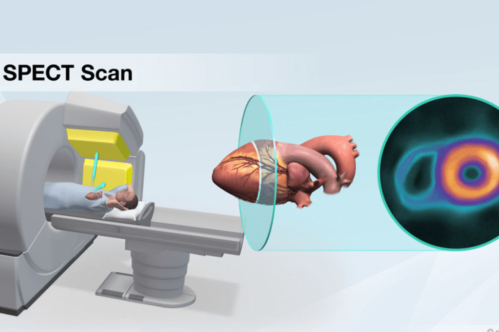
SPECT imaging is very useful for diagnosing, but it has some big downsides or SPECT scan disadvantages. One major issue is that almost 30% of SPECT scans have low image resolution. This can lead to wrong diagnoses.
We will look at the disadvantages of SPECT scans. These include the risk of radiation exposure and the hassle of long scan times. SPECT scans are great for learning about the body’s functions. But, knowing their limits is key for doctors and patients.

SPECT imaging helps us understand the body’s inner workings. It’s key in fields like oncology, cardiology, and neurology. It uses a radioactive tracer that goes to specific areas of the body.
SPECT scans find gamma rays from a radioactive tracer. This tracer is picked for the body function being studied. For example, it’s great for spotting bone problems or checking heart health.
SPECT shows how the body works, unlike MRI or CT scans that show what it looks like. MRI is good at showing soft tissues, but SPECT tells us about metabolic activity. This is key for diagnosing diseases, like tumors.
SPECT is used in many medical areas. In cardiology, it checks heart blood flow and health. In neurology, it looks at brain blood flow and helps with Alzheimer’s diagnosis. Here’s a table of some SPECT uses:
| Medical Specialty | Application of SPECT |
| Oncology | Tumor detection and assessment of metastasis |
| Cardiology | Myocardial perfusion and viability assessment |
| Neurology | Cerebral blood flow assessment and diagnosis of neurodegenerative diseases |
Knowing how SPECT works helps us see its value in medicine. It also shows its limits in some cases.

Understanding SPECT’s technical limits is key to its use in medical diagnostics. We’ll look at SPECT’s inherent constraints, what affects its image quality, and how it stacks up against other high-resolution imaging methods.
SPECT imaging gives us functional info about the body. But, it has low image resolution compared to some other methods. This can make SPECT images less clear.
The main reason for this is the tech used to detect gamma rays. SPECT scanners use collimators to track gamma rays. The type of collimator used greatly affects the image quality. For example, parallel-hole collimators are common but have their own set of limitations.
Several things impact SPECT image quality. These include the gamma ray energy, the collimator type, and the algorithms used. Higher energy gamma rays can penetrate better but might lose some resolution due to scatter.
The choice of radiotracer is also key. It affects how well SPECT images turn out. Some radiotracers are better suited for SPECT, improving image quality.
| Factor | Impact on Image Quality |
| Collimator Type | Affects resolution and sensitivity |
| Gamma Ray Energy | Influences penetration and scatter |
| Radiotracer | Determines gamma ray emission characteristics |
Comparing SPECT to MRI or CT scans shows SPECT’s SPECT imaging limitations in resolution. MRI, for example, has much higher resolution and better soft tissue contrast. It’s better for some diagnostic tasks.
But SPECT has its own strengths, like functional imaging and insights into physiological processes. The right imaging modality depends on the clinical question at hand.
SPECT imaging involves radiation, which is something we need to think about carefully. We use SPECT scans for diagnosis. It’s important to know the risks and how we reduce them.
The dose from a SPECT scan changes based on the tracer and the procedure. “A typical SPECT scan’s effective dose is between 2 to 20 mSv,” a study says. We aim to keep the dose low while getting good images.
A cardiac SPECT stress test might have a dose of about 10 mSv. Bone scans might have a dose of around 15 mSv. Knowing these doses helps us weigh the benefits against the risks.
Long-term radiation exposure is a big concern, mainly for those who have many scans. We keep track of how much radiation patients have had. This helps us decide if more scans are safe.
The International Commission on Radiological Protection (ICRP) says the risk of health problems from radiation is linked to the dose. We think about this when deciding on more scans.
Children and pregnant women need extra care because they are more sensitive to radiation. We use special methods to lower their exposure. Sometimes, we choose other imaging options instead.
In kids, we use smaller doses and special protocols to cut down radiation. “Lower doses are key for kids to avoid too much radiation,” say pediatric nuclear medicine guidelines.
By understanding the risks and taking steps to lessen them, we make sure SPECT scans are safe for everyone.
SPECT technology has a big drawback: it takes a long time to scan. This affects how many patients can be seen and how quickly emergencies are handled. The long wait can make patients uncomfortable and limits how many scans can be done in a short time.
SPECT scans can last for hours. This is because they need to take pictures from many angles around the patient’s body. The long wait can make patients tired and uncomfortable.
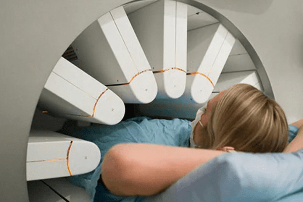
To get these images, a gamma camera moves around the patient. It takes pictures from different sides, then makes a 3D image. While this gives important info, the time it takes is a big problem.
The long scan times of SPECT imaging slow down how many patients can be seen. With each scan taking hours, there’s a limit to how many can be done in a day. This can cause delays and longer waits for patients.
| Imaging Modality | Typical Scan Time | Patient Throughput |
| SPECT | 1-3 hours | Limited |
| CT Scan | 5-30 minutes | High |
| MRI | 15-90 minutes | Moderate |
In emergencies, quick diagnosis is key. SPECT’s long scan times can slow down getting a diagnosis and treatment. This can hurt patient outcomes. Faster imaging options might be better for urgent cases.
But SPECT is great for certain tests, like heart and cancer checks. We’re trying to make SPECT scans faster without losing quality.
Getting a SPECT scan can be tough for patients because of comfort and compliance problems. The scan needs patients to stay very quiet and not move for a long time. This can make it hard for patients to feel comfortable and can also affect the scan’s quality.
Physical discomfort during the scan is a big worry. SPECT scans can last from 15 to 60 minutes or more. Patients have to lie very quietly on a table, which can be hard, even for those without muscle problems.
Some patients feel claustrophobia and anxiety because of the scanner’s closed space. This can make it hard for them to stay calm and quiet. Their anxiety can make them uncomfortable and might even ruin the scan’s quality if they move.
Movement artifacts happen when patients move during the scan. Even a little movement can make the images blurry. It’s very important for patients to stay very quiet and not move to get good images.
Pediatric and geriatric patients face special challenges. Kids find it hard to stay quiet for a long time and might need help or even sedation. Older patients might feel more pain and have trouble understanding what’s happening, making the scan harder for them.
In short, making patients comfortable and helping them follow instructions is key for SPECT scans to work well. By understanding and fixing these problems, doctors can make the scan better for patients and get clearer images.
When we compare SPECT and PET imaging, we see big differences. These differences are mainly in how well they can diagnose diseases.
PET scans are better at finding and identifying diseases than SPECT scans. This is because of the technical differences between them.
Here’s a table that shows how SPECT and PET compare in sensitivity and specificity:
| Characteristics | SPECT | PET |
| Sensitivity | Lower | Higher |
| Specificity | Lower | Higher |
| Typical Use Cases | Cardiac stress tests, bone scans | Oncology, neurology, cardiology |
PET scans are better at showing how cells work than SPECT scans. This is very helpful in finding and studying tumors.
F-FDG PET is a key tool for finding and tracking cancer. It does this by showing where cells use a lot of sugar.
PET scans are better than SPECT scans in many areas. This includes oncology, neurology, and cardiology. PET’s better sensitivity and specificity help doctors find and track diseases early.
Here’s a list of areas where PET is better than SPECT:
SPECT has its limits in nuclear medicine, mainly when compared to MRI. MRI is better at showing soft tissues and detailed anatomy. We’ll look at how MRI beats SPECT in these areas, like soft tissue contrast, anatomical detail, and brain scans.
SPECT struggles to show soft tissues clearly. But MRI shines here, making soft tissues stand out. This is why MRI is often the go-to for soft tissue issues, like musculoskeletal injuries or soft tissue tumors.
SPECT’s images aren’t as sharp as MRI’s. MRI’s high-resolution pictures help spot problems and understand complex body structures. This is key for neurosurgical planning and oncological assessments.
For brain scans, MRI is way ahead of SPECT. MRI can show brain details and catch small issues. SPECT can show brain activity, but MRI offers more, making it better for brain health checks. For example, MRI is essential for stroke or brain tumors.
Even though SPECT has its uses, MRI is better for soft tissue, anatomy, and brain scans. MRI’s better images make it the top choice for these areas.
It’s important to know the costs of SPECT imaging to see its value in medical care. The costs go beyond just buying the equipment. They also include ongoing expenses like keeping the facility running, making radiotracers, and moving them around.
Buying a SPECT scanner is very expensive. But the costs don’t stop there. Places need to keep their equipment in good shape and update it to meet rules. These costs can make it hard for some healthcare places to afford.
Radiotracers for SPECT imaging are special and pricey to make. They have to be made often because they don’t last long. Also, moving these radioactive materials safely adds to the cost.
Patients sometimes struggle to get their SPECT imaging covered by insurance. The billing process can be complicated. This can lead to delays or claims being denied, making things harder for patients financially.
Looking at how much SPECT imaging costs compared to its benefits is key. Even though it’s not the cheapest option, it’s worth it. It gives doctors important information to help decide on treatments and can lead to better health outcomes.
The economic side of SPECT imaging is complex. By understanding these costs, healthcare providers and patients can make better choices about using SPECT in different medical situations.
SPECT facilities are not spread out evenly. This makes it hard for patients, mainly in rural or underserved areas, to get them. It can also delay diagnosis and treatment for many health issues.
The spread of SPECT facilities affects how easy it is for people to get them. Cities usually have more SPECT imaging because of their big hospitals. But, rural areas struggle because they have fewer facilities.
Even with SPECT facilities, scheduling and wait times can be big problems. The demand for SPECT scans is high, and there’s not enough room. This means patients might have to wait longer than they should for a diagnosis.
Long waits can be really tough for patients. To help, many hospitals are using new scheduling systems. They’re also working to make their processes more efficient to cut down wait times.
Rural areas face big challenges when it comes to SPECT imaging. Patients there have to travel far to get these scans. This can make getting a diagnosis and treatment take longer. Plus, there are fewer doctors in these areas to help.
To fix these problems, healthcare is looking at new ideas. They’re thinking about using mobile SPECT units and telemedicine. These ideas could help more people in rural areas get the imaging they need.
In summary, SPECT imaging is very useful, but it’s not always easy to get. We need to work on making it more available and accessible. This is key to making sure everyone gets the healthcare they deserve.
SPECT scans face many technical and interpretive challenges. These issues affect how well SPECT imaging works for diagnosis. Knowing about these problems helps us use SPECT scans better in medical care.
False positives and false negatives are big issues in SPECT imaging. False positives can cause extra tests and worry for patients. False negatives can mean late diagnosis and treatment. The quality of the scan, the tracer used, and the interpreter’s skill all play a part.
To lower these errors, we need to focus on a few things. For example, using top-notch equipment and following the best image-taking methods can help. This can cut down on mistakes.
| Factor | Impact on False Positives | Impact on False Negatives |
| Equipment Quality | High-quality equipment reduces false positives | Improves detection, reducing false negatives |
| Radiotracer Characteristics | Specificity of the radiotracer affects false positive rates | Sensitivity of the radiotracer impacts false negative rates |
| Interpreter Expertise | Experienced interpreters can reduce false positives through accurate image analysis | Skilled interpreters are better at detecting abnormalities, reducing false negatives |
Reading SPECT images needs a lot of skill. Interpreter variability can cause different results. It’s key to keep professionals learning and updated with new methods and tools.
Many issues can mess with SPECT scan accuracy. These include artifacts from body parts and movement, and limits in detail. Knowing these problems helps us find ways to lessen their effects. For example, using special correction methods and keeping patients calm during scans.
By tackling these issues, we can make SPECT scans more reliable. This will help us give better care to patients.
SPECT has its limits in certain clinical areas. It struggles to detect some tumors and assess neurological conditions well.
In oncology, SPECT faces challenges. It’s not as accurate as PET/CT in finding tumors. This is because SPECT can miss small tumors or those with low activity.
Some main issues are:
This shows we need other imaging methods for cancer diagnosis.
In neurology, SPECT has its own set of problems. It’s not as good as MRI or PET for some brain diseases. It also struggles to catch early brain function changes.
For heart imaging, SPECT has its own hurdles. It’s not as detailed as MRI or angiography for heart disease. It’s used for heart blood flow imaging but has its limits.
Some of these challenges are:
SPECT also has its own set of problems in muscle and bone imaging. It’s good for bone health but not as detailed as MRI for soft tissues.
Some main issues are:
SPECT imaging has its challenges, like technical issues, radiation risks, and patient comfort. These problems can affect how well SPECT works in some cases. Yet, SPECT is a key tool in nuclear medicine, helping doctors diagnose and treat patients.
We need to look at both the good and bad sides of SPECT imaging. Improving SPECT technology is important. This includes better detectors and image processing. By understanding SPECT’s limits and strengths, doctors can use it more effectively.
The world of nuclear medicine is always changing. It’s important to consider the pros and cons of SPECT imaging. This way, patients get the best care for their needs.
SPECT scans have several drawbacks. They have low image quality, pose a risk of radiation exposure, take a long time, and are not always accurate in some cases.
PET scans are more sensitive and specific than SPECT, which is important for metabolic and some cancer-related imaging.
SPECT scans expose patients to ionizing radiation, which can harm them, more so with repeated doses. The dose depends on the procedure and the tracer used.
Yes, children, pregnant women, and people with certain health issues are more at risk from SPECT scans’ radiation.
MRI offers better soft tissue contrast and detail than SPECT. This makes MRI a better choice for some diagnostic needs, like in the brain and muscles.
Patients might feel uncomfortable, anxious, or claustrophobic during SPECT scans. This discomfort can cause movement artifacts, affecting image quality.
SPECT facilities might be scarce in some areas. Scheduling issues or long wait times can also limit access to SPECT imaging.
SPECT imaging comes with high costs for equipment, facilities, tracers, and staff. Insurance and reimbursement policies can also affect its accessibility.
No, SPECT scans are not always the first choice. Other imaging methods like MRI or CT might be better for certain conditions.
Yes, there’s ongoing research to enhance SPECT technology. This includes better image reconstruction, tracer design, and instrumentation to overcome current limitations.
Rarely, patients might react to the tracers used in SPECT scans. Symptoms can include nausea, headaches, or rashes.
SPECT’s accuracy and interpretation challenges, like false results and variability, can affect its reliability and confidence in clinical use.
Subscribe to our e-newsletter to stay informed about the latest innovations in the world of health and exclusive offers!