
Getting the thyroid gland checked right is key to good health. MRI and ultrasound help a lot in checking how well the thyroid is working.
MRI gives a detailed look at the thyroid, showing homogeneous signal intensity in healthy parts. A thyroid gland MRI helps doctors tell the difference between good and bad tissue. Liv Hospital focuses on patients, making sure they get the best MRI and ultrasound scans. They highlight 12 important measurements to keep the thyroid healthy.
Key Takeaways
- Advanced MRI and ultrasound imaging are key for checking thyroid health.
- Knowing the normal measurements of the thyroid gland is vital for diagnosis.
- Liv Hospital offers patient-centered care with top-notch imaging.
- MRI gives a detailed look at the thyroid gland’s structure.
- Healthy thyroid tissue shows homogeneous signal intensity.
Thyroid Gland Anatomy and Physiological Significance

The thyroid gland is shaped like a butterfly and is key to keeping our metabolism balanced. Knowing its anatomy is vital for doctors to diagnose thyroid diseases. It helps them understand what the images show.
Anatomical Location and Structure
The thyroid gland sits in the neck, in front of the trachea. It has two lobes joined by an isthmus. The size can vary based on age, gender, and where you live.
Knowing the normal measurements of the thyroid gland is important. It helps doctors tell if something is wrong. The gland’s location and shape are key to its function and balance in the body.
Physiological Function and Importance
The thyroid gland makes hormones that control how our cells grow and work. These hormones are vital for our health. The gland’s work is closely watched by the body to keep hormone levels just right.
It’s important to understand how the thyroid gland works. This knowledge helps doctors diagnose and treat thyroid problems. Problems with hormone levels can cause serious health issues.
Normal Variations in Thyroid Morphology
Healthy people’s thyroid glands can look different. Age, gender, and ethnicity can affect its size and shape. For example, women’s glands are often bigger than men’s, and they change with age.
It’s important to know these normal variations. This helps doctors avoid mistakes and give the right treatment. They must think about these differences when they look at thyroid images and check how well the gland works.
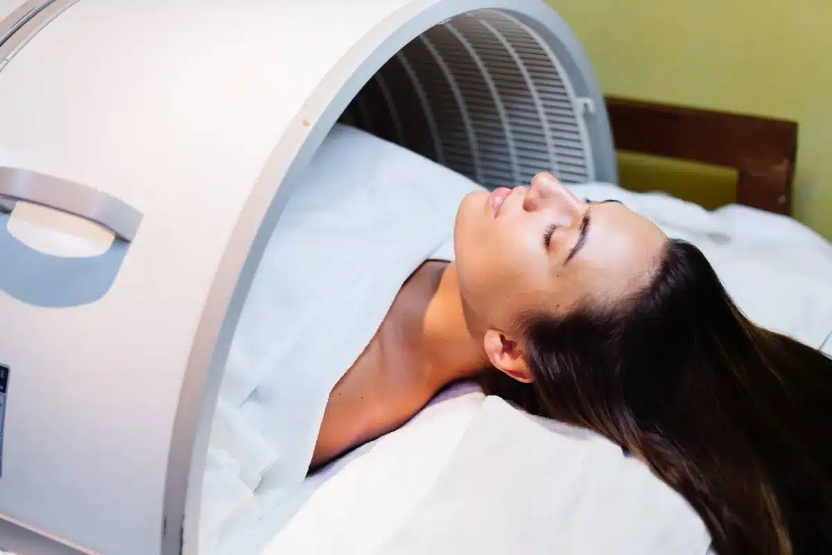
Thyroid Gland MRI: Advanced Imaging Technique

MRI is a detailed way to look at the thyroid gland. It uses magnetic fields and radio waves to create clear images. This helps doctors see the gland’s structure and function, which is key for diagnosing and tracking thyroid issues.
MRI Physics and Signal Generation
MRI physics is key to making thyroid gland images. It works by aligning hydrogen nuclei in the body with a strong magnetic field. When a radiofrequency pulse is applied, these nuclei send out signals. These signals are then used to make detailed images.
The process of making MRI images is complex. It involves magnetic fields and radio waves interacting with body tissues. For thyroid imaging, knowing these details is vital for getting the best images and understanding them correctly.
Optimal Sequences for Thyroid Visualization
For the best thyroid gland images, certain MRI sequences are used. T1-weighted images show the gland’s shape and where it is in relation to other parts of the body. T2-weighted images are better for spotting problems in the gland because they show tissue differences. Fat-suppressed sequences also help by making some lesions or problems more visible.
- T1-weighted sequences for anatomical detail
- T2-weighted sequences for detecting abnormalities
- Fat-suppressed sequences for enhanced lesion visibility
Normal Homogeneous Signal Intensity Patterns
A normal thyroid gland looks the same on MRI images. It is usually the same intensity as the muscles around it on T1-weighted images. This uniform look means the gland is likely healthy.
Knowing what a normal thyroid gland looks like on MRI is important. Any changes from this normal pattern could mean there’s a problem, like thyroiditis or nodules. This would need more investigation.
6 Essential MRI Measurements of Normal Thyroid Gland
MRI gives detailed measurements of the thyroid gland. It helps check if it’s the right size and volume. Getting these measurements right is key to diagnosing and tracking thyroid problems.
Right and Left Lobe Dimensions
The thyroid gland has two lobes, the right and left, joined by the isthmus. Each lobe is usually 4 to 6 cm long, 1.3 to 1.8 cm wide, and 0.8 to 1.8 cm thick. These sizes can change based on age, gender, and thyroid health.
Isthmus Width and Thickness
The isthmus connects the two lobes. It’s normally 1.2 to 2.0 cm wide, and its thickness should be under 3 mm. Changes in the isthmus size can suggest thyroid problems.
Parenchymal Volume Calculation Methods
To find the thyroid gland’s volume, we measure both lobes and the isthmus. The ellipsoid formula is used: length × width × depth × 0.52 for each lobe. The total volume is the sum of both lobes and sometimes the isthmus too.
Knowing these measurements is vital for doctors to check thyroid health. Any changes from normal can mean there’s a thyroid issue that needs looking into.
Ultrasound as the Primary Thyroid Assessment Tool
Thyroid ultrasonography is now the top choice for checking the thyroid. It’s better than other methods because it’s non-invasive and doesn’t use harmful radiation. It also gives clear pictures of the thyroid’s structure.
Technical Principles of Thyroid Ultrasonography
Ultrasound uses sound waves to show the thyroid gland in detail. A transducer sends and receives sound waves, making images in real-time. High-frequency transducers (7-15 MHz) are best for thyroid pictures because they show details well.
Ultrasound uses B-mode and Doppler techniques. B-mode shows the gland’s shape, and Doppler checks blood flow. This mix helps fully check the thyroid’s shape and blood flow.
Advantages Over Other Imaging Modalities
Ultrasound beats other methods for thyroid checks in many ways. It’s:
- Non-invasive and painless
- Free from harmful radiation
- Good at showing thyroid details
- Can check blood flow in the gland
- Helps with needle biopsies
- Easy to get and not as expensive as an MRI or a CT
Standardized Scanning Protocol
Having a set protocol is key to good thyroid ultrasound tests. It includes:
- Putting the patient in a lying down position with the eck up
- Using a high-frequency transducer
- Scanning both thyroid lobes from top to bottom
- Measuring the lobes and isthmus thickness
- Looking at the gland’s texture and finding nodules
- Checking blood flow with Doppler
Ultrasound is the main tool for checking the thyroid isthmus. A normal thickness is up to 3 mm. By sticking to a set protocol, doctors can get accurate and consistent results. This helps spot problems and track thyroid diseases.
6 Critical Ultrasound Measurements of Normal Thyroid
Getting accurate ultrasound measurements of the thyroid gland is key to diagnosing and tracking thyroid issues. These measurements tell us about the gland’s size, shape, and any problems it might have.
Longitudinal Lobe Measurements (4-6 cm Range)
The length of the thyroid lobe is a main measurement during an ultrasound. It usually falls between 4 to 6 cm. Normal thyroid ultrasound measurements show that each lobe should be about 4“6 cm long. This helps doctors check if the thyroid is healthy.
Anteroposterior and Transverse Dimensions
Doctors also check the gland’s width and thickness. These measurements help figure out the gland’s volume and size. The width is measured at its biggest point, not along the length.
To get a full picture, ultrasound measurements include the gland’s length, width, and thickness. They also look at the isthmus’s thickness. These details are vital for a complete thyroid health check.
Thyroid Isthmus Thickness (Normal up to 3 mm)
The isthmus, which links the thyroid’s two lobes, is another important area to examine. It should be no thicker than 3 mm. If it’s thicker, it could mean there’s a problem with the gland.
Knowing these critical ultrasound measurements of the normal thyroid gland is vital for doctors. They help diagnose and manage thyroid issues. By combining these measurements with other tests, doctors can get a clear view of thyroid health.
Normal Thyroid Gland Volume and Calculation Methods
Knowing the normal size of the thyroid gland is key to a correct diagnosis. The size of the thyroid gland is important for checking its health and finding any problems.
Standard Volume Ranges by Age and Gender
The size of the thyroid gland changes with age and gender. It grows until adulthood and then stays the same.
| Age Group | Male Volume (mL) | Female Volume (mL) |
| Children (6-10 years) | 4-6 | 4-5 |
| Adolescents (11-18 years) | 8-12 | 7-10 |
| Adults | 15-25 | 12-18 |
Mathematical Formulas for Volume Estimation
Ultrasound and math formulas can estimate the thyroid gland’s volume. The ellipsoid model is the most used. It calculates each lobe’s volume and adds them together.
Ellipsoid Formula: Volume = Length × Width × Depth × 0.52
The total volume is the sum of both lobes and the isthmus. This method gives a good estimate of the gland’s size.
Clinical Significance of Volume Measurements
Accurate thyroid gland volume measurements are very important in healthcare. They help diagnose issues like goiter, nodules, and thyroiditis. Watching how the volume changes helps see if treatments are working.
In summary, knowing about thyroid gland volume and how to calculate it is essential for doctors. It helps them make better decisions for their patients with thyroid problems.
Demographic Variations in Thyroid Dimensions
Thyroid gland sizes vary across different ages, genders, and ethnicities. Knowing these variations is key to diagnosing and treating thyroid issues.
Age-Related Changes in Thyroid Size
The thyroid gland grows with age. Research shows it gets bigger from childhood to adolescence and then might shrink a bit in older age. A study in the Journal of Clinical Endocrinology and Metabolism found thyroid volume peaks in late teens.
Here’s a summary of age-related changes in thyroid dimensions:
| Age Group | Average Thyroid Volume (mL) |
| Children (6-12 years) | 6-8 |
| Adolescents (13-18 years) | 10-12 |
| Adults (19-65 years) | 12-15 |
| Elderly (66+ years) | 10-12 |
Gender Differences in Normal Measurements
Gender affects thyroid gland size. Men generally have larger thyroid glands than women. A study in the European Journal of Endocrinology found men’s thyroid volumes are 15-20% bigger than women’s.
“The thyroid gland size is generally larger in males compared to females, which is an important consideration in clinical assessments.”
Geographic and Ethnic Variations
Where you live and your ethnicity also impact thyroid gland size. People in iodine-deficient areas often have bigger thyroid glands. A study in iodine-deficient regions found larger thyroid volumes than in iodine-sufficient areas.
These differences show why age, gender, and background matter when checking thyroid gland sizes. Healthcare providers can make better diagnoses and treatment plans by understanding these factors.
Comparative Analysis: Thyroid Gland X-Ray, CT, and Nuclear Imaging
Many imaging methods are used to check thyroid health. Each has its own benefits and drawbacks. X-ray, CT scans, and nuclear medicine give different kinds of information about the thyroid gland.
X-Ray Limitations for Thyroid Assessment
X-rays aren’t very good for looking at the thyroid gland. The thyroid gland X-ray can’t show the gland’s details or find small problems. But it can help see how thyroid growth affects nearby areas, like the trachea.
CT Imaging of the Thyroid Region
CT scans are better at showing the thyroid gland and what’s around it. CT scans can show the gland’s shape, find nodules, and see how big thyroid disease is. They’re great for checking if a goiter goes below the sternum and for looking at lymph nodes in thyroid cancer.
Nuclear Medicine Applications
Nuclear medicine, like thyroid scintigraphy, is key to checking thyroid function and finding thyroid problems. Nuclear medicine helps see how well the thyroid works and where the active parts are. It’s best for telling different thyroid nodules apart and for checking thyroiditis.
In summary, while X-rays have their limits, CT scans and nuclear medicine are very helpful. Knowing what each method can do is key to a good thyroid check-up.
Clinical Applications of Thyroid Measurements
In clinical settings, thyroid measurements are key for checking thyroid health and planning care. They help doctors make the right decisions. This is important for diagnosing and treating thyroid issues.
Distinguishing Normal from Pathological Findings
Thyroid measurements help tell normal from abnormal thyroid conditions. By comparing values to normal ranges, doctors spot problems that might mean thyroid disease.
- Normal thyroid sizes change with age and gender. It’s important to consider these when checking thyroid health.
- Conditions like goiter or thyroid nodules show up when measurements are off from normal.
Monitoring Thyroid Disease Progression
Regular thyroid checks are key for tracking thyroid disease and how well treatment works. This helps doctors adjust treatment plans for the best results.
Key aspects of disease monitoring include:
- Watch for changes in thyroid size and shape over time.
- Seeing how well treatments, like medicine or surgery, work.
- Finding problems early.
Surgical Planning and Post-Treatment Assessment
Thyroid measurements are also vital for planning surgery and checking how well it went. Before surgery, they help surgeons know what to do. After, they show whether the surgery was a success.
As thyroid specialists say, “Precise thyroid measurements are essential for both planning surgery and checking up after.”
Using thyroid measurements in these ways helps doctors give better, more tailored care to patients with thyroid issues.
Conclusion: Standardized Approach to Thyroid Imaging
A standardized approach to thyroid imaging is key for accurate and comparable measurements. It helps healthcare professionals determine normal thyroid size and spot any issues. This is important for all imaging types and settings.
Standardizing thyroid imaging is critical for both doctors and researchers. It makes sure diagnoses and comparisons are reliable. This helps improve patient care and our understanding of thyroid disorders.
Using the same thyroid imaging protocols helps doctors share information better. This leads to a more effective treatment plan. As thyroid imaging gets better, sticking to a standard approach will keep patient care top-notch.
FAQ
What are the normal dimensions of the thyroid gland on ultrasound?
The thyroid gland’s size on ultrasound is usually between 4 to 6 cm long. Its width is less than 2 cm. The thickness varies by lobe. The isthmus, the part in the middle, is up to 3 mm thick.
How is thyroid gland volume calculated?
To find the thyroid gland’s volume, you multiply its length, width, and thickness. Then, you multiply by 0.52 for each lobe. Adding both lobes’ volumes gives you the total volume.
What is the normal volume range for the thyroid gland?
The thyroid gland’s volume changes with age and gender. Adult males usually have a larger volume than females. The average volume is about 12 to 20 mL.
How does thyroid gland size change with age?
The thyroid gland size can grow until adulthood and then might shrink with age. Hormonal changes and where you live can affect these changes.
What are the advantages of using ultrasound for thyroid assessment?
Ultrasound is the best tool for checking the thyroid gland. It’s non-invasive, doesn’t use radiation, and shows the gland’s details well. It also helps guide biopsies.
How does MRI compare to ultrasound for thyroid gland evaluation?
MRI gives detailed images of the thyroid gland and nearby areas. It’s great for checking large goiters or thyroid cancers. But it’s more expensive and harder to get than an ultrasound.
What is the role of CT imaging in thyroid assessment?
CT imaging is good for checking the thyroid gland when it might be growing into the chest. It also helps find swollen lymph nodes or cancer spread.
Are there gender differences in normal thyroid gland measurements?
Yes, males usually have larger thyroid glands than females. These differences are important when setting normal volume ranges.
How do geographic or ethnic factors influence thyroid gland size?
Where you live and your ethnicity can affect thyroid gland size. These factors help set the right volume ranges for different groups.
What is the significance of homogeneous signal intensity on MRI?
A uniform signal on MRI means the thyroid gland looks normal. Changes in the signal might mean there’s a problem, like thyroiditis or cancer.
How is thyroid gland volume used clinically?
Measuring thyroid gland volume helps track disease, see if treatments work, and plan surgeries.
What are the limitations of X-rays for thyroid gland assessment?
X-rays aren’t good for looking at the thyroid gland because they can’t show soft tissue details well.
References:
- Bozdag, Z., et al. (2018). Normal size and volume of the thyroid gland in Turkish children measured by ultrasonography. Clinical Radiology and Ultrasound, 9(6), 24-30. https://pmc.ncbi.nlm.nih.gov/articles/PMC6291476/


