Medical imaging is key in today’s medicine. It helps doctors diagnose and treat many health issues. At LivHospital, we use top-notch imaging tech for precise diagnoses and treatment plans. Different types of body scans let doctors see inside the body, helping them care for patients better.
Many imaging methods are used, like X-ray, CT, MRI, ultrasound, and PET scans.
Key Takeaways
- Medical imaging is key for diagnosing and treating many health issues.
- Different body scans are used to check on organs or find diseases.
- Liv Hospital uses advanced imaging tech for accurate diagnoses.
- Many imaging methods are available, including X-ray, CT, MRI, ultrasound, and PET scans.
- Our hospital is dedicated to using the latest tech for the best health care.
The Evolution of Medical Imaging Technology
Medical imaging has changed a lot, starting with X-rays in 1895. New technologies keep coming, making it easier to see inside the body. These tools help doctors make better decisions for patients.
Historical Development of Diagnostic Imaging
Wilhelm Conrad Röntgen found X-rays in 1895. This led to radiography, the first way to see inside the body. Later, CT scans, MRI, ultrasound, and PET scans were developed. Each has its own way of working and is used for different things.
A medical expert said, “Medical imaging has changed a lot. It helps doctors find and treat problems better than before.”
“New imaging technologies have changed patient care. They help find problems early and treat them more precisely.”
How Modern Scanning Technologies Work
Today’s scanning tech works in different ways to show what’s inside the body. CT scans use X-rays for detailed images. MRI uses magnetic fields and radio waves for soft tissue.
Understanding these techs is key to seeing their value. Scan technology has gotten better, giving more detailed info and better patient care.
Ultrasound uses sound waves for live images, great for checking on babies and organs. PET scans show how active cells are, helping spot and track cancer. This variety lets doctors pick the best tool for each patient.
Types of Body Scans Used in Modern Medicine
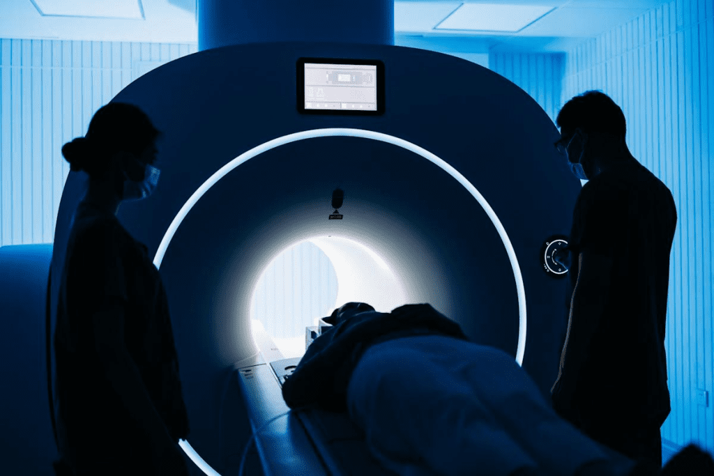
Modern medicine uses many body scans to find and treat health problems. Doctors use these scans to diagnose, track, plan, and monitor health. This includes checking on pregnancies.
Overview of Diagnostic Imaging Categories
Diagnostic imaging has several types, each with its own benefits and drawbacks. The main scans are X-ray, Computed Tomography (CT), Magnetic Resonance Imaging (MRI), Ultrasound, and Positron Emission Tomography (PET). Each scan has a special role in patient care.
X-ray scans are often used for bone and lung issues. MRI scans are great for soft tissues, like the brain and joints. Knowing these types helps doctors pick the best scan for each patient.
How Physicians Determine the Appropriate Scan Type
Doctors look at many things to choose the right scan. They consider the patient’s history, symptoms, and the condition being checked. For example, an X-ray scan is often first for bone fractures because it shows bones well.
- The patient’s overall health and any contraindications for certain imaging techniques
- The specific condition or disease being diagnosed or monitored
- The need for detailed imaging of soft tissues or bone structures
- The availability of imaging equipment and the patient’s ability to undergo the scan
By thinking about these things, doctors can pick the best scan. This ensures patients get accurate diagnoses and effective treatments. Choosing the right scan is key to good patient care and the best results.
X-Ray Scans: The Foundation of Medical Imaging
X-ray scans have been key in medical imaging for many years. They quickly show what’s inside the body. This is because they can easily see bones and some soft tissues.
How X-Ray Technology Captures Internal Structures
X-rays send electromagnetic waves through the body to make images. Different tissues absorb these waves at different rates. This creates contrast, letting us see inside the body.
Bones absorb more X-rays than soft tissues. So, bones show up white on the image, and soft tissues appear gray.
Common Applications in Orthopedics and Pulmonary Medicine
In orthopedics, X-rays help find fractures, dislocations, and bone issues. For lung and chest problems, like pneumonia or tumors, X-rays are also key. They’re fast and cheap, making them a first choice for many doctors.
Radiation Safety and Modern X-Ray Advancements
X-ray scans do involve radiation, but modern tech has made them safer. Digital X-rays need less radiation, which is better for patients. There are also strict rules to use X-rays wisely and keep radiation low.
| Advancements | Benefits |
| Digital X-ray Technology | Reduced radiation dose, faster image acquisition |
| Improved Image Processing | Enhanced image quality, better diagnostic accuracy |
| Radiation Safety Protocols | Minimized radiation exposure, safer for patients |
These updates have made X-rays safer and more useful in medicine. As tech keeps improving, X-rays will stay a critical tool in healthcare.
CT Scans: Detailed Cross-Sectional Visualization
CT scans have changed medical imaging by showing detailed views of the body’s inside. This method combines many X-ray images from different angles. It creates a full picture of the body’s internal parts.
The Science Behind Computed Tomography
CT scans use X-rays and computer tech to show the body’s inside. An X-ray machine rotates around the body to take images from various angles. A computer then puts these images together to show detailed cross-sections of the body.
CT scans work because different tissues absorb X-rays at different rates. This lets CT scans tell apart soft tissue, bone, and other body parts. This skill makes CT scans great for finding many medical problems.
Critical Applications in Cancer and Vascular Disease Detection
CT scans are key in finding and diagnosing cancer and vascular diseases. In cancer, CT scans spot tumors and check their size and location. They also see how well treatments are working. For vascular diseases, CT scans show blood vessels and find problems like aneurysms or blockages.
“CT scans are invaluable in the early detection of cancer, allowing for timely intervention and improved patient outcomes.”
Dr. Jane Smith, Oncologist
| Condition | CT Scan Application | Benefits |
| Cancer | Tumor detection and monitoring | Early detection, treatment planning |
| Vascular Disease | Blood vessel visualization | Detection of aneurysms or blockages |
Preparation and What to Expect During a CT Scan
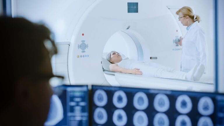
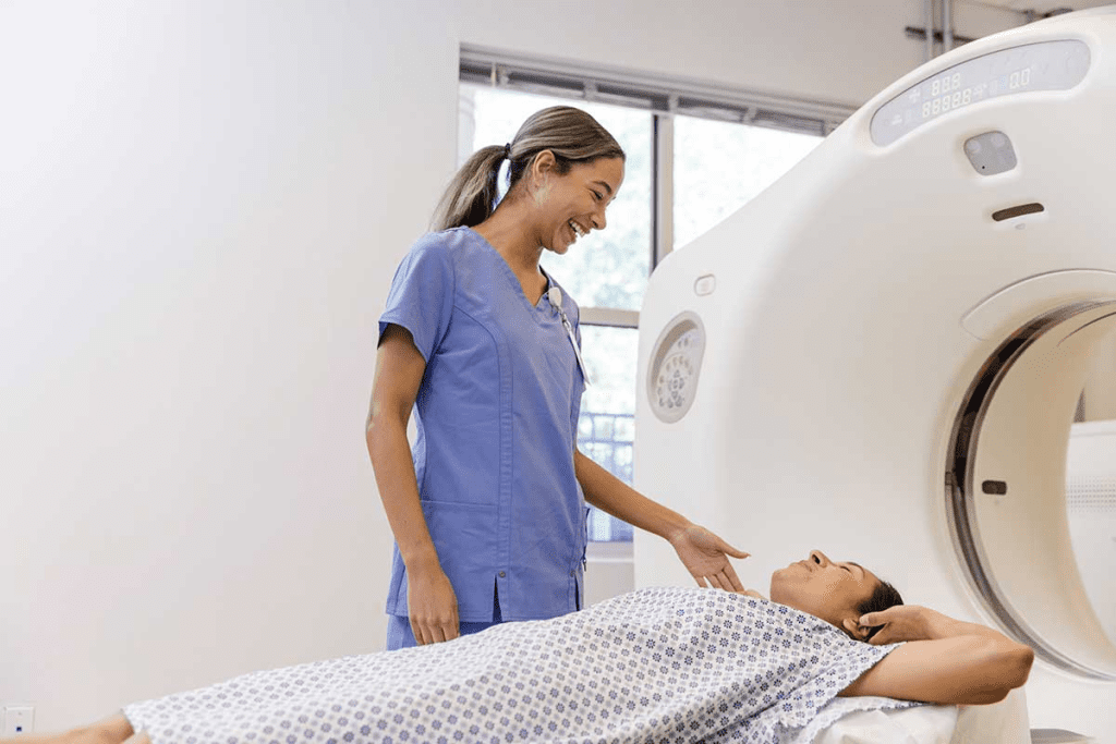
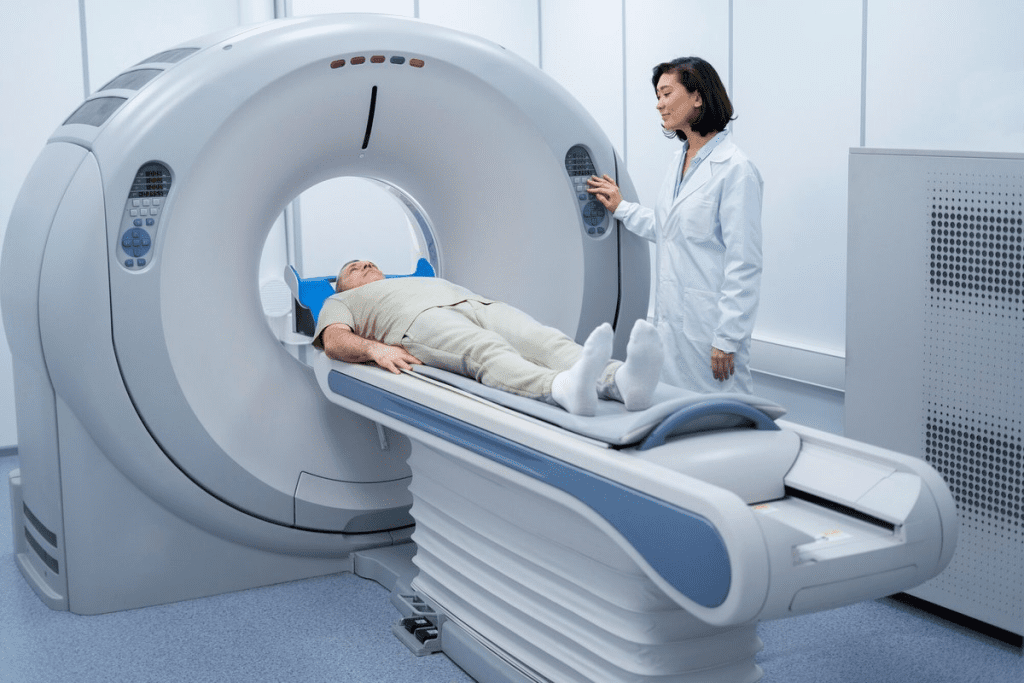
Before a CT scan, remove metal objects and jewelry and wear a hospital gown. You might need to fast or drink a contrast agent to improve image quality. During the scan, you lie on a table that slides into a big, doughnut-shaped machine. The scan is usually quick, lasting just a few minutes.
It’s important to stay very quiet during the scan for clear images. The CT scanner will move around you, taking the needed images. You might be asked to hold your breath to avoid blurry images.
Knowing what to expect during a CT scan helps patients prepare. This reduces anxiety and makes the experience smoother.
MRI Scans: Superior Soft Tissue Imaging
MRI scans have changed medical imaging by showing soft tissues in great detail. They are key in modern medicine, giving a safe choice instead of other methods.
The Physics of Magnetic Resonance Imaging
MRI uses strong magnets and radio waves to show soft tissues clearly. It aligns hydrogen atoms in the body with a magnetic field. Then, radio waves disturb them to create detailed images.
MRI machines vary in strength, measured in Tesla (T). Higher strengths mean clearer images. Common strengths are 1.5T and 3T, each for different exams.
Key Applications for Brain, Spine, and Joint Visualization
MRI scans are great for the brain, spine, and joints because they show soft tissues well. They help find issues like:
- Brain tumors and neurological problems
- Spinal cord injuries and diseases
- Joint injuries, like ligament and tendon tears
These clear images help doctors diagnose and plan treatments better, helping patients.
Patient Experience and Considerations for MRI Procedures
Getting an MRI scan is a special experience for patients. They lie very quietly in a possibly tight space. But, open MRI machines are there for those who need them.
Patients should:
- Take off metal items, like jewelry and some clothes
- Tell their doctor about metal implants or fear of tight spaces
- Follow any special instructions from their doctor
We know MRI can seem scary, but our team works hard to make it comfortable for you.
| Imaging Technique | Radiation | Soft Tissue Detail | Common Applications |
| MRI | No | Excellent | Brain, spine, joints |
| CT Scan | Yes | Good | Cancer, internal injuries |
| X-Ray | Yes | Poor | Bones, lung conditions |
This table shows MRI’s special benefits, like seeing soft tissues without radiation.
Ultrasound Scans: Non-Invasive Sound Wave Imaging
We use ultrasound scans as a non-invasive tool in many medical areas. They use sound waves to show pictures of inside organs and tissues. This helps doctors without using harmful radiation.
Creating Real-Time Images
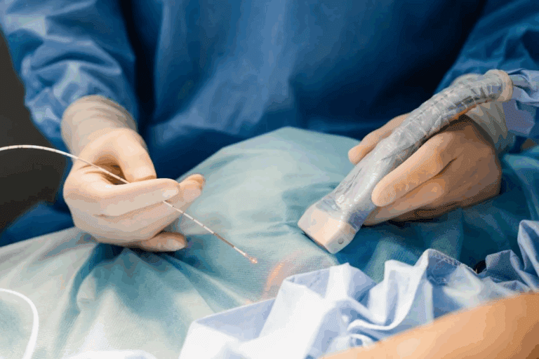
Sonography makes images live through a transducer. It sends and gets sound waves. These waves hit internal parts and turn into images on a screen.
The steps are:
- Apply gel on the skin for sound wave transmission
- Use a transducer to send and get sound waves
- Change sound waves into electrical signals
- Make images from signals on a screen
Beyond Pregnancy Monitoring
Ultrasound scans are not just for pregnancy. They help in many medical fields, like:
- Abdominal imaging for gallbladder and liver issues
- Vascular imaging for blood flow and diseases
- Musculoskeletal imaging for muscle and tendon injuries
- Thyroid and parathyroid gland imaging for gland disorders
Advantages of Radiation-Free Imaging
Ultrasound scans are safe because they don’t use radiation. This makes them good for all patients, even pregnant women. They are also non-invasive, making them a top choice for many tests.
The good things about ultrasound scans are:
- Safe and non-invasive
- No radiation exposure
- Live images
- Used in many medical areas
Ultrasound scans are key in today’s medical imaging. They help a lot with patient care, working with other scans and tests.
PET Scans: Visualizing Metabolic Activity
PET scans are a big step forward in medical imaging. They let us see how active different parts of the body are. This is really helpful for finding and tracking diseases like cancer.
The Science of Positron Emission Tomography
PET scans use tiny amounts of radioactive tracers to show how the body works. These tracers build up in busy areas, like tumors. Then, they send out signals that the scanner can pick up.
The most used tracer is FDG (Fluorodeoxyglucose). It goes into cells based on how much they use glucose. This helps PET scans spot busy areas, like tumors.
Role in Cancer Detection and Treatment Monitoring
PET scans are great for finding cancer and checking how treatments are working. They show how active different areas of the body are. This helps doctors see if a treatment is effective early on.
“PET scans have revolutionized the field of oncology by enabling clinicians to assess the metabolic activity of tumors, thereof guiding treatment decisions.”
Hybrid Imaging: PET-CT and PET-MRI Advancements
Combining PET with CT and MRI has led to new imaging methods. PET-CT mixes PET’s metabolic info with CT’s detailed pictures. This makes diagnosing better.
PET-MRI takes it further by adding MRI’s clear pictures of soft tissues. It’s a powerful tool for checking complex areas.
How Body Scans Improve Early Disease Detection and Outcomes
Body scans are key in finding diseases early. New scan technologies help us spot diseases sooner. This leads to better treatment results.
Impact on Cancer Survival Rates
Body scans greatly help in fighting cancer. They help find cancer early, which makes treatments work better. For example, a study showed that lung cancer screening with CT scans cut down on deaths.
We use many scans to find cancer early. These scans help us see tumors and track how they change with treatment.
Role in Preventive Medicine and Screening Programs
Body scans are important for preventing and screening diseases. They help doctors find health problems before symptoms show up. This means we can treat problems early.
Using scans in screening programs has cut down on late-stage cancer diagnoses. Finding diseases early helps improve treatment results and saves money on healthcare costs.
Integration with Modern Treatment Pathways
Scans play a big role in today’s treatments. They help us understand how serious a disease is, plan treatments, and check how well they work. For example, PET-CT scans help see if chemotherapy and radiation are working.
By combining scans with other treatments, we can give care that fits each patient’s needs. This approach has changed healthcare, making treatments better and improving patients’ lives.
| Scan Type | Primary Use | Benefits |
| CT Scan | Cancer staging, vascular disease | Detailed cross-sectional imaging, quick procedure |
| MRI | Soft tissue imaging, neurological disorders | High-resolution images, no radiation |
| PET Scan | Cancer detection, metabolic activity | Functional imaging, early disease detection |
Comparing Radiation Exposure and Safety Across Scan Types
Different medical imaging techniques have varying levels of radiation exposure. It’s essential to compare their safety profiles. We use various imaging modalities to diagnose and treat medical conditions. But, it’s important to understand the radiation risks.
Ionizing Radiation Levels in Common Imaging Techniques
Medical imaging techniques like X-rays and CT scans involve exposure to ionizing radiation. MRI and ultrasound are radiation-free. The level of radiation exposure varies significantly across these modalities.
For example, a typical chest X-ray delivers a much lower dose of radiation compared to a CT scan of the abdomen.
To better understand these differences, let’s examine the relative radiation levels of common imaging techniques:
| Imaging Technique | Relative Radiation Level | Effective Dose (mSv) |
| Chest X-ray | Low | 0.1 |
| CT Abdomen/Pelvis | High | 10-20 |
| MRI | None | 0 |
| Ultrasound | None | 0 |
Assessing the Risks and Benefits of Radiation-Based Imaging
When evaluating the safety of radiation-based imaging, we must consider both the benefits and the risks. The benefits include accurate diagnoses and effective treatment planning. The risks involve the possibility of radiation-induced harm, such as increased cancer risk.
Key considerations in the risk-benefit analysis include:
- The necessity of the imaging procedure
- The radiation dose and its risks
- Alternative imaging options
By carefully weighing these factors, healthcare providers can minimize radiation exposure while maximizing diagnostic benefits. This balanced approach ensures that patients receive the necessary care while minimizing risks associated with hospital scans and other imaging modalities.
Conclusion: The Future of Medical Imaging in Healthcare
Medical imaging technologies keep getting better, helping doctors diagnose and treat patients more effectively. At Liv Hospital, we’re always looking to improve our care with the latest technology.
New tools like AI and machine learning are making image analysis faster and more accurate. These tools are changing radiology scans, helping doctors find and treat diseases better.
Today, we have many types of scans that have changed how we diagnose and treat diseases. As we go forward, we’ll see even more advanced scans that will help patients even more.
We’re committed to using the latest in medical imaging to give our patients the best care. Our focus on innovation and quality keeps us at the top in medical imaging.
FAQ
What are the different types of body scans used in medical imaging?
Medical imaging includes X-ray, CT, MRI, Ultrasound, and PET scans. Each has its own use and benefits.
How do doctors determine which type of scan is best for a patient’s condition?
Doctors look at symptoms, medical history, and the condition. They choose the best scan based on these factors.
Are body scans safe, and what are the risks associated with radiation exposure?
Body scans are safe when used right. But, some like X-rays and CT scans have radiation risks. We follow strict safety to reduce these risks.
What is the difference between a CT scan and an MRI scan?
CT scans use X-rays for bones and lungs. MRI scans show soft tissues like organs and tendons.
Can I undergo an MRI scan if I have metal implants or devices?
It depends on the metal type. Some implants are safe for MRI. Always tell your doctor about implants before an MRI.
How do PET scans help in cancer detection and treatment?
PET scans show where cancer is active. They help track treatment and disease growth. They’re key in cancer management.
Are there any alternatives to scans that involve radiation?
Yes, Ultrasound and MRI are radiation-free. Ultrasound uses sound waves, and MRI uses magnetic fields. They’re good when avoiding radiation is important.
How do body scans contribute to early disease detection and improved outcomes?
Scans help find diseases early. This leads to quicker treatment and better results. They also track disease and treatment progress.
What advancements are being made in medical imaging technology?
New tech includes better scanners and hybrid imaging. These advances improve accuracy, reduce radiation, and expand uses.
How can I prepare for a body scan, and what can I expect during the procedure?
Preparation varies by scan type. You might need to remove jewelry or follow a diet. During the scan, you’ll lie on a table that slides into the scanner. Stay calm and stay in place while images are taken. The process varies by scan type.
References
- Bushberg, J. T., Seibert, J. A., Leidholdt, E. M., & Boone, J. M. (2011). The Essential Physics of Medical Imaging (3rd ed.). Lippincott Williams & Wilkins. https://www.ncbi.nlm.nih.gov/books/NBK459455/


