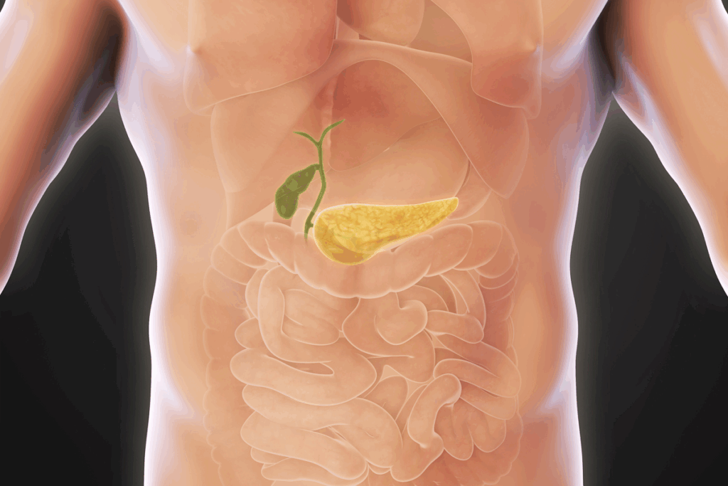
Diagnostic imaging is key in spotting gallbladder problems. An abdominal ultrasound is a safe test that uses sound waves to see inside the body, including the gallbladder.What does an unhealthy gallbladder ultrasound look like? We explain the difference between normal vs abnormal images and what to look for.
Experts look for signs of trouble in ultrasound images. Liv Hospital’s team will help you understand the differences between normal and abnormal pictures.

Ultrasound technology has changed how we check for gallbladder problems. It’s a non-invasive way to see if the gallbladder is healthy.
Ultrasound uses sound waves to make images of the gallbladder. A transducer sends and gets these sound waves, showing them on a screen. It’s non-invasive and painless, making it a top choice for checking the gallbladder.
When getting a gallbladder ultrasound, you lie on a table. A gel is put on your belly to help sound waves move. The transducer is moved around to get pictures of the gallbladder from different sides.
Ultrasound is the best choice for gallbladder imaging because it’s safety, effective, and shows real-time images. It doesn’t use radiation, which is safer for pregnant women and kids.
It’s also great at finding gallstones, inflammation, and other gallbladder problems. Its ability to show things in real-time helps doctors see how the gallbladder works and find any issues.
To get the best images, patients often need to fast for up to 12 hours before the ultrasound. This makes the gallbladder easier to see.
Other steps might include avoiding certain foods or medicines that could mess with the images. Following your doctor’s instructions is key to getting the best results from the ultrasound.

Knowing what a normal gallbladder looks like on ultrasound is key for spotting problems early. A normal ultrasound image shows the gallbladder’s health. It helps doctors tell if something is wrong.
A healthy gallbladder looks like a pear on ultrasound. Its wall is thin, under 3 mm thick. The inside of the gallbladder is dark because it doesn’t reflect sound waves.
Here are the normal sizes for a gallbladder on ultrasound:
The inside of a healthy gallbladder is dark, as mentioned before. The wall is brighter than the liver but not too thick. The liver helps doctors check the gallbladder’s brightness.
Some changes in the gallbladder on ultrasound are normal and don’t mean trouble. These include:
It’s important to know these normal changes when looking at gallbladder ultrasounds. This helps avoid mistakes in diagnosis.
The look of a gallbladder on ultrasound can tell us a lot about its health. It can show us if there are problems that need medical help.
There are several signs that can mean a gallbladder is not healthy. Knowing these signs is key for the right diagnosis and treatment.
Wall thickening is a big sign of a sick gallbladder. A normal gallbladder wall is under 3 mm thick. If it’s thicker, it might mean inflammation or disease.
Wall thickening can be caused by various factors, including:
Seeing abnormal fluid around the gallbladder is another warning sign. This fluid, called pericholecystic fluid, can mean inflammation or infection.
Pericholecystic fluid can be associated with:
Changes in the gallbladder’s shape and size can also be a sign of trouble. A healthy gallbladder is pear-shaped and smooth.
Any deformities or big size changes can mean there’s something wrong inside.
| Indicator | Normal Finding | Abnormal Finding |
| Wall Thickness | Less than 3 mm | Greater than 3 mm |
| Pericholecystic Fluid | Absent | Present |
| Gallbladder Shape | Pear-shaped, smooth outline | Deformed, irregular outline |
Spotting these signs on a gallbladder ultrasound is vital for diagnosing and treating gallbladder disease well.
Ultrasound images show gallstones clearly, which is key for diagnosis. Gallstones are common in the gallbladder. Ultrasound is a main tool for finding them.
Gallstones look like bright echogenic foci in the gallbladder on ultrasound. Their brightness comes from their dense material. The size and number of stones differ from person to person.
“Ultrasound is very good at showing gallstones,” say doctors. Gallstones often cause posterior acoustic shadowing, a key sign.
Acoustic shadowing makes the area behind the stone look darker. This is a telltale sign of gallstones. The shadow’s size depends on the stone’s size and type.
Ultrasound is great for finding gallstones but hard to tell their types. The stone’s density and shadowing can give hints.
“Knowing the type of gallstone on ultrasound helps in choosing the right treatment,” studies show.
In summary, ultrasound gives important info on gallstones. Knowing these details helps in accurate diagnosis and treatment.
Ultrasound is key for spotting gallbladder sludge and polyps. These conditions show up differently on ultrasound and have unique health effects.
Gallbladder sludge looks like thick, bright material inside the gallbladder on ultrasound. It settles at the bottom and doesn’t block sound like stones do. It’s often linked to bile stasis, like from fasting or IV nutrition.
Gallbladder polyps are growths that stick out into the gallbladder. They show up as small, bright spots on the ultrasound. Polyps are different from stones because they stick to the wall and don’t move with the body. They can be harmless or cancerous, and ultrasound helps figure out what they are.
| Characteristic | Polyps | Gallstones |
| Attachment | Attached to the gallbladder wall | Free within the gallbladder lumen |
| Mobility | Non-mobile | Mobile with patient repositioning |
| Acoustic Shadowing | Typically absent | Usually present |
Not every polyp needs more tests. But, polyps over 1 cm are more likely to be cancer and usually need more checks. Doctors also look at the patient’s age, symptoms, and risk for cancer.
In short, ultrasound is great for finding gallbladder sludge and polyps. Knowing how they look on ultrasound helps doctors diagnose and treat them right.
It’s key to know the differences between normal and abnormal gallbladder ultrasounds for correct diagnosis. This section offers a detailed comparison. It focuses on important diagnostic criteria and common mistakes.
Several criteria are used to check gallbladder ultrasounds. These include the gallbladder’s size, wall thickness, and any issues like stones or sludge.
Normal Gallbladder Measurements:
| Characteristic | Normal | Abnormal |
| Wall Thickness | < 3 mm | ≥ 3 mm |
| Gallstones | Absent | Present |
| Sludge | Absent | Present |
Even with modern ultrasound tech, mistakes can happen. Issues include mistaking artifacts for real problems or missing small issues.
Common pitfalls:
Let’s look at some examples to show the differences in gallbladder ultrasounds.
Case 1: A normal gallbladder ultrasound with a thin wall and clear lumen.
Case 2: An abnormal ultrasound showing gallstones and thickened walls.
These examples stress the need for careful and accurate ultrasound readings.
Ultrasound imaging is key in spotting acute cholecystitis, a serious gallbladder inflammation. It’s important to catch it early to avoid serious issues.
Ultrasound techs look for certain signs of inflammation in patients with acute cholecystitis. These include:
These signs help doctors know if someone has acute cholecystitis and what to do next.
Murphy’s sign is a tool used during ultrasound. It checks if the patient is tender when the probe touches the gallbladder. If they are, it means they likely have acute cholecystitis.
Ultrasound findings and Murphy’s sign together make for a more accurate diagnosis.
If acute cholecystitis isn’t treated, it can cause serious problems. Some of these include:
| Complication | Description |
| Gangrene | Necrosis of the gallbladder wall, potentially leading to perforation. |
| Perforation | A hole in the gallbladder wall, allowing bile to leak into the abdominal cavity. |
| Abscess Formation | A collection of pus around the gallbladder, requiring drainage. |
Spotting it early with ultrasound is key to avoiding these serious issues.
Ultrasound has its limits when it comes to diagnosing gallbladder problems. It’s a great first step, but sometimes it’s not enough. This is when we need to use other imaging methods.
Ultrasound might not work well in some cases. For example, if the gallbladder is hidden by gas in the bowel. Or if we need a closer look at the gallbladder wall or nearby tissues.
Obesity and bowel gas can make it hard to get clear ultrasound images. In these situations, we need other imaging methods to get a clear picture.
CT scans give us a detailed view of the gallbladder and its surroundings. They’re great for spotting serious problems like gallbladder perforation or gangrene. CT scans can also find other causes of pain that ultrasound might miss.
MRI shows the gallbladder and bile ducts in high detail. It’s good for finding blockages or stones in the bile ducts. MRI can also tell us more about gallbladder tumors or masses.
HIDA scans check how well the gallbladder works. They’re useful for spotting cystic duct obstruction, a common cause of acute cholecystitis. HIDA scans give us functional info that other scans can’t.
New technologies are making gallbladder imaging better. Contrast-enhanced ultrasound helps us see gallbladder problems more clearly. Other new methods, like advanced MRI sequences, can better identify gallbladder lesions.
These new tools are helping doctors make more accurate diagnoses. They might even mean we need fewer invasive tests. As technology keeps getting better, we’ll be able to diagnose and treat gallbladder diseases even more effectively.
Understanding gallbladder ultrasound results is key. It involves knowing what’s normal and what’s not. This helps doctors spot problems early.
Looking at ultrasound images, doctors check for wall thickening and fluid buildup. They also look at shape and size changes. Gallstones, sludge, or polyps are signs of trouble. Knowing these signs and the patient’s symptoms is important for a correct diagnosis.
Doctors use ultrasound results to decide on next steps. They combine what they see on the ultrasound with the patient’s symptoms. This helps them create a treatment plan that fits the patient’s needs.
Knowing how to read gallbladder ultrasound images is critical. It helps doctors diagnose and treat gallbladder issues. By accurately interpreting these images, doctors can give the best care to patients with gallbladder problems.
An unhealthy gallbladder on ultrasound may show signs such as wall thickening. It may also have abnormal fluid collections or changes in shape and size. Gallstones, sludge, or polyps can also be seen.
Gallstones on ultrasound appear as highly echogenic structures. They are seen within the gallbladder lumen. They often have acoustic shadowing.
Acoustic shadowing is a sign of gallstones. It shows that the stone is dense enough to block ultrasound waves. This creates a shadow behind it.
Sludge on ultrasound appears as low-level echoes within the gallbladder lumen. It often layers dependently. It can be distinguished from stones by its lack of acoustic shadowing and mobility.
Polyps larger than 1 cm or those with suspicious features may need further investigation. This could include additional imaging or biopsy.
Ultrasound signs of acute cholecystitis include gallbladder wall thickening and pericholecystic fluid. Inflammatory changes and a positive Murphy’s sign during the examination are also signs.
Ultrasound may be limited by factors such as bowel gas or patient body habitus. It may also be limited by complex pathology. Additional imaging techniques like CT, MRI, or HIDA scans may be needed.
Normal gallbladder ultrasounds show a typical appearance and measurements. Abnormal ultrasounds may display signs such as wall thickening, gallstones, or inflammatory changes.
CT, MRI, and HIDA scans provide additional information. They are used when ultrasound is inconclusive or insufficient. They offer detailed images of the gallbladder and surrounding structures.
Gallbladder ultrasound results should be interpreted in the context of the patient’s clinical presentation and medical history. They should be interpreted with other diagnostic findings to provide an accurate diagnosis and guide treatment.
National Institute of Diabetes and Digestive and Kidney Diseases. (2017, November). Diagnostic tests for gallbladder disease. National Institutes of Health. https://www.niddk.nih.gov/health-information/diagnostic-tests/gallbladder-disease
Subscribe to our e-newsletter to stay informed about the latest innovations in the world of health and exclusive offers!
WhatsApp us