
It’s important to spot unilateral optic nerve edema early. At Liv Hospital, we focus on telling true edema from pseudo-edema. We do this with careful checks. What causes unilateral optic nerve edema? This guide covers 5 dangerous causes, symptoms, and essential treatment options.
Swelling in the optic disc can happen for many reasons. These include optic neuritis, optic neuritis, and compressive optic neuropathy. We promise to give you the best care by checking your eyes fully.
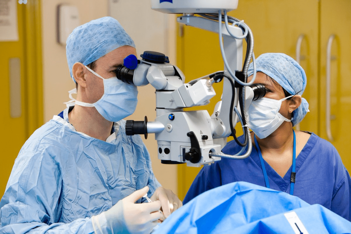
We look closely to find out why the edema is happening. This way, we can treat you right. We want to give you top-notch care, no matter where you’re from.
Key Takeaways
- Unilateral optic nerve edema needs careful checks.
- Swelling in the optic disc can be caused by many things.
- It’s key to tell true edema from pseudo-edema through detailed checks.
- Full eye exams are vital for managing the condition well.
- Finding it early is key to getting the right treatment.
Understanding Unilateral Optic Nerve Edema

Unilateral optic nerve edema is when the optic disk in one eye swells. This swelling can happen for many reasons. It’s important because it can cause vision problems and serious issues if not treated right.
Definition and Pathophysiology
Unilateral optic nerve edema means the optic nerve head in one eye swells. This swelling often comes from blocked axonal transport. It can cause the swelling of the optic disk, which is a key sign of many eye and brain problems.
The swelling happens because axoplasmic material builds up in the optic nerve head. This can be due to high pressure in the brain, among other reasons. Knowing why it happens helps doctors diagnose and treat it better.
Unilateral vs. Bilateral Presentation
Papilledema usually shows up in both eyes, but sometimes it’s only in one. Finding out if it’s in one eye or both is key. The reasons for papilledema, like high brain pressure, can cause swelling in one or both eyes.
Knowing if it’s in one eye or both is important for figuring out what’s wrong and how to fix it. Doctors need to look closely and do tests to find out why it’s happening.
Clinical Significance
Unilateral optic nerve edema is serious because it can mean there’s something wrong deep inside. Finding and treating it quickly is key to avoiding lasting eye damage. Doctors need to think about many possible reasons, like ischemic conditions or inflammatory disorders.
Understanding how serious it is helps doctors focus on the right cases. It also helps them explain things to patients and plan the best treatment. A thorough check-up is always needed.
Clinical Presentation and Key Symptoms

Unilateral optic nerve edema shows up in many ways. Patients see changes in their vision that can really affect their life. These symptoms can be quite a challenge.
Visual Acuity Changes
One big worry for those with unilateral optic nerve edema is visual acuity changes. They might see things as blurry or dim. In some cases, vision loss can be quite severe.
These vision changes can last for a while or go away. It all depends on why the edema happened in the first place.
Visual Field Defects
Visual field defects are another key symptom. Patients might see transient visual obscurations or lasting field defects. These can mess with their side or central vision.
Associated Symptoms
Patients with unilateral optic nerve edema often have headaches and other symptoms. They might experience brief vision loss or dimming. This usually happens when they change their posture.
It’s important to understand these symptoms to diagnose and treat unilateral optic nerve edema well. We need to look at the whole picture to help our patients.
Ophthalmoscopic Examination Techniques
Diagnosing unilateral optic nerve edema relies on good ophthalmoscopic exams. A detailed ophthalmoscopic exam is key to spotting optic disc edema. This condition is marked by swelling of the optic disc.
Techniques like direct ophthalmoscopy, indirect ophthalmoscopy, and slit lamp biomicroscopy are used. Each method gives a different view of the optic disc and its surroundings. This helps in diagnosing optic nerve edema.
Direct Ophthalmoscopy
Direct ophthalmoscopy gives a close-up look at the optic disc. It’s great for spotting small changes linked to disc edema.
Indirect Ophthalmoscopy
Indirect ophthalmoscopy shows a wider view of the fundus. It’s good for seeing the optic disc and retina together. This helps in understanding how big optic disc edema is and any related retina changes.
Slit Lamp Biomicroscopy
Slit lamp biomicroscopy uses a strong lens for a detailed look at the optic disc. It’s best for checking how swollen the disc is and finding early signs of optic nerve edema.
Here’s a comparison of these methods in a table:
Technique | Magnification | Field of View | Usefulness in Diagnosing Optic Disc Edema |
Direct Ophthalmoscopy | High | Narrow | Excellent for detailed examination of the optic disc |
Indirect Ophthalmoscopy | Lower | Wide | Useful for assessing the extent of optic disc edema and retinal changes |
Slit Lamp Biomicroscopy | High | Stereoscopic view | Ideal for evaluating disc swelling and subtle signs of optic nerve edema |
In summary, a detailed ophthalmoscopic exam is vital for diagnosing and treating unilateral optic nerve edema. Knowing the strengths of each method helps doctors make better care plans for their patients.
Characteristic Findings of Unilateral Optic Nerve Edema
Unilateral optic nerve edema shows clear signs that help in early detection and treatment. Spotting these signs early is key to managing the condition well. Knowing these signs is vital for diagnosing and treating unilateral optic nerve edema.
Early Signs of Disc Edema
The first signs of disc edema include slight changes in the optic disc. These include elevation of the optic disc and vascular tortuosity. A detailed ophthalmoscopic exam can spot these changes.
Look for signs like hyperemia of the optic disc. This could mean edema is starting.
Advanced Disc Edema Findings
As disc edema gets worse, more obvious signs appear. These include more elevation of the optic disc and vascular tortuosity. Also, peripapillary retinal folds become more noticeable.
The optic disc looks swollen, with blurry edges. There might also be hemorrhages.
Vascular Changes
Vascular changes are a key feature of unilateral optic nerve edema. We see vascular tortuosity and dilated capillaries on the optic disc. These can cause retinal hemorrhages and exudates.
Peripapillary Changes
Peripapillary changes are also important. These include retinal folds and peripapillary hemorrhages. These signs show how severe the edema is and its effect on the retina. We check these to gauge the condition’s severity.
In summary, knowing the signs of unilateral optic nerve edema is critical for diagnosis and treatment. A detailed ophthalmoscopic exam is essential to catch these signs early. This allows for timely action.
Differentiating True Edema from Pseudo-Edema
Telling true optic nerve edema from pseudo-edema is key for right diagnosis and treatment. Pseudo-edema looks like true edema but comes from different causes. It’s important to tell them apart.
Optic Disc Drusen
Optic disc drusen often cause pseudo-edema. These are calcified spots in the optic disc that look like swelling. They’re usually harmless but can link to other issues.
To spot optic disc drusen, we use tools like ultrasound or OCT. These help find the calcifications in the optic disc.
Congenital Anomalies
Certain optic disc anomalies, like a tilted disc or hypoplasia, can look like optic disc oedema. These are found through detailed eye exams.
Knowing the patient’s history and doing a full eye check are key to telling these apart from real edema.
Myelinated Nerve Fibers
Myelinated nerve fibers can also look like swelling edematous optic disc. This happens when myelination goes past the lamina cribrosa, unlike normal eyes.
To tell myelinated nerve fibers from true edema, we look at the myelination’s details and how it affects the optic disc and retina.
Condition | Characteristics | Diagnostic Techniques |
Optic Disc Drusen | Calcified deposits within the optic disc | Ultrasound, OCT |
Congenital Anomalies | Tilted or hypoplastic optic disc | Comprehensive ophthalmologic examination |
Myelinated Nerve Fibers | Myelination anterior to the lamina cribrosa | Ophthalmoscopy, OCT |
In conclusion, telling true optic nerve edema from pseudo-edema needs a deep understanding of the mimicking conditions. It also requires the right diagnostic tools.
Diagnostic Approach to Unilateral Optic Nerve Edema
To diagnose unilateral optic nerve edema, we need a detailed look at the patient’s history, physical exam, and neurological check. This approach is key to finding the cause of unilateral disc edema and choosing the right treatment.
Clinical History Taking
Starting with the patient’s history is the first step in diagnosing optic nerve edema. We gather info on symptoms, like when they started, how long they’ve lasted, and any changes. It’s important to ask about headaches, eye pain, or any brief vision problems.
We also ask about past health issues, like high blood pressure, diabetes, or vascular diseases. Any recent injuries, infections, or inflammatory diseases are noted. Knowing what medications or supplements the patient takes can help find the cause of optic nerve head swelling.
Clinical History Element | Relevance to Diagnosis |
Onset and duration of symptoms | Helps determine the acute or chronic nature of the condition |
Associated symptoms | May indicate underlying causes or complicating factors |
Medical history | Reveals potentially risky factors or underlying conditions |
Medication and supplement history | Can identify possible causes or contributing factors |
Physical Examination
A detailed physical exam is vital for checking swelling of the optic disk. We test visual acuity and check pupillary reactions. Fundoscopic exams, direct and indirect ophthalmoscopy, let us closely look at the optic disc and retina.
The exam also checks the patient’s visual fields for any defects. We look at the eye and surrounding area for signs of injury, infection, or other issues.
Neurological Assessment
Neurological assessment is a key part of diagnosing unilateral optic nerve edema. We check cranial nerve function, focusing on the optic nerve. Looking for signs of increased intracranial pressure, like papilledema, is important.
A thorough neurological exam helps find possible causes of optic nerve edema, like compressive lesions or demyelinating diseases. It also helps decide if more neuroimaging studies are needed.
By combining history, physical exam, and neurological assessment, we get a full picture of the patient’s condition. This helps guide further testing to find the cause of unilateral disc edema.
Imaging and Diagnostic Tests
Healthcare experts use various tests to diagnose unilateral optic nerve edema. These tests help understand the condition and find the cause.
Optical Coherence Tomography (OCT)
Optical Coherence Tomography (OCT) is a non-invasive test that shows detailed images of the retina and optic disc. It’s great for spotting optic disc swelling and checking the retinal nerve fiber layer’s thickness. This helps doctors see how severe optic nerve head edema is.
Fluorescein Angiography
Fluorescein Angiography helps check swelling of the optic nerve head. It involves injecting a dye that makes blood vessels in the retina and optic disc show up. This test shows vascular changes linked to optic nerve edema, like leakage or blockage.
Neuroimaging Studies
Neuroimaging studies, like MRI and CT scans, are key for finding why one optic nerve swells. They help spot problems inside the brain that might be pressing on or harming the optic nerve, causing edematous optic disc.
By looking at OCT, fluorescein angiography, and neuroimaging results together, doctors get a full picture of the patient’s situation. They can then plan the best treatment.
Common Causes of Unilateral Optic Nerve Edema
Several conditions can lead to unilateral optic nerve edema. These include vascular, inflammatory, and compressive disorders. Knowing these causes is key for proper diagnosis and treatment.
Anterior Ischemic Optic Neuropathy
Anterior ischemic optic neuropathy (AION) is a main cause of unilateral optic nerve edema, mainly in older adults. It happens when the optic nerve head doesn’t get enough blood. This is often due to vascular problems like giant cell arteritis or atherosclerosis.
Key features of AION include:
- Sudden vision loss
- Optic disc edema
- Visual field defects
Condition | Characteristics | Typical Patient Profile |
AION | Sudden vision loss, optic disc edema | Older adults, often with vascular risk factors |
Optic Neuritis | Painful vision loss, often with eye movement pain | Young adults, more common in females |
Compressive Optic Neuropathy | Gradual vision loss, may have proptosis or diplopia | Variable, depending on the cause of compression |
Optic Neuritis
Optic neuritis is an inflammatory condition of the optic nerve. It can cause unilateral optic nerve edema. It’s often linked to multiple sclerosis and presents with painful vision loss, worse with eye movement.
“The clinical profile of optic neuritis includes acute vision loss, often accompanied by pain on eye movement, and is more common in young adults, specially females.”
Compressive Optic Neuropathy
Compressive optic neuropathy is caused by mechanical pressure on the optic nerve. This can be due to tumors, aneurysms, or other space-occupying lesions. Vision loss is gradual and may be accompanied by symptoms like proptosis or diplopia.
Infiltrative Diseases
Infiltrative diseases, like lymphoma or sarcoidosis, can also cause unilateral optic nerve edema. These diseases infiltrate the optic nerve. They often present with systemic symptoms along with visual disturbances.
It’s important to identify the cause of unilateral optic nerve edema for proper management. A thorough diagnostic approach is needed. This includes clinical history, examination, and imaging studies to determine the cause and guide treatment.
Age-Related Considerations in Diagnosis
It’s important to understand how age affects diagnosing unilateral optic nerve edema. Different conditions are more common in different age groups. This means the diagnosis can change based on how old a person is.
Pediatric Presentations
In kids, unilateral optic nerve edema is often linked to optic neuritis or tumors. Children might show signs like vision problems, headaches, or other symptoms. These signs depend on the cause of the edema.
Young Adults
Young adults often face optic neuritis as a cause of unilateral optic nerve edema. This is linked to diseases like multiple sclerosis. It’s key to do a detailed neurological check-up in this age group.
Older Adults (Over 50)
For those over 50, anterior ischemic optic neuropathy (AION) is a common reason for unilateral optic nerve edema. It’s often tied to high blood pressure and diabetes. Older adults might also face other issues like tumors or systemic diseases affecting the optic nerve.
Knowing the patient’s age helps us focus on the most likely causes. This makes diagnosing and treating unilateral optic nerve edema more effective.
Conclusion
Unilateral optic nerve edema is a serious issue that needs quick action and detailed checks to find the cause. We’ve talked about the signs, how to diagnose it, and what might cause it. This includes optic disc edema and swelling of the optic disc.
Spotting unilateral optic nerve edema early is key to saving vision and helping patients. Knowing the signs and using tools like optical coherence tomography helps us care for these patients well. This includes those with optic nerve head edema.
Managing this condition well means understanding it fully and its causes. A detailed check-up is vital to choose the right treatment. This helps improve how patients do after treatment.
FAQ
What is unilateral optic nerve edema?
Unilateral optic nerve edema is when the optic disc in one eye swells. It can happen for many reasons. These include problems like anterior ischemic optic neuropathy, optic neuritis, and compressive optic neuropathy.
What are the common symptoms of unilateral optic nerve edema?
Symptoms can include blurry vision, blind spots, headaches, and brief vision problems. The exact symptoms depend on the cause.
How is unilateral optic nerve edema diagnosed?
Doctors use several steps to diagnose it. They take a detailed medical history, do a physical exam, and look at the eye with an ophthalmoscope. They might also use tests like optical coherence tomography (OCT) and neuroimaging studies.
What is the difference between true edema and pseudo-edema of the optic disc?
True edema is actual swelling of the optic disc. Pseudo-edema looks like swelling but is caused by other issues. These can include optic disc drusen, congenital problems, or myelinated nerve fibers.
What are the characteristic findings of unilateral optic nerve edema on ophthalmoscopy?
Early signs include slight changes in the optic disc. As it gets worse, you might see changes in blood vessels and around the optic disc.
What imaging tests are used to evaluate unilateral optic nerve edema?
Doctors use optical coherence tomography (OCT) to look at the optic disc and retina. They also do fluorescein angiography to check blood vessels. Neuroimaging studies help find causes like problems inside the brain.
What are the common causes of unilateral optic nerve edema in different age groups?
Causes vary with age. In kids, optic neuritis is common. Young adults might have demyelinating diseases. For people over 50, anterior ischemic optic neuropathy is more common.
How does the diagnostic approach differ for unilateral versus bilateral optic nerve edema?
The steps are similar, but unilateral edema needs a closer look to find the cause. Bilateral edema might point to a bigger problem inside the brain.
Can unilateral optic nerve edema be a sign of a serious underlying condition?
Yes, it can be a sign of serious issues like tumors or aneurysms. Getting a quick and thorough diagnosis is very important.
What is the role of optical coherence tomography (OCT) in diagnosing unilateral optic nerve edema?
OCT gives detailed images of the optic disc and retina. It helps measure swelling, spot structural changes, and track how the condition changes over time.
References
National Center for Biotechnology Information. Evidence-Based Medical Guidance. Retrieved from https://www.ncbi.nlm.nih.gov/pmc/articles/PMC3120262/


