Last Updated on November 26, 2025 by Bilal Hasdemir

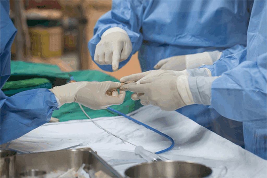
At Liv Hospital, we are leaders in vascular interventional radiology procedures, a field that uses minimally invasive techniques to treat blood vessel problems. Our internationally recognized team employs the latest methods to make these procedures safer, faster, and more effective for patients.
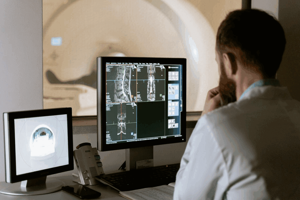
Vascular interventional radiology procedures rely on advanced imaging technologies, such as X-rays, CT scans, and ultrasounds, to guide treatments with precision. By minimizing the need for traditional surgery, these procedures reduce recovery time and improve patient comfort while addressing a wide range of vascular conditions.
At Liv Hospital, our expertise in vascular interventional radiology procedures ensures patients receive top-quality care. From diagnosis to treatment, our team combines innovation, skill, and patient-centered service to deliver optimal outcomes with minimal risk.
Key Takeaways
- Minimally invasive treatments for blood vessel conditions
- Advanced imaging methods for precise treatment
- Internationally recognized vascular interventionists
- Evidence-based techniques for safer treatments
- Faster recovery times for patients
- Personalized care for improved patient outcomes
The Evolution of Vascular Interventional Radiology

Vascular interventional radiology has grown from a simple diagnostic tool to a complex treatment field. We’ve seen big steps forward, making care better and outcomes better. Vascular and interventional radiologists now help diagnose and treat vascular issues with new, less invasive methods.
From Diagnostic to Therapeutic Applications
The change from just looking at problems to actually fixing them has been huge. At first, it was all about looking at blood vessels. But now, thanks to new tech and skills, we can treat many vascular problems. Techniques in interventional radiology let us do this with great accuracy and little risk.
We use live images to guide small tools through the body. This helps us find and fix vascular diseases accurately. It’s changed how we treat these conditions, making treatments safer and more effective for patients.
Technological Advancements in Imaging Techniques
New tech has driven the growth of vascular interventional radiology. Better imaging tools like digital subtraction angiography and ultrasound help us see and treat vascular issues better. These tools also let us try new vascular intervention methods, broadening what we can do.
| Imaging Technique | Application | Benefits |
| Digital Subtraction Angiography | Diagnostic imaging of vascular structures | High-resolution images, real-time visualization |
| Ultrasound | Guidance for vascular access and interventions | Non-invasive, real-time imaging, reduced radiation exposure |
| Magnetic Resonance Angiography | Diagnostic imaging of vascular structures | High-resolution images, no radiation exposure |
These tech improvements have made patient care and results much better. Vascular interventional radiology is now a key part of modern vascular care. As we keep improving and learning, we expect to see even better ways to diagnose and treat vascular diseases.
Understanding Vascular Interventional Radiology Procedures

Vascular interventional radiology procedures have changed how we treat vascular conditions. They use a minimally invasive method. This approach helps diagnose and treat many vascular diseases, making patients recover faster and better.
Minimally Invasive Approach to Vascular Conditions
These procedures are done without open surgery. This means less harm to the patient and fewer risks. Quick recovery is another benefit.
Procedures like angiography and angioplasty are used to fix blockages and other issues. We use small punctures in the groin or arm to reach the problem area. Advanced imaging guides us, ensuring precise treatment with little harm to nearby tissues.
The Role of Advanced Imaging in Guiding Treatments
Advanced imaging is key in vascular interventional radiology. Tools like fluoroscopy, ultrasound, and MRI help us see the blood vessels in real-time. This precision is vital for our treatments.
Imaging makes our treatments more accurate and safe. It lets us see the blood vessels clearly. This helps us avoid complications and ensure the best results.
| Imaging Technique | Application in Vascular Interventional Radiology |
| Fluoroscopy | Real-time imaging for guiding catheters and wires during procedures |
| Ultrasound | Used for vascular access and to guide certain interventions |
| MRI | Provides detailed images of vascular anatomy for pre-procedure planning |
By using minimally invasive methods and advanced imaging, we can treat many vascular conditions. This improves patient outcomes and quality of life.
The Specialized Training of Vascular Interventionists
To become a vascular interventionist, one must start a journey of detailed training. This training covers both diagnostic imaging and therapeutic techniques. These specialists are key in diagnosing and treating vascular diseases.
The training of these experts is complex, with a lot of education and practical experience. The path to becoming a vascular interventionist is tough. It makes sure these doctors can give the best care to their patients.
Educational Pathways for Vascular and Interventional Radiologists
The journey for vascular and interventional radiologists starts with a solid medicine foundation. This is usually a medical degree. Then, they get specialized training in radiology, covering both diagnostic and interventional areas.
We outline the typical educational pathway as follows:
| Stage | Description | Duration |
| Medical School | Obtaining a medical degree | 4 years |
| Residency in Radiology | Specialized training in radiology | 4-5 years |
| Fellowship in Interventional Radiology | Advanced training in interventional radiology | 1-2 years |
Skills and Expertise Required in the Field
Vascular interventionists need a mix of technical skills, knowledge, and personal traits. They must be good at using complex imaging tools and reading images to guide their treatments.
Key skills include:
- Proficiency in diagnostic imaging techniques
- Expertise in minimally invasive therapeutic procedures
- Ability to interpret complex medical images
- Strong communication and patient care skills
The training of vascular interventionists is not just about technical skills. It’s also about developing the judgment and expertise needed for top-notch patient care.
Benefits of Interventional Vascular Procedures Over Traditional Surgery
Interventional vascular procedures have changed how we treat vascular diseases. They offer many benefits over traditional surgery. These include better patient outcomes and faster recovery times. We’ll look at why vascular IR procedures are now the top choice for many.
Reduced Recovery Time and Hospital Stays
Vascular IR procedures have a big advantage: they reduce recovery time. Unlike traditional surgery, which can take weeks or even months, vascular IR procedures are quick. This is great for older patients or those with health issues.
For example, patients having an angiography or angioplasty can often go home the same day. This is much faster than traditional surgery. It makes patients happier and saves money too.
Lower Complication Rates and Surgical Risks
Vascular IR procedures have fewer complications than traditional surgery. They are less invasive, which means less damage and risk of infection. This has led to fewer serious problems for patients.
Studies show IR procedures are less painful and have shorter recovery times. They also have lower risks compared to traditional surgery. This is very important for patients at high risk for surgery complications.
Cost-Effectiveness and Patient Satisfaction
Vascular IR procedures are also more cost-effective. They reduce the need for long hospital stays and lower the risk of complications. This makes them cheaper and more appealing to patients and healthcare providers.
We think vascular IR procedures are a great choice because of their cost-effectiveness, quick recovery times, and lower risks. As the field grows, we expect to see even more ways to improve patient care.
Procedure 1: Angiography and Angioplasty
Vascular interventional radiology uses angiography and angioplasty to see and treat blood vessel diseases. These steps are key in finding and fixing problems with blood vessels.
Diagnostic and Therapeutic Applications
Angiography lets us see inside blood vessels. It spots blockages and other issues. Angioplasty uses a balloon to open up blocked or narrowed vessels, improving blood flow.
These methods are vital for treating diseases like peripheral artery disease (PAD) and coronary artery disease (CAD). They help us give full care to patients with vascular diseases.
The Procedure Process and Technology
Angiography and angioplasty start with a local anesthetic and a small cut to reach the blood vessel. A catheter is then put in and guided to the right spot with imaging like fluoroscopy.
Next, contrast dye is used to see the blood vessels through angiography. If there’s a blockage, a balloon is inflated to widen the vessel. Sometimes, a stent is placed to keep it open.
Patient Outcomes and Recovery
Most patients see big improvements in blood flow and feel better after angiography and angioplasty. The local anesthetic makes the procedure almost painless.
Recovery times differ, but most can get back to normal in a few days. We watch patients closely after the procedure to catch any issues and help them heal well.
| Procedure | Recovery Time | Complications |
| Angiography | 1-2 days | Low risk of bleeding or vascular damage |
| Angioplasty | 2-5 days | Risk of restenosis or stent thrombosis |
Knowing the benefits and risks of angiography and angioplasty helps patients make smart choices about their vascular health. We aim to give top-notch care and support every step of the way.
Procedure 2: Vascular Stent Placement
Vascular stents are mesh tubes that keep blood vessels open. This helps restore blood flow. Vascular and interventional radiologists use stents to treat many vascular conditions.
Types of Stents and Their Applications
There are different stents for various needs. Bare-metal stents are simple and effective. Drug-eluting stents release medicine to prevent the vessel from narrowing again. The right stent depends on the patient’s condition and the blockage’s location.
The Stent Placement Procedure
The procedure is minimally invasive and done under local anesthesia. Advanced imaging guides the stent to the blockage. The stent expands to open the vessel and keeps it open.
This procedure is quick, and many patients can go home the same day.
Post-Procedure Care and Long-Term Results
Patients are watched closely after the procedure. We guide them on post-procedure care, including medication and lifestyle changes. Long-term, patients often see improved blood flow and reduced symptoms.
Regular follow-ups are key to monitor the stent and blood vessel health.
Understanding vascular stent placement helps patients make informed choices. As vascular & interventional radiology specialists, we aim to provide top-notch care and guidance.
Procedure 3: Embolization Techniques for Vascular Abnormalities
Embolization is a key method in vascular interventional radiology. It treats many vascular problems, like aneurysms and arteriovenous malformations. This method blocks blood flow to certain areas. It helps control bleeding or cut off blood to tumors.
As interventionist radiologists, we use embolization. It offers patients a less invasive treatment. This can greatly improve their life quality.
Treating Aneurysms and Arteriovenous Malformations
Aneurysms and arteriovenous malformations (AVMs) are complex issues. Embolization is very effective in treating them. It stops blood flow to the problem area.
For aneurysms, it fills the sac with materials to prevent rupture. For AVMs, it shrinks the malformation. This makes it less likely to cause problems.
“Embolization is key in treating vascular issues,” says a leading expert. It’s a less invasive option than surgery. It also reduces risks and shortens recovery times.
Embolic Materials and Delivery Methods
The success of embolization depends on the materials and how they’re delivered. There are many embolic agents, like coils, particles, and liquid agents. The right material depends on the condition, location, and desired outcome.
- Coils: Used for aneurysm treatment and vessel occlusion.
- Particles: Employed for tumor embolization and AVM treatment.
- Liquid embolic agents: Utilized for complex AVMs and certain tumors.
Case Studies and Success Rates
Many studies show embolization’s success in treating vascular issues. For example, a study on intracranial aneurysms treated with coils had high success and low complications. Embolizing AVMs also reduces hemorrhage risk and improves outcomes.
The success of embolization comes from the interventionist radiologist‘s skill and new materials and systems. As these improve, we’ll see even better results and more uses in vascular interventional radiology.
Procedure 4: Thrombolysis and Thrombectomy for Deep Vein Thrombosis
Thrombolysis and thrombectomy are new treatments for deep vein thrombosis (DVT). They are less invasive and are changing how we treat vascular health. DVT is when a blood clot forms in the deep veins, usually in the legs.
If not treated, DVT can cause serious problems. These include pulmonary embolism and post-thrombotic syndrome.
Identifying and Accessing Clots
The first step is finding and getting to the clot. We use ultrasound and venography to locate it. Then, we make a small incision or puncture near the clot to access it.
Mechanical and Pharmacological Approaches
There are two main ways to treat DVT: mechanical thrombectomy and pharmacological thrombolysis. Mechanical thrombectomy uses devices to remove the clot. Pharmacological thrombolysis uses drugs to dissolve it.
We pick the best method based on the patient’s condition and the clot’s size and location.
Prevention of Post-Thrombotic Syndrome
Thrombolysis and thrombectomy help prevent post-thrombotic syndrome (PTS). PTS causes chronic pain, swelling, and skin ulcers in the affected limb. By removing or dissolving the clot, we restore normal blood flow.
Thrombolysis and thrombectomy are key treatments for DVT. They offer a minimally invasive solution with great benefits. Understanding these procedures helps patients make better care choices.
Procedure 5: Vascular Access Procedures
Vascular access is key for treatments like chemotherapy, dialysis, and nutritional support. It helps healthcare providers give therapies that boost patient outcomes and quality of life.
Central Venous Catheter Placement
Central venous catheters are for long-term access. They let doctors give medications, fluids, and blood products. Placing these catheters is a common task in vascular radiology. It needs precise imaging to place them right and avoid problems.
Benefits of Central Venous Catheters:
- Long-term access for therapies
- Reduced need for repeated venipunctures
- Ability to deliver high-volume or irritant therapies
Port Placement for Long-Term Therapy
Ports are implantable devices for long-term access. They’re great for patients on long-term chemotherapy or treatments. Ports are placed under imaging, ensuring they’re in the right spot and lowering complication risks.
Dialysis Access Creation and Maintenance
For patients with end-stage renal disease, dialysis access is vital. Vascular access procedures like arteriovenous fistulas and grafts are key. They keep dialysis access working and life-saving therapy going.
| Type of Access | Description | Advantages |
| Arteriovenous Fistula | Surgical connection between an artery and vein | Long-term durability, lower complication rates |
| Arteriovenous Graft | Surgical implantation of a synthetic graft between an artery and vein | Faster maturation time compared to fistulas |
| Central Venous Catheter | Catheter placed into a large vein for temporary or long-term access | Immediate use, useful for bridging or in patients with limited vascular options |
Vascular access procedures are essential in healthcare. They allow for life-saving treatments and better patient outcomes. As vascular and interventional radiologists, we’re key in creating and keeping vascular access. This ensures patients get the care they need.
Procedure 6: Inferior Vena Cava (IVC) Filter Placement
Vascular radiologists often suggest IVC filter placement to prevent pulmonary embolism in certain patients. IVC filters catch blood clots that could travel to the lungs. This is a big risk for people with deep vein thrombosis (DVT).
The procedure involves putting a filter device into the inferior vena cava. This vein carries blood from the lower body to the heart.
Indications and Patient Selection
Doctors decide on IVC filter placement based on specific reasons. These include patients at high risk of pulmonary embolism but can’t take blood thinners. This might be due to bleeding risks or recent surgery.
Other reasons include patients with DVT that doesn’t go away with blood thinners and those with big clots.
We look at each patient’s situation to see if an IVC filter is right for them. We check the risk of pulmonary embolism, DVT, and overall health.
Filter Types and Placement Techniques
There are different IVC filters, like permanent and retrievable ones. The type chosen depends on the patient’s condition and how long they’ll need the filter. Retrievable filters are best when the risk is temporary, as they can be taken out later.
Putting in an IVC filter is a small procedure done under imaging. This is usually ultrasound or fluoroscopy. We make a small cut, usually in the groin or neck, and guide the filter into place with a catheter.
Retrieval Procedures and Long-Term Management
Patients with retrievable IVC filters can have them removed when the risk goes away. This removal is also done under imaging and involves using a catheter to grab and pull out the filter.
Managing patients with IVC filters long-term means watching for problems like filter movement or breakage. We also check if the filter is needed anymore and plan for its removal.
In summary, IVC filter placement is a key way to prevent pulmonary embolism in high-risk patients. With careful selection, precise placement, and proper management, we can greatly reduce this dangerous condition’s risk.
Procedure 7: Uterine Fibroid Embolization
Uterine fibroid embolization (UFE) is a new, less invasive way to treat fibroids. As vascular interventionists, we’ve seen how it improves our patients’ lives.
Non-Surgical Alternative for Fibroid Treatment
UFE is a non-surgical option for fibroid treatment. It avoids the need for surgery. This method targets fibroids through the arteries that feed them.
By injecting particles into these arteries, we cut off the fibroids’ blood supply. This causes them to shrink over time. It reduces symptoms like heavy bleeding and pain, and it keeps the uterus intact.
The Embolization Process and Materials
The process starts with a small puncture in the groin or wrist. We use imaging to guide a catheter to the uterine arteries. Then, we inject embolic materials to block blood flow to the fibroids.
The type of embolic material used depends on the fibroids’ size and location, and the patient’s health. Our skills in techniques in interventional radiology make sure the procedure fits each patient’s needs.
Recovery and Quality of Life Improvements
UFE has a quick recovery time compared to surgery. Most women can go back to their normal activities in a week or two. The decrease in fibroid size and symptoms greatly improves their quality of life.
Because of these benefits, UFE is a top choice for many women with symptomatic fibroids. It’s a less invasive and effective solution.
Conclusion: The Future of Vascular Interventional Radiology
Vascular interventional radiology is changing fast. New imaging and procedures are making care better. They offer non-invasive ways to treat complex vascular issues.
The future looks bright for vascular interventional radiology. Research and development are making treatments better and more options available. We’ll see more precise and effective procedures.
As vascular radiology grows, we’ll see more advanced technologies. This will improve patient care and results. New techniques and tools will be key in the future of vascular treatments.
Vascular interventional radiology is set to change how we manage vascular diseases. It’s becoming more important in healthcare. We’re excited to see how it will evolve with new technology and techniques.
FAQ
What is vascular interventional radiology?
Vascular interventional radiology is a medical field. It uses small procedures to treat blood vessel diseases. This approach is less invasive than traditional surgery.
What are the benefits of vascular interventional radiology procedures?
These procedures have many advantages. They lead to faster recovery times and fewer complications. They also cost less than traditional surgery.
What is angiography and angioplasty?
Angiography shows blood vessels through imaging. Angioplasty uses a balloon to widen narrowed blood vessels.
What is vascular stent placement?
Vascular stent placement uses a small device to keep a blood vessel open. This improves blood flow and reduces the chance of narrowing again.
What are embolization techniques used for?
Embolization techniques treat vascular problems like aneurysms. They block or reduce blood flow to the affected area.
What is thrombolysis and thrombectomy?
Thrombolysis and thrombectomy dissolve or remove blood clots. This restores blood flow and lowers the risk of complications.
What is vascular access?
Vascular access creates or maintains blood vessel access. It’s used for dialysis, chemotherapy, or medication.
What is IVC filter placement?
IVC filter placement places a filter in the inferior vena cava. It prevents blood clots from reaching the lungs, reducing pulmonary embolism risk.
What is uterine fibroid embolization?
Uterine fibroid embolization is a non-surgical method. It blocks blood flow to fibroids, reducing their size and symptoms.
What kind of training do vascular interventionists receive?
Vascular interventionists get specialized training. They learn through education and hands-on experience. This prepares them for their role.
How do advanced imaging techniques contribute to vascular interventional radiology?
Advanced imaging, like fluoroscopy and ultrasound, is key. They guide procedures, ensuring accurate diagnosis and treatment.
What is the future of vascular interventional radiology?
The future looks bright. Advances in technology and techniques will improve patient care. It will also offer more treatment options.


