Last Updated on November 27, 2025 by Bilal Hasdemir
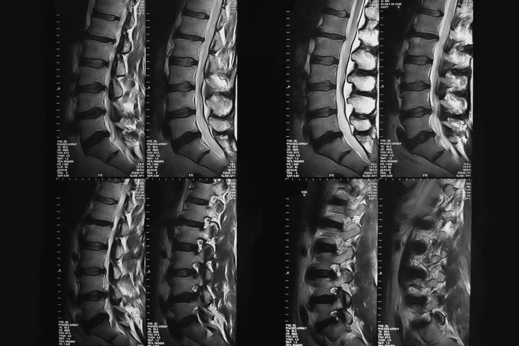
At Liv Hospital, we use advanced imaging like bone scintigraphy for accurate diagnoses. A radionuclide bone scan is a special imaging method. It shows where bone metabolism is not normal.
We use a radioactive tracer to find different bone issues, like cancer and fractures. The whole process can take up to four hours. This includes the time for the tracer to spread and the actual scan. Wondering what can a bone scan show? See 7 essential conditions, including cancer and arthritis, revealed by bone scintigraphy.
Key Takeaways
- Bone scintigraphy detects areas of abnormal bone activity.
- It helps diagnose various bone conditions, including cancer and fractures.
- The scan process involves using a radioactive tracer.
- At Liv Hospital, we utilize advanced imaging technologies.
- Bone scans provide complete care for patients.
Understanding Bone Scintigraphy: The Science Behind Bone Scans
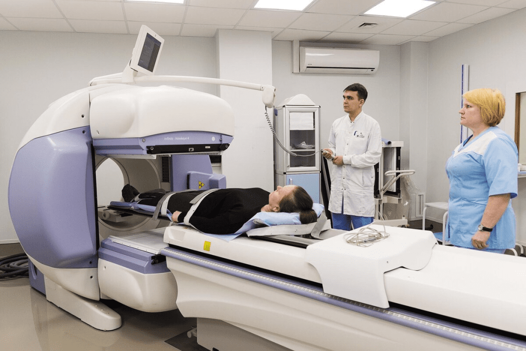
Bone scintigraphy uses radioactive isotopes to show what’s happening in our bones. It’s key for spotting and tracking bone problems.
How Radioactive Isotopes Help Visualize Bone Activity
Radioactive isotopes, or radionuclides, help see bone activity in bone scintigraphy. Injected into the body, they go to active bone areas, like diseased or injured spots. Technetium-99m methylene diphosphonate (Tc-99m MDP) is the top choice because it sticks to bone well.
Tc-99m MDP helps find bone issues like cancer, fractures, and infections. It shows where bone activity is off, helping doctors see how bad the disease is and if treatments are working.
The Role of Gamma Rays in Bone Imaging
Gamma rays from the isotopes are caught by a gamma camera. These rays are like X-rays but with different strengths. The camera makes images of the skeleton, showing bone activity.
Gamma cameras are super sensitive to these rays. This lets them spot tiny changes in bone activity. It makes bone scintigraphy great for catching and tracking bone diseases early.
How Bone Scintigraphy Machines Work
Bone scintigraphy machines, or gamma cameras, catch gamma rays from the isotopes in bones. They move around the patient, taking pictures from different sides. Then, they make detailed bone images, showing where things are off.
Today’s gamma cameras can take very clear pictures. A study in Nature shows how better technology has made bone scans more accurate.
Advancements in Nuclear Imaging Technology
Nuclear medicine, including bone scintigraphy, has made big strides. Better isotopes, cameras, and image tech have made scans more precise and detailed.
- Hybrid imaging, like SPECT/CT, mixes bone scan info with CT scans. This gives a fuller picture of bone problems.
- New treatments using radionuclides are being tested. They might help treat some bone diseases.
These advances show how bone scintigraphy is becoming more important in diagnosing and treating bone health issues.
The Bone Scan Procedure: What to Expect
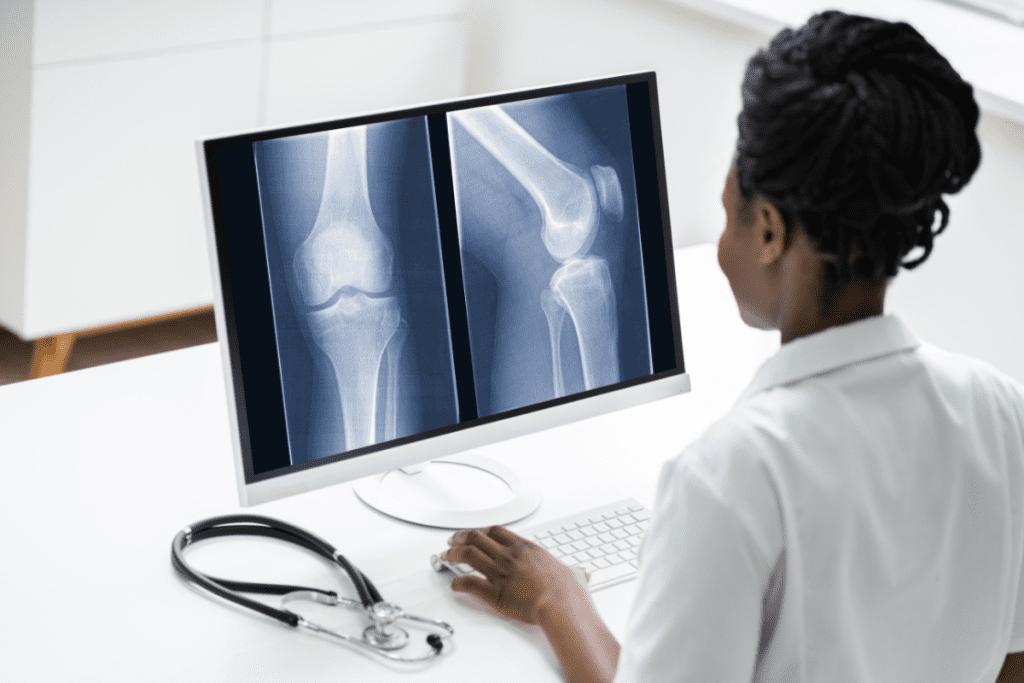
If you’re set for a bone scan, you might have questions. Knowing what to expect can ease your mind and make things go smoother.
Before Your Bone Scan: Preparation Guidelines
Getting ready for a bone scan is easy. To get the best results, follow these steps:
- Drink lots of water before the scan to help the tracer move through your body.
- Take off any jewelry or metal items that could mess with the scan.
- Tell your doctor about any meds you’re taking. Some might need to be changed before the scan.
- Wear comfy clothes and be ready to stay very quiet during the scan.
Arriving a bit early is smart. It lets you fill out any needed papers and get ready before the scan starts.
During and After the Procedure: Timeline and Sensations
The bone scan usually takes a few hours. But the actual scanning time is much shorter. Here’s what happens:
- You’ll get an injection of a tiny bit of radioactive tracer. It will spread through your body for a while.
- During the scan, you’ll lie on a table. A gamma camera will take pictures of your bones.
- The scan itself is painless and usually lasts 30-60 minutes.
After the scan, you can go back to your usual activities. The tracer will leave your body in your urine and feces over a few days. Keep drinking water to help flush it out.
Some people might feel a little rash or itch at the injection site. But this is rare. If you have any worries or feel something odd after the scan, call your doctor right away.
Knowing what to expect from your bone scan can make you feel more ready and confident. It’s an important step in your care.
What Can a Bone Scan Show? An Overview of Diagnostic Capabilities
Bone scans are a key tool for diagnosing bone issues. They help us see how bones are working. This lets us find and treat many bone problems.
Bone scans can spot bone metastases from cancers like breast, lung, and prostate. They also find primary bone tumors and other bone issues.
Identifying Regions of Decreased Bone Activity
Looking at areas where bone activity is low can show us certain conditions. We search for spots where bone activity is not normal. This can point to different bone disorders.
Some important things to look for include:
- Detecting bone infarctions, where bone tissue dies because of no blood supply.
- Finding areas of bone necrosis, caused by trauma or infection.
- Seeing patterns of low bone activity in metabolic bone diseases.
By studying these areas, we understand the condition better. Then, we can plan the right treatment.
Bone Metastases: How Bone Scans Detect Cancer Spread
Finding bone metastases is key in fighting cancer. Bone scans help a lot in this fight. Bone metastases happen when cancer cells move from a main tumor to the bones. This is common in cancers like breast, lung, and prostate.
Breast, Lung, and Prostate Cancer Metastasis Patterns
Different cancers spread to the bones in different ways. Breast cancer often spreads to the ribs, spine, and pelvis. Lung cancer can go anywhere in the skeleton, but often hits the spine, ribs, and long bones. Prostate cancer usually goes to the pelvis, spine, and ribs, causing bone growth.
Monitoring Cancer Treatment Response with Sequential Scans
Sequential bone scans help check how well the treatment is working. By comparing scans, doctors can see if the cancer in the bones is getting better, staying the same, or getting worse. This helps them change treatment plans if needed.
If a scan shows fewer or less intense lesions, it means treatment is working. But if new lesions appear or old ones get worse, it means treatment might not be working.
Bone scans are very important in cancer care. They help find bone metastases and check how well treatment is working. They give a full-body view, making them essential for patients with cancers that often spread to bones.
Primary Bone Cancers Revealed Through Scintigraphy
Bone scintigraphy is key in finding primary bone cancers. It shows where and how big these cancers are. This is very important for spotting osteosarcoma, a common bone cancer.
Osteosarcoma and Other Primary Bone Malignancies
Osteosarcoma is a fast-growing bone cancer. It usually happens in long bones like the femur and tibia. Scintigraphy helps find it by showing where bone activity is high, which means tumor growth.
Scintigraphy also finds Ewing’s sarcoma and chondrosarcoma. These cancers show up as abnormal bone activity in nuclear images.
Early Detection Advantages of Nuclear Imaging
Finding bone cancers early is key for better treatment and outcomes. Nuclear imaging, like scintigraphy, helps a lot. It spots cancers early, even before symptoms or big bone changes happen.
It also checks the whole body for other cancers or disease spots. This is important for planning right treatment.
Benefits of Early Detection through Scintigraphy:
- Improved treatment outcomes due to timely intervention
- Enhanced ability to detect multifocal disease or metastases
- Better patient prognosis through early initiation of appropriate therapy
| Type of Cancer | Common Locations | Characteristics |
| Osteosarcoma | Long bones (femur, tibia, humerus) | Aggressive, high-grade malignancy |
| Ewing’s Sarcoma | Pelvis, ribs, long bones | Highly malignant, often with systemic symptoms |
| Chondrosarcoma | Pelvis, femur, humerus, scapula | Malignant cartilage tumor, variable grade |
In conclusion, bone scintigraphy is a vital tool for finding primary bone cancers. It helps detect cancers early and gives a full picture of the disease. This makes it a key part of cancer care today.
Detecting Hidden Fractures: When X-Rays Aren’t Enough
Bone scans are key in finding fractures that X-rays miss. X-rays are great for spotting bone injuries, but they can’t catch all types. This includes hairline or stress-related fractures.
Stress Fractures and Microfractures Visualization
Stress fractures and microfractures are hard to spot with X-rays. These tiny bone cracks happen from too much stress or sudden activity. Bone scans are better at showing these because they highlight active bone areas.
Key characteristics of stress fractures detectable by bone scans include:
- Increased uptake of the radioactive tracer at the fracture site
- Often appears as a focal area of intense activity
- It can be detected even when X-rays appear normal
Traumatic Injuries with Delayed Radiographic Changes
Some injuries might not show up on X-rays right away, even if they’re big. Bone scans can help by showing bone activity that points to a fracture. This is helpful when patients have pain, but X-rays look fine.
The following table shows how X-rays and bone scans differ in finding fractures:
| Diagnostic Feature | X-ray | Bone Scan |
| Sensitivity for Stress Fractures | Low to Moderate | High |
| Detection of Microfractures | Difficult | Effective |
| Radiation Exposure | Low | Low (using modern tracers) |
| Time to Positive Findings | Immediate if visible | Can be positive before X-ray |
Bone scans help doctors understand a patient’s condition better, even when X-rays don’t show much. This leads to more accurate diagnoses and better treatment plans for hidden fractures.
Bone Infections: Identifying Osteomyelitis and Septic Arthritis
Diagnosing bone infections like osteomyelitis and septic arthritis is tough. But bone scans are a big help. They show us where the bone is most active, which often means there’s an infection.
Bone scans are great for finding osteomyelitis, an infection in the bone. It can come from bacteria or fungi. The scan spots areas where the bone is working too hard, showing that an infection is present.
Characteristic Patterns of Bone Infection
When we look at bone scans for infections, we search for characteristic patterns. Osteomyelitis shows up as a hot spot on the scan, where the infection is. For more on bone scans, check out RadiologyInfo.org.
Septic arthritis, though, shows up as increased activity around a joint. This is because of the inflammation it causes. By looking at these patterns, we can spot and track bone infections accurately.
Bone scans are key in finding infections early and seeing how treatments are working. They’re a big help in managing bone infections.
Metabolic Bone Diseases: Insights from Bone Scans
Bone scans give us a special look at metabolic bone diseases. They help us understand conditions like Paget’s disease and osteomalacia. These diseases mess with bone metabolism, causing changes that bone scans can spot.
Metabolic bone diseases include many disorders that harm bone health. Paget’s disease and osteomalacia are two examples. If not treated well, they can really hurt a patient’s life quality.
Paget’s Disease and Osteomalacia Patterns
Paget’s disease makes bones break down and rebuild too much. This can cause deformities and serious problems. Bone scans show the special patterns of Paget’s disease, highlighting areas where bone activity is high.
Osteomalacia softens bones because of vitamin D problems. Bone scans can find where bones are less dense and less active.
| Condition | Bone Scan Findings | Clinical Implications |
| Paget’s Disease | Increased bone activity, characteristic “hot spots” | Potential for deformities, fractures, and neurological complications |
| Osteomalacia | Decreased bone density, “cold spots” | Increased risk of fractures, bone pain, and muscle weakness |
Monitoring Treatment Response in Metabolic Conditions
Bone scans are great for diagnosing and tracking metabolic bone diseases. They help doctors see how treatment is working. This way, they can change plans to better manage the disease.
“The use of bone scans in monitoring metabolic bone diseases represents a significant advancement in the management of these conditions, allowing for more precise and effective treatment strategies.”” Expert in Nuclear Medicine
For example, in Paget’s disease, bone scans show if bisphosphonate therapy is working. They look for less bone activity. In osteomalacia, scans track how vitamin D helps improve bone density and activity.
Thanks to bone scans, doctors can give patients more tailored and effective care. This is a big step forward in treating metabolic bone diseases.
Arthritis and Inflammatory Joint Conditions
Arthritis and inflammatory joint conditions can be checked with bone scans. These scans show detailed images of bone activity. Bone scintigraphy is great because it spots areas of high bone activity linked to inflammation.
Distinguishing Arthritis from Malignancy on Bone Scans
It can be hard to tell arthritis from cancer on bone scans. Both can show up as hot spots. But, arthritis usually shows up in a symmetrical way in joints. Cancer might show up more randomly or unevenly.
We look at the whole picture, including other tests, to make a correct diagnosis.
Limitations of Bone Scans in Arthritis Diagnosis
Bone scans are good for finding arthritis and other joint problems. But, they’re not perfect. They can show up positive for many things, not just arthritis.
So, we need to look at the scan results with other tests and what the doctor knows. This helps us make sure we’re right.
Knowing these things helps us use bone scans better. This leads to better care for patients with arthritis and joint problems.
Conclusion: The Future of Bone Scanning in Modern Diagnostics
Bone scintigraphy is key in diagnosing and tracking bone issues. At Liv Hospital, we use top-notch imaging to help our patients. This ensures they get the best care possible.
The future of bone scanning looks bright. New tech in nuclear imaging is making bone scans more accurate and useful. As we keep improving, bone scans will play an even bigger role in treating bone problems.
By focusing on bone scintigraphy, we can make treatments better. We aim to give top-notch care to those who need it. As a leader in healthcare, we’re committed to leading in diagnostic innovation. This way, our patients get the best treatment available.
FAQ
What is a bone scan, and how does it work?
A bone scan, also known as bone scintigraphy, is a test that uses a small amount of radioactive material. It helps see how active the bones are. The test works by injecting a radioactive isotope into the blood. This isotope is then absorbed by the bones, allowing a special camera to capture images of bone activity.
Does a bone scan show arthritis?
Yes, a bone scan can show arthritis. But it’s not always clear-cut. It can spot inflammatory joint conditions, like arthritis, by showing where the bones are more active. Yet, telling arthritis apart from other conditions, like cancer, needs careful scan results.
What cancers can a bone scan detect?
Bone scans can spot cancers that have spread to the bones, like breast, lung, and prostate cancer. They also help find primary bone cancers, such as osteosarcoma.
Can a bone scan detect bone cancer?
Yes, bone scans can find primary bone cancers, like osteosarcoma, and cancers that have spread to the bones. They’re great for spotting abnormal bone activity that might mean cancer.
How is a bone scan used to monitor cancer treatment response?
By doing bone scans at different times, doctors can see how well cancer treatment is working. This helps them check if the treatment is effective and make better care plans.
Can a bone scan detect fractures that are not visible on X-ray?
Yes, bone scans can find fractures that X-rays can’t see, like stress fractures and microfractures. They’re really helpful for spotting injuries that don’t show up on X-rays right away.
What is the role of gamma rays in bone imaging?
Gamma rays are key in bone imaging. They let doctors detect radioactive isotopes in bones. The gamma rays from these isotopes are caught by a camera, making images of bone activity.
How do bone scintigraphy machines work?
Bone scintigraphy machines, or gamma cameras, detect gamma rays from radioactive isotopes in bones. They make images of bone activity, helping doctors see where bones might be acting strangely.
What are the limitations of bone scans in diagnosing arthritis?
While bone scans can spot arthritis, they’re not always sure. The results need careful thought, and more tests might be needed to confirm a diagnosis.
What can a bone scan reveal about metabolic bone diseases?
Bone scans can give clues about metabolic bone diseases, like Paget’s disease and osteomalacia. They show specific bone activity patterns. They also help track how well treatment is working for these conditions.
References
- Adams, C. (2023). Bone scan. In StatPearls. StatPearls Publishing. https://www.ncbi.nlm.nih.gov/books/NBK531486/
- Magdy, O., et al. (2024). Bone scintigraphy based on deep learning model and its application for bone metastases diagnosis. Scientific Reports, 14, Article 73991. https://www.nature.com/articles/s41598-024-73991-8
- Lee, J. W., et al. (2020). Clinical use of quantitative analysis of bone scintigraphy for arthritis disease evaluation. Frontiers in Medicine, 7, Article 576458. https://www.ncbi.nlm.nih.gov/pmc/articles/PMC7761348/


