At Liv Hospital, we know how worried you might be if your PET scan shows lymph nodes lighting up. This is often because of increased metabolic activity.
Metabolic activity can be from cancer or other non-cancerous reasons. Many patients ask, what causes lymph nodes to light up on PET scan”it can be due to cancer, infections, or inflammation. It’s important to figure out the exact cause to ensure the right treatment. We use advanced imaging to help with this.
We have the latest technology to check why lymph nodes light up on PET scans. This ensures our patients get the best care for their needs.
Key Takeaways
- PET scans help identify areas of increased metabolic activity in lymph nodes.
- Lymph nodes can light up due to both malignant and benign causes.
- Accurate diagnosis is key for effective treatment planning.
- Advanced imaging techniques help tell the difference between causes.
- Liv Hospital is dedicated to patient-centered care with the latest technology.
Understanding PET Scans in Lung Cancer Diagnosis
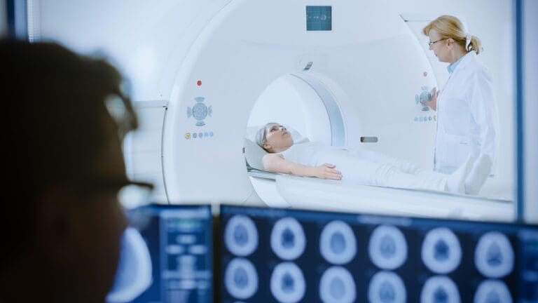
PET scans are key in lung cancer diagnosis. They help doctors see how far the cancer has spread. This info is vital for choosing the right treatment.
Basic Principles of PET Imaging
PET scans use a special tracer that cancer cells take up more of. This makes cancer cells show up on scans. It helps doctors find and tell apart cancer from non-cancer cells.
Role in Lung Cancer Evaluation
PET scans are important for lung cancer. They help see if cancer has spread to lymph nodes. This info is key for accurate staging and treatment planning.
PET scans have many benefits in lung cancer diagnosis. They:
- Improve staging accuracy
- Help find metastatic disease
- Let doctors track how well treatments are working
Advantages Over Conventional Imaging
PET scans have big advantages over CT scans or MRI. They show how tissues are working, not just what they look like.
| Imaging Modality | Primary Information Provided | Key Benefit in Lung Cancer |
| PET Scan | Metabolic Activity | Identifies cancerous tissues based on metabolic rate |
| CT Scan | Anatomical Details | Provides detailed images of structures and organs |
| MRI | Soft Tissue Detail | Excellent for visualizing soft tissue abnormalities |
Using PET scans with other imaging helps doctors understand lung cancer better. This leads to better treatment plans.
The Lymphatic System and Its Importance in Cancer
Understanding the lymphatic system is key to knowing how cancer spreads. It’s a vital part of our immune defense. It filters out harmful substances and helps fight infections.
Structure and Function of Lymph Nodes
Lymph nodes are small, bean-shaped structures in our body. They act as filters, trapping pathogens and abnormal cells, including cancer cells. They filter lymph fluid and are key sites for immune responses.
Lymph nodes have a vital function. They filter lymph fluid and store lymphocytes, a type of white blood cell. When cancer cells metastasize, they often travel through the lymphatic system. Lymph nodes can become sites where cancer cells accumulate.
Lymphatic Drainage of the Lungs
The lungs have a rich network of lymphatic vessels. This network is key for removing waste and transporting immune cells. Understanding this is essential for knowing how lung cancer spreads to lymph nodes.
Lymphatic drainage from the lungs can go through various pathways. It can go to the hilar, mediastinal, and sometimes directly to the supraclavicular lymph nodes. The pattern of drainage is important for predicting lung cancer spread.
Significance in Cancer Spread
Lymph nodes play a critical role in cancer spread. When cancer cells are found in lymph nodes, it means the cancer has started to metastasize. This can change treatment plans and prognosis.
In lung cancer, the status of lymph nodes is a key factor in staging. The presence of cancer in lymph nodes can signify a more advanced stage. This often requires more aggressive treatment strategies.
| Lymph Node Group | Role in Lung Cancer | Significance |
| Hilar Lymph Nodes | First station for lymphatic drainage from the lungs | Early involvement indicates local spread |
| Mediastinal Lymph Nodes | Further drainage pathway | Indicative of more advanced disease if involved |
| Supraclavicular Lymph Nodes | Distant lymph node group | Involvement signifies distant metastasis |
What Causes Lymph Nodes to Light Up on PET Scan
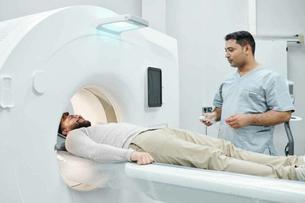
Lymph nodes lighting up on PET scans is due to their metabolic activity. This activity is measured by how much fluorodeoxyglucose (FDG) they take in. When lymph nodes are active, they use more glucose and FDG, making them visible on scans.
Metabolic Activity and FDG Uptake
FDG uptake shows how much glucose cells are using. Cells that are very active, like cancer cells, take in more FDG. This is why lymph nodes show up as “lit up” or hypermetabolic on PET scans.
Factors influencing FDG uptake include:
- Cancerous infiltration
- Inflammatory processes
- Infection
- Granulomatous diseases
Standardized Uptake Value (SUV) Measurement
The Standardized Uptake Value (SUV) measures FDG uptake in tissues. SUVmax is the highest value in a certain area, showing how active lymph nodes are. A higher SUVmax means more activity.
Significance of SUV Thresholds
SUV thresholds help tell if lymph nodes are benign or malignant. While there’s no one standard, higher values often mean cancer. Here’s a table showing typical SUVmax values and what they mean.
| SUVmax Value | Implication |
| < 2.5 | Typically benign |
| 2.5 – 4.0 | Suspicious, may require further evaluation |
| > 4.0 | Highly suggestive of malignancy |
Knowing these thresholds is key to understanding PET scans. It helps doctors make better decisions.
Malignant Causes of Lymph Node Illumination
It’s important to know how lung cancer spreads to lymph nodes. This knowledge helps in treating the disease effectively. Lung cancer spreading to lymph nodes affects how serious the cancer is and the patient’s outlook.
Primary Lung Cancer Metastasis
Lung cancer can spread to lymph nodes through the lymphatic system. This means cancer cells move from the main tumor to nearby lymph nodes. Experts say, “Lymph node metastasis is a key factor in lung cancer staging and prognosis.”
“Lymph node involvement is a key indicator of disease severity and guides treatment decisions.”
We will look into how this spread happens and its effects on lung cancer patients. For more on lung cancer, check out Cedars-Sinai’s lung cancer page.
Patterns of Lymphatic Spread in Lung Cancer
The way lung cancer spreads through lymph nodes depends on the tumor’s location and type. Common patterns include:
- Spread to hilar lymph nodes
- Involvement of mediastinal lymph nodes
- Metastasis to supraclavicular lymph nodes
Knowing these patterns is key for accurate cancer staging and treatment planning. Accurate staging shows how far the disease has spread.
Cellular Characteristics of Malignant Nodes
Malignant lymph nodes have certain cell features, such as:
- Increased metabolic activity
- Higher glucose uptake (measured by FDG-PET scans)
- Alterations in cellular morphology
These features help tell malignant nodes apart from benign ones. We use advanced imaging like PET scans to check these characteristics.
Benign Conditions That Cause Lymph Node Activity on PET
When we look at PET scans for lung cancer, we need to know about benign conditions. These conditions can make lymph nodes look active, even if they’re not cancer. This can lead to mistakes in diagnosis.
Inflammatory Processes
Inflammatory conditions often cause lymph nodes to show up on PET scans. Sarcoidosis and other diseases can make lymph nodes take up more FDG. It’s important to think about these when we look at PET scans to avoid mistakes.
Infections
Infections can also make lymph nodes light up on PET scans. This includes bacterial, viral, or fungal infections. For example, tuberculosis can make lymph nodes show up a lot, making it hard to tell if lung cancer has spread. A study on the National Center for Biotechnology Information website shows how important it is to think about infections when we look at PET scans.
Granulomatous Diseases
Granulomatous diseases create granulomas in response to certain things. This can make lymph nodes show up on PET scans, like in sarcoidosis. It’s key to know the patterns and match them with the patient’s history to tell if it’s benign or cancer.
Post-Treatment Inflammation
Inflammation after treatment can also make lymph nodes look active on PET scans. This can look like cancer, but it’s not. It’s important to look at the treatment history and other scans to make sure we’re not making a mistake.
Hilar and Mediastinal Lymph Nodes in Lung Cancer

The status of hilar and mediastinal lymph nodes is very important in lung cancer. These nodes are key for cancer spread. Their involvement affects patient prognosis and treatment.
Anatomical Significance
Hilar lymph nodes are in the lung hilum. This is where the bronchi, blood vessels, and nerves enter and exit the lungs. Mediastinal lymph nodes are in the mediastinum, the chest cavity’s center. Both are vital for lung lymphatic drainage and common metastasis sites in lung cancer.
Staging Implications
The involvement of hilar and mediastinal lymph nodes is key in lung cancer staging. The TNM staging system uses these nodes to classify the disease. Accurate assessment is essential for treatment planning.
Prognostic Value
The presence or absence of metastasis in these lymph nodes is very important. Patients with lymph node involvement generally have a poorer prognosis. The number and location of involved nodes also provide valuable information.
| Lymph Node Group | Location | Significance in Lung Cancer |
| Hilar Lymph Nodes | Within the lung hilum | First station for lymphatic drainage; often involved in lung cancer |
| Mediastinal Lymph Nodes | In the mediastinum | Critical for staging; involvement affects prognosis and treatment |
Understanding hilar and mediastinal lymph nodes in lung cancer is key. It helps in accurate disease staging and prognosis. Their evaluation aids in creating treatment plans tailored to each patient’s needs.
Distant Lymph Node Metastasis in Lung Cancer
Distant lymph node metastasis is a key part of lung cancer’s growth. When lung cancer spreads, it often goes to lymph nodes in different parts of the body. Knowing how it spreads helps doctors stage the cancer and plan treatment.
Supraclavicular and Cervical Nodes
The involvement of supraclavicular and cervical lymph nodes in lung cancer is a big sign of advanced disease. These nodes are above the clavicle and in the neck. Cancer in these areas means the disease has spread beyond the chest, making treatment harder.
Research shows that lung cancer in supraclavicular lymph nodes means a worse outlook. Cancer in these nodes changes treatment plans. It often means moving from surgery to palliative care or more intense treatments.
Abdominal Lymph Nodes
Lung cancer can also spread to abdominal lymph nodes, though it’s less common. These nodes are in the abdomen and help filter out cancer cells. Cancer in these nodes means the disease is more advanced.
- Cancer in abdominal lymph nodes is found through scans like PET or CT scans.
- Treatment for these nodes includes systemic therapies.
- Patients with cancer in these nodes usually have a less favorable outlook.
Impact on Disease Staging
Distant lymph nodes, like supraclavicular, cervical, or abdominal, greatly affect lung cancer staging. Accurate staging is key for choosing the right treatment and understanding prognosis.
The TNM staging system says stage IV if cancer has spread to distant lymph nodes. This stage helps doctors decide on treatment and talk about the disease’s extent and future.
In summary, distant lymph node metastasis in lung cancer is complex and important for staging and treatment. Knowing how it spreads and its impact on prognosis is vital for the best care for lung cancer patients.
Lymphadenopathy in Lung Cancer Patients
Understanding lymphadenopathy is key for lung cancer staging and treatment. Lymphadenopathy means lymph nodes are enlarged. This can happen for many reasons, like cancer, infection, or inflammation.
Definition and Clinical Significance
Lymphadenopathy is when lymph nodes get too big. Doctors find this through touch, scans, or lab tests. In lung cancer, it shows if cancer has spread to lymph nodes.
This is important because it affects treatment choices and how well a patient will do. Knowing about lymphadenopathy helps doctors pick the right treatment, like surgery or chemo.
Correlation with PET Findings
PET scans are vital for checking lymphadenopathy in lung cancer. They use a special sugar to see how active lymph nodes are. If nodes light up a lot, they might be cancerous.
Linking lymphadenopathy with PET scans helps spot cancerous nodes. Research shows PET scans are very good at finding these nodes in lung cancer patients.
Distinguishing Benign from Malignant Enlargement
Telling if lymph nodes are swollen because of cancer or not is hard but very important. Swelling can be caused by many things, like infections or inflammation.
| Characteristics | Benign Lymphadenopathy | Malignant Lymphadenopathy |
| FDG Uptake on PET | Low to moderate | High |
| Lymph Node Size | Variable, often smaller | Often larger |
| Clinical Context | Associated with infection or inflammation | Associated with cancer spread |
Doctors use PET scans and other tests to tell if swelling is cancer or not. This helps them manage lung cancer patients better.
Sensitivity and Specificity of PET Scans for Lymph Node Evaluation
Understanding PET scans’ sensitivity and specificity is key for lung cancer lymph node evaluation. PET scans are vital in diagnosing and staging lung cancer, focusing on lymph nodes.
PET scans are great at spotting cancer in lymph nodes because they show where cells are most active. But, their accuracy can vary. This is because other issues like infections can also show up as active areas on scans.
False Positives and Their Causes
False positives happen when PET scans show cancer where there isn’t any. This can be due to:
- Inflammatory processes: Conditions like sarcoidosis can make lymph nodes show up as active.
- Infections: Viral or bacterial infections can cause lymph nodes to swell, looking like cancer on scans.
- Post-treatment changes: After treatments, lymph nodes might look active because of healing.
These false positives can lead to lung cancer being treated more aggressively than needed. It’s important to look at the whole picture when interpreting PET scan results.
False Negatives and Their Implications
False negatives, where PET scans miss cancer in lymph nodes, can also happen. This is often because:
- Small node size: Tiny lymph nodes might not show up on scans.
- Low metabolic activity: Some cancerous nodes might not take up enough FDG to be seen.
Missing cancer in lymph nodes can mean the disease is not treated fully. Knowing these limitations helps doctors make better decisions.
Limitations of Current Technology
PET scans are powerful, but they’re not perfect. New research is looking at better scanners and new tracers. Using PET with other scans like CT or MRI can also help.
Improving PET scans is important. We need to balance their high sensitivity with their specificity issues. This will help us better diagnose lung cancer, improving patient care.
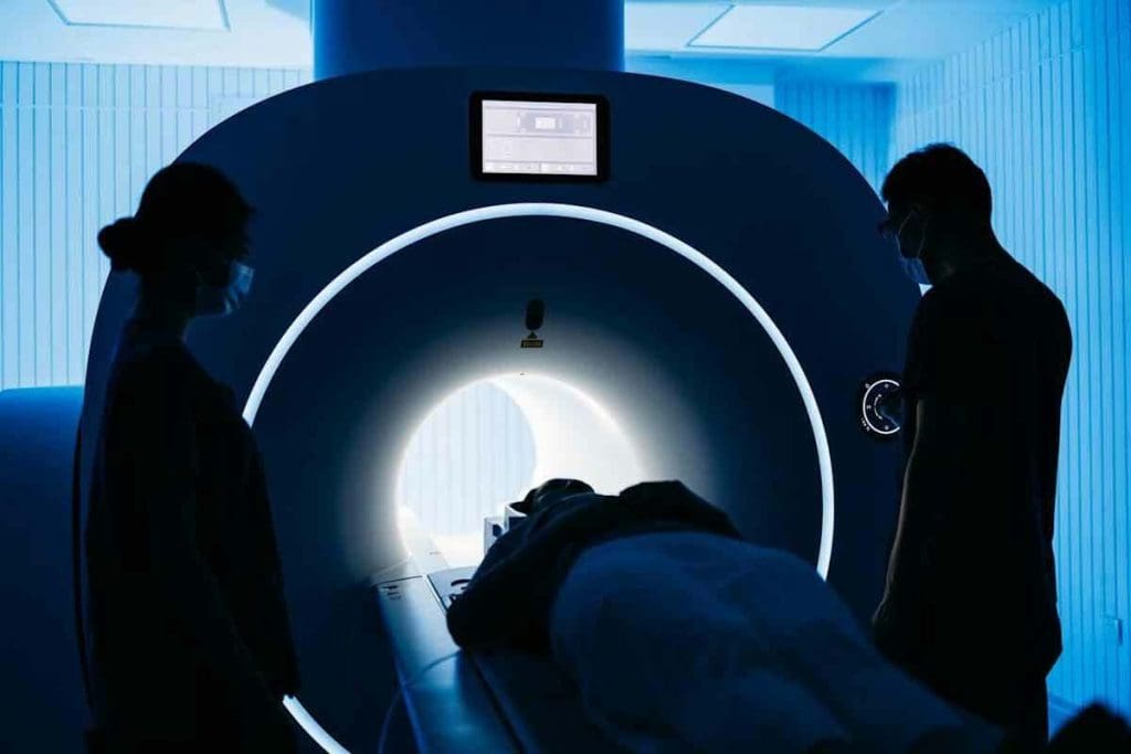
Integrated PET/CT Imaging in Lymph Node Assessment
In oncology, PET/CT imaging is key for checking lymph nodes. It mixes PET and CT scans’ strengths. This gives a better view than either scan alone.
Advantages Over Standalone PET
PET/CT imaging beats standalone PET scans in many ways. The main plus is better location accuracy. It combines PET’s function data with CT’s detailed anatomy. This helps doctors pinpoint lymph node issues more precisely.
This combo is super useful in tricky spots like the mediastinum and hilum. These areas are key for lung cancer staging.
Interpretation Challenges
Yet, reading PET/CT images can be tough. One big hurdle is telling benign from malignant lymph node activity. Some infections or inflammation can look like cancer on PET scans.
To get past these hurdles, doctors must link PET/CT findings with patient history, lab results, and other tests.
Reading PET/CT Images: What Clinicians Look For
Doctors check a few things when looking at PET/CT images. They look at the FDG uptake intensity in lymph nodes. A high Standardized Uptake Value (SUV) means more activity, which might mean cancer.
They also check the size and shape of lymph nodes on the CT part. Big or odd-shaped nodes might hint at cancer.
Clinical Correlation of PET Findings
Getting a correct diagnosis and treatment plan needs a detailed look at PET scan results. It’s key to match PET scan findings with clinical and pathological data. This helps us see how far lung cancer has spread and what treatment is best.
Importance of Multidisciplinary Approach
Working together is vital when linking PET scan results with other diagnostic data. We team up with radiologists, oncologists, and pathologists. This ensures PET scan results are seen in the light of the patient’s full clinical picture. This teamwork helps us make better decisions for patient care.
By combining PET scan data with clinical information, we can grasp tumor metabolism better. This helps us predict how treatment might work. Research shows that teamwork boosts accuracy and improves patient care in lung cancer.
Complementary Diagnostic Methods
Other tests are also key in confirming lung cancer diagnosis and staging. Computed Tomography (CT) scans give us detailed body images. Magnetic Resonance Imaging (MRI) helps us see soft tissue details. We use these tests to back up PET scan results and plan treatment fully.
PET/CT imaging has greatly improved lung cancer staging accuracy. It combines PET’s metabolic info with CT’s body details. This lets us pinpoint disease spread more accurately.
Role of Biopsy in Confirming PET Results
Biopsy is the top way to confirm cancer presence, even though PET scans are useful. We use biopsy results to check PET scan findings, mainly when PET results are unsure or don’t match other tests.
Biopsy is essential for accurate PET scan interpretation. By matching biopsy results with PET data, we get a clearer picture of the disease. This helps us make better treatment choices.
Conclusion: The Future of PET Imaging in Lung Cancer Lymph Node Evaluation
PET imaging is key in lung cancer diagnosis and staging, focusing on lymph nodes. It works best when combined with other imaging methods. This combination boosts accuracy in diagnosis.
New technologies and methods in PET imaging will make it even better for lung cancer. We look forward to clearer images, fewer false positives, and better patient care.
The future of PET imaging in lung cancer looks bright. Ongoing research aims to create new tracers and techniques. These advancements will lead to more accurate staging and treatment plans, benefiting patients.
By improving PET imaging and using it with other tools, we’re getting closer to fully understanding lung cancer. This will help doctors create more effective treatments. As a result, patient care will improve.
FAQ
What is a PET scan and how does it work in evaluating lung cancer?
A PET scan uses a special sugar molecule to find cancer cells in the body. Cancer cells use more sugar than normal cells, making them show up on the scan. This helps doctors see how far lung cancer has spread, including to lymph nodes.
What causes lymph nodes to light up on a PET scan in lung cancer patients?
Lymph nodes light up on a PET scan because of increased activity. This can be from cancer, inflammation, infections, or other diseases. The level of activity is measured by the Standardized Uptake Value (SUV). Higher SUV values mean cancer is more likely.
How does lung cancer spread to lymph nodes?
Lung cancer cells spread through the lymphatic system. They first go to nearby lymph nodes in the lungs and mediastinum. Then, they can move to more distant nodes. Knowing how cancer spreads helps doctors understand the disease’s stage and prognosis.
What is the significance of hilar and mediastinal lymph nodes in lung cancer?
Hilar and mediastinal lymph nodes are key in lung cancer staging. Their involvement shows how far the disease has spread. This information affects treatment plans and how well the patient might do.
Can benign conditions cause lymph nodes to light up on a PET scan?
Yes, conditions like inflammation, infections, and granulomatous diseases can make lymph nodes show up on PET scans. This can lead to false positives.
How accurate are PET scans in evaluating lymph nodes for lung cancer?
PET scans are very sensitive but can sometimes give false positives. To improve accuracy, doctors use PET/CT imaging and look at other clinical data.
What is the role of SUV measurements in evaluating lymph nodes on PET scans?
SUV measurements show how active lymph nodes are. Higher values mean cancer is more likely. But, the exact value that means cancer can vary.
How does PET/CT imaging compare to standalone PET in lymph node assessment?
PET/CT imaging is better than PET alone because it shows both function and anatomy. This helps doctors better understand lymph nodes and tell benign from malignant causes.
Why is a multidisciplinary approach important in interpreting PET findings?
A team approach ensures PET scan results are checked against other data. This reduces mistakes and helps make a complete diagnosis for better treatment planning.
What is lymphadenopathy, and how is it related to lung cancer?
Lymphadenopathy is when lymph nodes get bigger. It can be from cancer or other causes. In lung cancer, it means the disease is spreading. PET scans help see how active these nodes are.
Can distant lymph node metastasis occur in lung cancer, and what are the implications?
Yes, lung cancer can spread to distant lymph nodes. This includes nodes in the neck, chest, and abdomen. Such spread means the disease is more advanced, affecting staging and prognosis.
Reference
FDG PET-CT for Solitary Pulmonary Nodule and Lung Cancer


