Last Updated on November 27, 2025 by Bilal Hasdemir

Our trained staff professionals at Liv Hospital use advanced brain imaging techniques to ensure patient safety. A brain CT scan is key for quick checks in emergencies. It shows detailed brain images, helping spot injuries or issues like bleeding or stroke. Many patients often ask, what color is blood on a CT scan”it usually appears brighter than surrounding tissues, helping doctors quickly identify bleeding or other abnormalities.
Blood looks hyperdense (bright white) on a CT scan because it’s denser than brain tissue. This is true for fresh bleeding. Knowing how blood looks on a CT scan helps doctors make the right calls.
We’ll dive into why CT scans are important for brain health. We’ll look at how to spot odd brain images. By grasping blood’s appearance on a CT scan, doctors can act fast and right.
Key Takeaways
- CT scans are key in emergencies to spot brain issues quickly.
- Blood shows up as hyperdense (bright white) on a CT scan, mainly in fresh bleeding.
- Knowing blood’s look on a CT scan is key for correct diagnoses.
- CT scans help doctors spot odd brain images and treat them fast.
- Liv Hospital offers advanced brain imaging, focusing on patient safety.
The Fundamentals of Brain CT Scan Imaging

Learning about brain CT scans is key for correct diagnosis and treatment. We use CT scan technology to get detailed brain images. These images help us spot different brain problems.
How CT Scan Technology Creates Brain Images
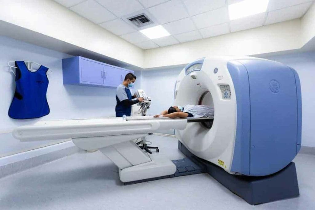
CT scans use X-rays and computers to make brain images. They rotate an X-ray source around the head. Then, a computer turns this data into images.
These images show us important details about the brain. They help us find issues like bleeding, fractures, and tumors. A normal brain CT scan shows clear brain structures and no unusual areas.
When Brain CT Scans Are Medically Indicated
Brain CT scans are needed in many situations, like when a quick diagnosis is required. They’re often used in emergencies to spot bleeding, check for head injuries, and find other urgent issues.
Medical guidelines say a brain CT scan is usually the first choice for patients with stroke symptoms, severe headaches, or head injuries.
Knowing when and how to use CT scans helps doctors make better choices. This leads to better care for patients.
Understanding CT Density Measurements and the Hounsfield Scale

CT density measurements are key to spotting and understanding body structures and issues. The Hounsfield scale helps measure tissue density. This is vital for reading CT scans right.
How Tissue Density Appears on CT Images
On CT images, tissue density shows up in shades of gray, white, or black. The Hounsfield scale gives each tissue a number based on its density. For example, bone is dense and looks white, while air is less dense and appears black.
Knowing how tissues look on CT scans is important for diagnosing diseases. A hyperdense spot might mean fresh blood, calcification, or other dense materials. This helps doctors spot hemorrhages, tumors, or other issues.
Differentiating Structures Based on Density Values
The Hounsfield scale lets doctors tell apart different structures and problems by their density. It helps in:
- Spotting hemorrhages and understanding them
- Telling apart different tumors or cysts
- Finding calcifications or bone pieces
- Checking organs and tissues for oddities
In a head CT scan, knowing the difference between gray and white matter is key. It helps diagnose conditions like ischemic stroke. The density measurements show how severe the injury or disease is.
In summary, knowing about CT density measurements and the Hounsfield scale is key to understanding CT scans. By understanding how tissues and structures show up on CT images, doctors can make better diagnoses and treatment plans.
What Color Is Blood on a CT Scan?
Understanding the color and density of blood on CT scans is key for accurate diagnosis and treatment. When we look at CT scans, it’s important to know how blood shows up. This is true, mainly when there’s a suspected hemorrhage.
Blood on a CT scan usually looks hyperdense. This means it shows up as bright white because it’s denser than the brain tissue around it. This is very clear in acute hemorrhages.
Why Fresh Blood Appears Hyperdense
Fresh blood looks hyperdense on CT scans because it’s denser than the brain tissue. Several things make this happen:
- The high hemoglobin content in blood
- The presence of clotting elements
- The relative density compared to brain parenchyma
These factors together make blood appear bright white on CT images because it has a higher attenuation coefficient than the surrounding tissues.
How Blood Density Changes During Clot Evolution
As a blood clot ages, its density on CT scans changes. At first, the clot is very dense. But as it matures, its density slowly goes down. This happens for a few reasons:
- Clot retraction and compaction
- Breakdown of hemoglobin
- Resorption of the clot
Knowing these changes is important for tracking the progress of hemorrhagic conditions. It also helps in seeing if treatments are working.
By understanding how blood looks on CT scans and how its density changes, doctors can make better diagnoses. They can also plan the right treatments.
Normal CT Scan of the Brain: Key Characteristics
A normal brain on a CT scan is a key reference for spotting problems. It’s important to know what a healthy brain looks like on CT images. This helps doctors find and diagnose issues accurately.
Normal Brain Anatomy on CT Images
A normal CT scan shows a symmetrical brain with clear grey and white matter. The grey matter is denser (brighter) than the white matter, which is less dense (darker). This difference is key for checking brain health.
Grey-White Matter Differentiation
The difference between grey and white matter is a key feature of a normal brain CT scan. Grey matter, with more neuronal cell bodies, is denser. White matter, with myelinated nerve fibers, is less dense. This distinction is important for diagnosing neurological conditions.
Ventricular System and CSF Spaces
The ventricular system, including the lateral, third, and fourth ventricles, should be symmetrical. The cerebrospinal fluid (CSF) spaces, like sulci and cisterns, should also be clear. Any asymmetry or abnormality in these areas can signal a problem.
| Structure | Normal Appearance on CT |
| Grey Matter | Dense (Brighter) |
| White Matter | Less Dense (Darker) |
| Ventricular System | Symmetrical, filled with CSF |
| CSF Spaces | Visible sulci and cisterns |
How to Read Head CT Scans: A Systematic Approach
Reading head CT scans needs a careful method to spot normal and abnormal changes. Doctors must be thorough in their checks to make sure they get the diagnosis right.
The ABC Method for CT Interpretation
The ABC method is a way to read head CT scans. It makes sure all important parts are checked. This method includes:
- Looking at anatomical structures for any oddities or imbalances.
- Examining the bone and soft tissue windows to see different tissue types.
- Checking for contrast enhancement if contrast was used during the scan.
Essential Anatomical Landmarks to Identify
Spotting key anatomical landmarks is key when reading head CT scans. Important structures to look at include:
- The ventricular system and its size, shape, and position.
- The grey-white matter differentiation to see if it’s normal or not.
- The basal cisterns and other CSF spaces for signs of effacement or hemorrhage.
Window Settings for Optimal Visualization
Changing the window settings on a head CT scan is important for clear views of different tissues. Common settings include:
- Brain window: Good for looking at brain tissue.
- Bone window: Best for checking bones and finding fractures or calcifications.
- Subdural window: Helps spot small subdural hemorrhages.
Learning how to read head CT scans well helps doctors make better diagnoses. This leads to better care for patients.
Identifying Hyperdense Findings on Brain CT Scans
Brain CT scans often show hyperdense findings. These are areas that look brighter or whiter than the rest of the brain. They can mean different things, like hemorrhage, calcifications, or contrast enhancement.
Blood Vessels and Calcifications vs. Hemorrhage
It’s hard to tell apart blood vessels, calcifications, and hemorrhage on brain CT scans. Blood vessels can look hyperdense because of contrast material or calcium in the walls. Calcifications, like in the choroid plexus or basal ganglia, also show up as hyperdense.
Hemorrhage looks hyperdense because of fresh blood. Its density changes as the clot ages. Acute hemorrhage is very dense, while older hemorrhage is less dense.
| Condition | Appearance on CT | Common Locations |
| Hemorrhage | Hyperdense (acute) | Various, depending on the type of hemorrhage |
| Calcifications | Hyperdense | Choroid plexus, basal ganglia, pineal gland |
| Blood Vessels with Contrast | Hyperdense | Throughout the brain, following vascular paths |
Contrast Enhancement vs. Natural Hyperdensity
Contrast enhancement happens when we give contrast material through an IV. It makes some structures or lesions stand out. It can look like natural hyperdensity from hemorrhage or calcification.
“The use of contrast material can significantly improve the detection and characterization of brain lesions on CT scans.” –
Radiology Expert
To tell contrast enhancement from natural hyperdensity, we look for specific patterns. Contrast enhancement often shows up around lesions or in blood vessels. Natural hyperdensity looks more uniform and isn’t related to contrast.
Being able to spot hyperdense findings on brain CT scans is key for diagnosis and treatment. Knowing what different hyperdense conditions look like helps us make accurate diagnoses. This leads to better care for our patients.
Abnormal Brain CT Scan: Common Pathological Findings
Abnormal brain CT scans can show serious conditions that need quick medical help. These scans are key for spotting and handling brain emergencies.
Intracranial Hemorrhage Patterns
Intracranial hemorrhage is a serious issue that shows up as bright white spots on a CT scan. The spot’s shape and where it is can tell us a lot about its cause and how bad it is. Acute hemorrhage looks bright, while older hemorrhages might look less bright or even darker.
Ischemic Changes and Infarcts
Ischemic changes happen when brain areas don’t get enough blood, often because a blood vessel is blocked. Early signs on a CT scan might include slight changes in brain appearance. Established infarcts show up as darker areas.
Mass Lesions and Space-Occupying Processes
Mass lesions, like tumors or abscesses, can push brain structures and raise pressure inside the skull. On a CT scan, these look different from normal brain areas. If a mass is pushing on brain structures, it’s a big deal.
Spotting these issues on a brain CT scan is vital for quick and right medical action. Doctors need to know how to spot different types of problems to care for patients best.
Types of Intracranial Hemorrhages on CT Images
CT scans are key in finding intracranial hemorrhages. These can be different types based on where they are and how they look. Knowing these types helps doctors diagnose and plan treatment better.
Epidural Hematomas: Biconvex Hyperdensity
Epidural hematomas show up as biconvex and hyperdense on CT scans. They happen between the dura mater and the skull, usually from trauma. Their biconvex shape makes them stand out from other hemorrhages.
Subdural Hematomas: Crescent-Shaped Collections
Subdural hematomas look like crescent-shaped collections on CT scans. They’re found between the dura mater and the arachnoid mater. They often come from trauma and can change in density.
Subarachnoid Hemorrhage: Blood in CSF Spaces
Subarachnoid hemorrhage shows up as blood in the cerebrospinal fluid (CSF) spaces around the brain. On CT, it looks like hyperdensity in the sulci and cisterns. It’s often a sign of aneurysm rupture or other blood vessel issues.
Intraparenchymal Hemorrhage: Blood Within Brain Tissue
Intraparenchymal hemorrhages happen inside the brain tissue. They can be caused by high blood pressure, vascular malformations, or tumors. On CT, they show up as hyperdense areas in the brain, with varying density and size.
In conclusion, knowing how to spot different types of intracranial hemorrhages on CT scans is very important. Each type has its own signs that help doctors diagnose and treat. By understanding these differences, we can help patients better.
Recognizing Mass Effect and Midline Shift on CT
Mass effect and midline shift on CT scans are important signs of serious health issues. They show increased intracranial pressure and brain herniation. These are life-threatening conditions that need quick medical help.
Signs of Increased Intracranial Pressure
Increased intracranial pressure (ICP) shows up on CT scans in several ways. A key sign is mass effect, where a lesion or hemorrhage presses on brain structures. This can cause brain swelling and herniation.
We look for signs such as:
- Compression of the ventricles or cisterns
- Displacement of brain structures
- Effacement of sulci
Measuring and Assessing Midline Shift
Midline shift is a key indicator of how severe the mass effect is. It’s measured by seeing how much brain structures, like the septum pellucidum, are off-center. A big midline shift means the patient’s condition is very serious and needs immediate help.
To measure midline shift, we follow these steps:
- Find the septum pellucidum on the CT scan.
- Draw a line from the front to the back of the skull, showing the true midline.
- Measure how far the septum pellucidum is from this line.
| Midline Shift Measurement | Clinical Implication |
| 0-5 mm | Mild displacement, monitor closely |
| 5-10 mm | Moderate displacement, consider intervention |
| >10 mm | Severe displacement, urgent intervention required |
Ventricular Compression and Effacement
Ventricular compression and effacement are also important signs on CT scans. When the ventricles are compressed, it can cause hydrocephalus. This makes the pressure inside the skull even worse.
Spotting these signs early is key to saving lives. By knowing how to spot mass effect, midline shift, and ventricular compression, doctors can act fast to help patients.
CT Scan Images of the Brain in Acute Stroke Assessment
When someone shows signs of acute stroke, CT scans are key. They help us quickly figure out the best treatment. We use these images to make fast, informed decisions.
Early Signs of Ischemic Stroke
Spotting ischemic stroke early is vital. On a CT scan, we look for hypodensity in the brain. This shows possible ischemic damage.
Some early signs include:
- Loss of gray-white matter differentiation
- Hypodensity in the affected vascular territory
- Swelling and effacement of sulci
Hemorrhagic vs. Ischemic Stroke Differentiation
Telling hemorrhagic from ischemic stroke is key. Treatment differs a lot. CT scans help us see if there’s blood.
Hemorrhagic strokes show up as hyperdense on CT scans. Ischemic strokes appear hypodense due to swelling and damage. Knowing the difference is essential for the right treatment.
Time-Sensitive Imaging Findings for Treatment Decisions
In acute stroke, time is critical. CT scans give us vital info for quick decisions. We look for signs that mean we need to act fast, like thrombolysis or mechanical thrombectomy.
Important findings include:
- Extent of ischemic changes
- Presence of hemorrhage
- Signs of mass effect or herniation
By quickly understanding these signs, we can start treatment fast. This might help improve the patient’s outcome.
Advanced CT Techniques for Enhanced Brain Visualization
We now have advanced CT technologies that give us detailed brain images. These help in diagnosing and treating brain disorders. These techniques are key in modern brain imaging, showing us the brain’s structure and any problems.
Vascular Assessment with CT Angiography
CT angiography is a powerful tool for seeing brain blood vessels. It uses a contrast agent to show detailed images of blood vessels. This helps find issues like aneurysms, stenosis, and malformations.
The benefits of CT angiography include:
- Rapid images, reducing patient wait time
- High-resolution views of small vessels
- Real-time assessment of vascular problems
CT Perfusion Studies in Stroke Evaluation
CT perfusion studies are key in acute ischemic stroke evaluation. They help find ischemia and see how much brain tissue might be saved.
| Parameter | Description | Clinical Significance |
| Cerebral Blood Flow (CBF) | Measures brain blood flow rate | Identifies ischemia areas |
| Cerebral Blood Volume (CBV) | Measures brain blood volume | Shows collateral circulation |
| Mean Transit Time (MTT) | Measures blood transit time through the brain | Indicates perfusion issues |
Dual-Energy CT Applications in Neuroimaging
Dual-energy CT technology captures images at two energy levels. This helps differentiate materials and tissues, improving lesion characterization and diagnostic accuracy.
Key applications of dual-energy CT in neuroimaging include:
- Improved detection of hemorrhage and calcifications
- Enhanced contrast enhancement visualization
- Potential contrast dose reduction
Using these advanced CT techniques, we can better diagnose and manage neurovascular conditions. This leads to better patient outcomes.
Conclusion: The Critical Role of CT in Rapid Diagnosis and Treatment
We’ve seen how CT scans are key in diagnosing brain issues like hemorrhages, strokes, and tumors. They offer quick and accurate images. These images help doctors make fast treatment plans.
At Liv Hospital, we aim for exceptional healthcare. We focus on international excellence, teamwork, and care that puts patients first. Our modern facilities and skills help us manage CT scans well. This improves patient results.
CT scans are vital for fast and clear images in neuroimaging. As medical tech gets better, CT scans will keep being essential. They support our goal of giving top healthcare solutions.
FAQ
What color is blood on a CT scan?
Fresh blood looks bright white on a CT scan. This is because it’s denser than brain tissue.
How do you differentiate between blood vessels, calcifications, and hemorrhage on a CT scan?
Blood vessels and calcifications might look white. But, their shape and location can tell them apart from hemorrhage. Hemorrhage is usually irregular and doesn’t follow blood vessel paths.
What are the key characteristics of a normal CT scan of the brain?
A normal scan shows clear brain anatomy. It should have distinct grey and white matter. The ventricles and CSF spaces should also look right.
How do you read head CT scans systematically?
Start with the ABC method. Look for key landmarks. Adjust settings for the best view. This ensures a complete check.
What are the common pathological findings on an abnormal brain CT scan?
You might see bleeding patterns, ischemic signs, tumors, or other growths. It’s important to spot and understand these.
How does blood density change over time on a CT scan?
Blood starts off dense, then gets less dense as it clots and resolves. This change is seen over time.
What is the role of CT scans in acute stroke assessment?
CT scans are key for spotting stroke early. They help tell if it’s bleeding or not. This info is vital for quick treatment.
How do advanced CT techniques enhance brain visualization?
New CT methods like angiography and perfusion studies give detailed views. They help doctors assess and manage brain issues better.
What are the signs of increased intracranial pressure on a CT scan?
Look for midline shift, ventricular compression, and changes in sulci and cisterns. These signs mean pressure is rising and need quick action.
How do you measure and assess midline shift on a CT scan?
Measure midline shift by checking how far structures are off center. This shows if there’s a big mass or pressure issue.
References
- Heit, J. J., & others. (2016). Imaging of intracranial hemorrhage. PMC (PubMed Central). https://pmc.ncbi.nlm.nih.gov/articles/PMC5307932/
- Zhou, H., et al. (2021). CT assessment of increased density of cerebral vessels: implications of hyperdense artery sign. Scientific Reports. https://www.nature.com/articles/s41598-021-85448-3


