Last Updated on November 26, 2025 by Bilal Hasdemir

Ultrasound technology has changed how we diagnose health issues. It’s a non-invasive way to see inside the body. Doctors use it to check on many health problems.
If you’re wondering what diseases can be detected by ultrasound, the list is broad. Ultrasound makes clear images of organs like the gallbladder, liver, and kidneys, helping detect conditions such as gallstones, liver disease, kidney stones, and cysts.
But can ultrasound find ulcers and other stomach problems? While it’s not the primary tool for ulcers, it can help identify inflammation, masses, or other issues that may point to underlying gastrointestinal concerns.
Understanding what diseases can be detected by ultrasound helps patients know what to expect and highlights the value of this safe and widely used imaging method.
Key Takeaways
- Ultrasound technology is a non-invasive diagnostic tool.
- It is used to visualize internal organs and structures.
- Ultrasound can detect various health issues, including gallbladder and liver conditions.
- The technology produces high-resolution images for accurate diagnoses.
- Its capabilities in detecting ulcers and gastrointestinal issues are being explored.
The Science and Technology Behind Ultrasound Imaging
Ultrasound imaging uses high-frequency sound waves to show internal body structures. This tool is non-invasive and has changed medical imaging. It lets doctors see organs and tissues without surgery or harmful radiation.
How Ultrasound Waves Create Medical Images
Ultrasound waves go into the body and bounce off structures. They then come back to the device as echoes. These echoes help create detailed images of the body’s inside.
Types of Ultrasound Equipment Used Today
There’s a wide range of ultrasound equipment out there. From small handheld devices to big machines with advanced features. Each one is made for different medical needs.
Evolution of Diagnostic Sonography
From Military Technology to Medical Breakthrough
Diagnostic sonography started in the military. But it has grown a lot for medical use. It has changed how doctors diagnose diseases.
Recent Technological Advancements
New tech in ultrasound technology has made images better. Now we have 3D and 4D imaging and contrast-enhanced ultrasound. These advancements have opened up more uses for ultrasound.
What Does an Ultrasound Show? Basic Principles
Understanding what an ultrasound shows starts with knowing how it works. Ultrasound imaging uses sound waves to create images inside the body. It sends high-frequency sound waves and catches the echoes to show what’s inside.
How Different Body Tissues Appear on Ultrasound
Ultrasound images show different tissues in various ways. Fluids like cysts or gallbladders look black because they don’t bounce sound waves back much. Bones and hard spots appear bright white because they bounce sound waves a lot. Soft tissues like the liver or spleen show up in shades of gray based on their density.
The Role of Sonographers in Image Acquisition
Sonographers are key in getting good ultrasound images. They use the equipment to get the best views of what’s inside. Their skill helps make sure the images are clear and useful for doctors.
How Radiologists Interpret Ultrasound Images
Radiologists study ultrasound images to find any problems. They look at the texture, size, and shape of organs and structures. They’re searching for anything that doesn’t look right, like lesions or inflammation.
Normal vs. Abnormal Findings
It’s important for radiologists to know what’s normal and what’s not. Normal findings match what’s expected for the area being looked at. Abnormal findings might include unexpected growths or changes in size or shape.
Documentation and Reporting Standards
Good documentation and reporting are key in ultrasound imaging. Reports should clearly describe what’s found, compare it to previous studies, and suggest what to do next.
| Tissue Type | Appearance on Ultrasound | Examples |
| Anechoic | Black | Fluid-filled structures (cysts, gallbladder) |
| Hyperechoic | Bright White | Bone, calcifications |
| Hypoechoic | Dark Gray | Some tumors, inflammation |
| Isoechoic | Same as surrounding tissue | Some lesions, normal variants |
Understanding Ulcers: Types and Characteristics
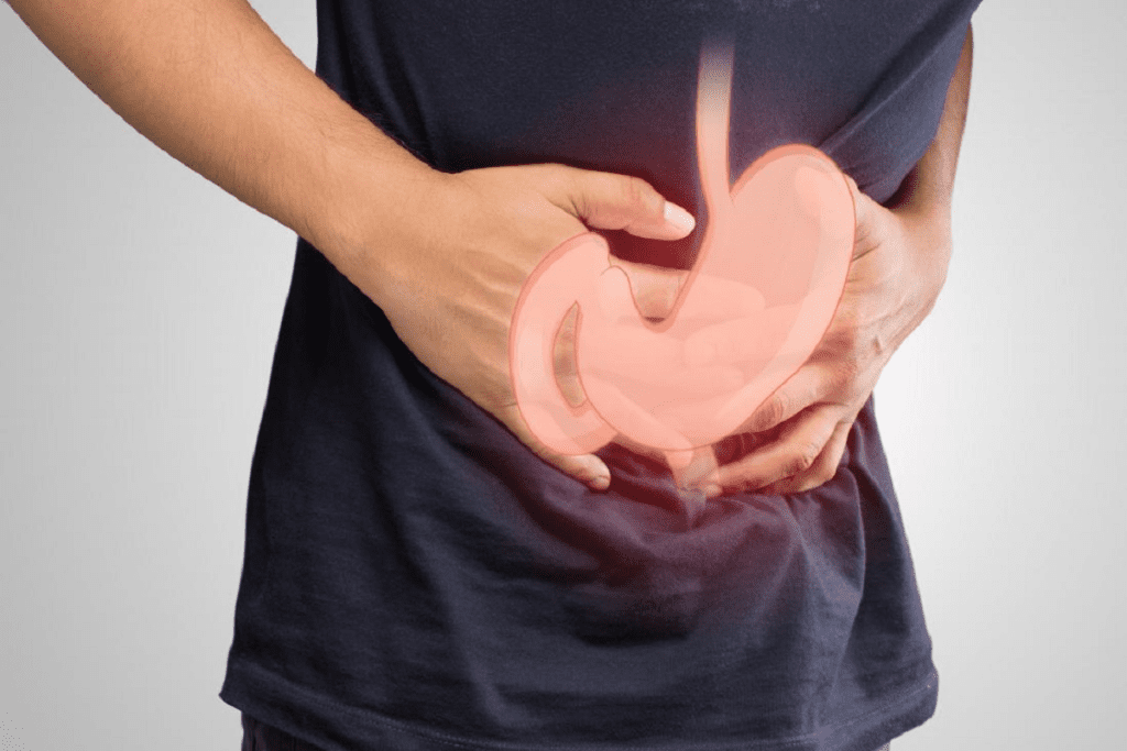
Ulcers are sores that form on the inside lining of the stomach and the upper small intestine. The main types are gastric, duodenal, peptic, and stress ulcers.
Gastric Ulcers: Location and Features
Gastric ulcers happen in the stomach lining. They are often caused by Helicobacter pylori infection or NSAIDs. Symptoms include stomach pain, nausea, and vomiting.
Duodenal Ulcers: Distinguishing Factors
Duodenal ulcers are in the duodenum, the first part of the small intestine. They are more common than gastric ulcers and linked to H. pylori infection. Symptoms include burning stomach pain that may feel better after eating.
| Type of Ulcer | Location | Common Causes |
| Gastric Ulcer | Stomach lining | H. pylori, NSAIDs |
| Duodenal Ulcer | Duodenum | H. pylori, NSAIDs |
| Peptic Ulcer | Stomach or duodenum | H. pylori, NSAIDs, stress |
| Stress Ulcer | Stomach | Severe illness, burns, trauma |
Peptic Ulcers: Causes and Presentation

Peptic ulcers are both gastric and duodenal ulcers. They are caused by H. pylori infection, NSAIDs, and stress. Symptoms include stomach pain.
Stress Ulcers: Risk Factors and Appearance
Stress ulcers happen in critically ill patients due to severe stress. They appear as multiple stomach mucosa erosions or ulcers.
Can an Ultrasound Detect Stomach Ulcers?
Using ultrasound to find stomach ulcers is complex. It depends on knowing what ultrasound can and can’t do. This imaging tech has improved a lot. It can now show many stomach problems, including ulcers.
Effectiveness of Ultrasound for Gastric Ulcer Detection
How well ultrasound finds gastric ulcers depends on a few things. These include the skill of the person doing the scan, the quality of the equipment, and the type of ulcer. High-resolution ultrasound might spot ulcers by looking at the stomach’s lining and finding any oddities.
Visualizing Ulcer-Related Complications
Ultrasound is great for spotting problems linked to stomach ulcers. This includes things like perforation and bleeding.
Perforation Detection
If an ulcer tears through, ultrasound can find fluid in the belly. This is a big warning sign. It means the ulcer has perforated, which is very serious and needs quick treatment.
Bleeding Assessment
Ultrasound isn’t the first choice for seeing bleeding from ulcers. But it can spot signs like blood in the stool or vomit. It does this by looking at the stomach and nearby areas.
Clinical Studies on Ultrasound Accuracy for Ulcers
Many studies have looked into how well ultrasound finds stomach ulcers. They say ultrasound is helpful but not always 100% accurate. More research is needed to know for sure how reliable it is for this job.
What Diseases Can Be Detected by Ultrasound
Ultrasound technology has changed how we diagnose diseases. It’s a non-invasive way to see many health issues. Doctors use it in different fields to check and track health problems.
Abdominal Conditions Visible on Ultrasound
Ultrasound is great for looking at the stomach area. It spots many problems in these organs.
Liver Disease and Gallstones
Ultrasound finds liver diseases like fatty liver and cirrhosis. It also sees gallstones, which cause pain.
Pancreatic Abnormalities
Ultrasound can find issues with the pancreas, like pancreatitis and tumors. It’s hard to see the pancreas, but skilled sonographers can get good images.
Cardiovascular Diseases Diagnosed with Ultrasound
Ultrasound is key for heart disease diagnosis. Echocardiography checks the heart’s function. It spots problems like valve issues and heart failure.
Reproductive System Disorders Identified by Sonography
Ultrasound is very helpful for women’s health. It finds issues like ovarian cysts, fibroids, and ectopic pregnancies.
Musculoskeletal Issues Evaluated Through Ultrasound
Musculoskeletal ultrasound looks at muscle, tendon, and ligament problems. It’s great for guiding treatments.
Ultrasound is a vital tool in many medical areas. It’s safe and can find many diseases. This has greatly helped patient care and results.
Abscess on Ultrasound: Detection and Characteristics
Ultrasound is a key tool in finding and understanding abscesses. It’s a non-invasive way to spot abscesses early. This is vital for quick treatment.
How Abscesses Appear in Ultrasound Images
Abscesses show up as dark spots with unclear edges on ultrasound. Their look changes based on their makeup and the tissue around them. Ultrasound lets doctors see abscesses live, helping them judge size, spot, and risks.
Differentiating Between Types of Infections
Ultrasound can tell infections apart by looking at their details. It can tell abscesses from other fluid or cysts. Knowing this helps doctors choose the right treatment.
Ultrasound-Guided Drainage Procedures
Ultrasound is great for guiding drainage. Ultrasound-guided drainage is a safe way to put in drainage tubes. It lowers the chance of problems.
Technique and Equipment
The method uses ultrasound to watch the abscess while putting in the tube. Special tools, like clean ultrasound probes and tubes, make the process safe and effective. For more on treatments,
Patient Outcomes and Recovery
Drainage with ultrasound helps patients heal faster and with fewer problems. Seeing the abscess live makes the procedure more likely to succeed.
| Procedure | Success Rate | Recovery Time |
| Ultrasound-Guided Drainage | 90% | 3-5 days |
| Surgical Drainage | 85% | 7-10 days |
What Can an Ultrasound of the Abdomen Show?
An abdominal ultrasound is a detailed test that checks many organs in the belly. It uses sound waves to create clear images inside the body. This helps doctors find and track many health issues without surgery.
Comprehensive Assessment of Liver and Gallbladder
This test is great for looking at the liver and gallbladder. It can spot liver problems like cirrhosis and fatty liver. It also checks for gallstones and inflammation in the gallbladder.
Pancreatic Visualization and Common Findings
Ultrasound can see the pancreas and find issues like pancreatitis and cysts. Even though the pancreas is hard to see, new ultrasound tech makes it easier.
Kidney and Bladder Evaluation
The kidneys and bladder are also checked by ultrasound. It can find kidney stones, cysts, and tumors. It also spots bladder problems like thickening and masses.
Intestinal Abnormalities Detectable by Ultrasound
Ultrasound can find problems in the intestines, like:
- Bowel wall thickening, which may indicate inflammation or infection
- Intussusception, a condition where one part of the intestine telescopes into another
- Obstruction, which can be suggested by the presence of dilated bowel loops
Bowel Wall Thickening
Bowel wall thickening is a big sign of issues like inflammatory bowel disease and infections.
Intussusception and Obstruction
Intussusception and obstruction are serious and need quick medical help. Ultrasound can spot these by showing signs like the “target sign” in intussusception.
| Organ/Structure | Common Abnormalities Detected by Ultrasound |
| Liver | Cirrhosis, fatty liver, liver masses |
| Gallbladder | Gallstones, cholecystitis |
| Pancreas | Pancreatitis, pancreatic cysts, tumors |
| Kidneys | Kidney stones, cysts, tumors, signs of kidney disease |
| Bladder | Thickening of the bladder wall, masses |
| Intestine | Bowel wall thickening, intussusception, obstruction |
An abdominal ultrasound is a key tool in medicine. It checks many parts of the belly and helps doctors find and treat problems.
Limitations of Ultrasound in Ulcer Detection
Ultrasound is a helpful tool for finding ulcers, but it’s not perfect. Its ability to accurately diagnose ulcers can be affected by several factors.
Factors Affecting Ultrasound Image Quality
Several things can make ultrasound images less clear. This makes it harder to spot ulcers. These include:
- Operator skill level
- Equipment quality
- Patient-related factors
When Ultrasound Might Miss Gastric Ulcers

Ultrasound might not catch all gastric ulcers. This is true for small ulcers or those in hard-to-see spots.
Patient-Related Challenges in Abdominal Scanning
Patients can also affect how well ultrasound works. Two main issues are:
Body Habitus Considerations
People with a higher BMI can be harder to scan. This is because more tissue gets in the way, weakening the ultrasound signal.
Bowel Gas Interference
Bowel gas can also be a problem. It can cause distortions in the ultrasound image. This makes it tough to see ulcers clearly.
In summary, ultrasound is useful but has its limits. Knowing these can help doctors make better choices when diagnosing ulcers.
Comparing Ultrasound to Other Diagnostic Tools for Ulcers
Ultrasound is one of several tools used to find ulcers. It has its own strengths and weaknesses compared to others. The right tool depends on the situation, the patient, and the ulcer’s location and complications.
Ultrasound vs. Endoscopy: Pros and Cons
Endoscopy is top for finding gastric ulcers because it lets doctors see the ulcer and take tissue samples. But, it’s more invasive than ultrasound and not for everyone. Ultrasound is non-invasive and can check for ulcer complications like penetration or abscesses. Here’s what sets ultrasound and endoscopy apart:
- Ultrasound is non-invasive, while endoscopy is more invasive.
- Endoscopy lets doctors take biopsies, but ultrasound doesn’t.
- Ultrasound can spot complications and look at the tissue around the ulcer.
Ultrasound vs. CT Scans for Gastrointestinal Issues
CT scans give a full view of the belly and can find many issues, like ulcers, holes, and abscesses. But, they use radiation and might not be as easy to get as ultrasound. Ultrasound is safer for people who should avoid radiation, like pregnant women. Here’s how ultrasound and CT scans compare:
- CT scans show more of the belly.
- Ultrasound is safer for some patients.
- CT scans can find more kinds of problems.
Ultrasound vs. MRI for Abdominal Diagnosis
MRI gives detailed pictures of soft tissues and can help diagnose some belly issues. But, it’s pricier and harder to get than ultrasound. Ultrasound is often the first choice for belly problems because it’s easy to get and safe.
Clinical Decision-Making: Choosing the Right Diagnostic Method
Choosing between ultrasound and other tools depends on the situation, the patient, and what the doctor thinks might be wrong. Doctors might use more than one method to make sure they have the right diagnosis. They need to think about each tool’s good and bad points to decide the best one.
Preparing for an Abdominal Ultrasound Examination
To get the best results from an abdominal ultrasound, patients need to prepare well. Following the right steps can make the images clearer. This helps doctors make more accurate diagnoses.
Pre-Examination Instructions for Optimal Results
Before your ultrasound, there are a few things to do. These steps help ensure the best possible images.
- Fasting: You might need to fast before the test. This empties your stomach and intestines.
- Dietary Restrictions: Stay away from foods and drinks that can make you uncomfortable.
- Medication Management: Tell your doctor about any medicines you’re taking.
What to Expect During the Ultrasound Procedure
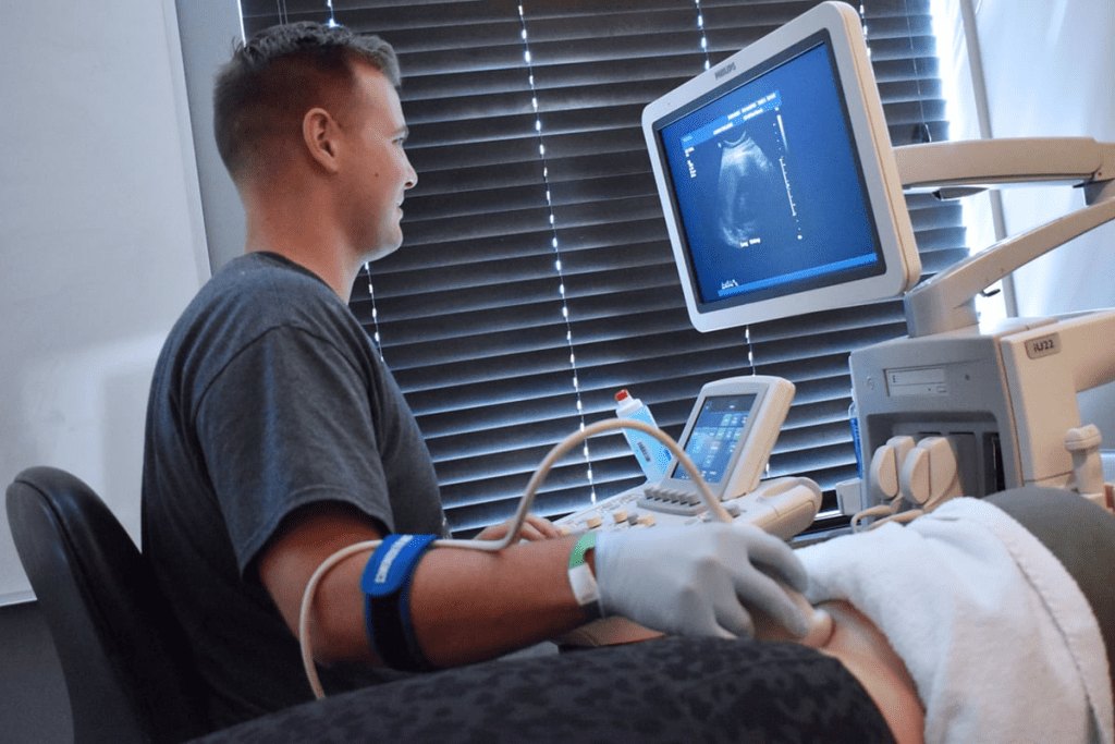
A sonographer will apply gel to your abdomen during the test. They use a transducer to take pictures of your organs. The whole process is usually painless and doesn’t hurt.
Post-Examination Information and Next Steps
After the test, you can usually go back to your normal activities. But, your doctor might tell you to do something different.
Results Timeline
How long it takes to get your ultrasound results can vary. Sometimes, you might get quick results. But, detailed reports might take a few hours or days.
Follow-Up Recommendations
Based on what the ultrasound shows, your doctor might suggest more tests or treatments. It’s important to follow these steps. They help manage any health issues you might have.
Advanced Ultrasound Technologies for Gastrointestinal Diagnosis
Advanced ultrasound technologies are changing how we diagnose gastrointestinal issues. They give us more detailed and accurate results. This has greatly improved how ultrasound helps in diagnosing and managing these conditions.
High-Resolution Ultrasound Capabilities
High-resolution ultrasound is a key tool in diagnosing the gut. It offers enhanced image quality. This lets us see mucosal layers and wall structures better. It’s great for checking how severe a disease is and if treatments are working.
Contrast-Enhanced Ultrasound for Improved Detection
Contrast-enhanced ultrasound (CEUS) uses agents to show blood flow and tissue better. It’s really good at spotting gastrointestinal lesions and finding complications from diseases.
3D and 4D Ultrasound Applications in GI Medicine
3D and 4D ultrasound have made diagnosing the gut even better. They give us detailed three-dimensional images and real-time four-dimensional assessments. This helps us understand complex gut anatomy and diseases better.
Volume Rendering Techniques
Volume rendering in 3D ultrasound creates detailed images from many two-dimensional slices. It’s super useful for seeing how big a disease is and planning surgeries.
Real-Time Assessment Benefits
4D ultrasound lets us see things in real-time. This is great for checking how the gut moves and works. It’s super helpful in diagnosing and checking if treatments are working.
Safety and Benefits of Ultrasound Compared to Other Imaging Methods
Ultrasound is safe and easy to use. It’s a key tool in medicine because of its many benefits.
Non-Invasive Nature: Patient Comfort and Accessibility
Ultrasound is non-invasive. This makes it comfortable for patients. It’s also good for many people, even those who can’t have other tests.
Radiation-Free Imaging: Safety Profile
Ultrasound doesn’t use radiation. This makes it safer than X-rays or CT scans. It’s great for pregnant women, kids, and those needing many tests.
Cost-Effectiveness and Availability Advantages
Ultrasound is cost-effective and easy to find. It’s cheaper than MRI or CT scans. This makes it more affordable for many people.
- Non-invasive and painless
- No radiation exposure
- Cost-effective
- Wide availability
- Real-time imaging capabilities
Ultrasound is a valuable tool in medicine. It’s safe and shows what’s inside the body well.
When to Seek Medical Attention for Suspected Ulcers
If you think you might have an ulcer, it’s important to know when to see a doctor. Ulcers can hurt a lot and can get worse if not treated.
Common Symptoms That Suggest Ulcer Presence
Ulcer symptoms can be different for everyone. They often include stomach pain, feeling sick, and throwing up. Sharp, burning pain in the upper abdomen is a common symptom. Spotting these signs early can help you get medical help fast.
Warning Signs That Require Immediate Medical Care
Some symptoms need urgent care. These include severe abdominal pain, throwing up blood, or having black tarry stools. These could mean you have a bleeding ulcer, which is a serious emergency.
Effectively Communicating Symptoms to Healthcare Providers
To get a correct diagnosis, it’s key to tell your doctor about your symptoms clearly. Keeping a symptom journal can be very helpful.
Symptom Tracking Methods
- Note the timing and severity of symptoms
- Record any factors that relieve or exacerbate symptoms
- List any medications or foods that seem to trigger symptoms
Questions to Ask Your Doctor
Having a list of questions can make your doctor’s visit more productive. Ask about what might be causing your symptoms, tests you’ll need, and treatment options.
| Symptom | Description | Action |
| Abdominal Pain | Sharp, burning pain in the upper abdomen | Seek medical evaluation |
| Vomiting Blood | Presence of blood in vomit | Immediate medical attention |
| Black Tarry Stools | Dark, tarry stools indicating gastrointestinal bleeding | Immediate medical attention |
The Future of Ultrasound in Gastrointestinal Diagnostics
Ultrasound technology is changing how we diagnose stomach and intestinal problems. New advancements are making it better at finding issues.
Emerging Ultrasound Technologies for Improved Detection
New tools are being added to ultrasound machines. These include high-resolution imaging and contrast-enhanced ultrasound. They help spot stomach and intestinal problems better.
Artificial Intelligence Integration in Image Interpretation
Artificial intelligence (AI) is changing how we read ultrasound images. AI finds patterns and problems that humans might miss. This makes diagnoses more accurate.
Point-of-Care and Portable Ultrasound Advancements
New portable and point-of-care ultrasound devices are making it easier to get checked. Doctors can now do ultrasound tests right at the bedside or in other places.
Telemedicine Applications
Ultrasound is now part of telemedicine. This means doctors can review ultrasound images from far away. It helps get diagnoses and treatments faster.
Home-Based Monitoring Possibilities
Technology is also making it possible to monitor health at home with ultrasound. This could help people with long-term stomach and intestinal issues get checked more often and easily.
| Technology | Application | Benefit |
| High-Resolution Ultrasound | Improved image quality | Better diagnostic accuracy |
| Artificial Intelligence | Enhanced image interpretation | Increased detection of abnormalities |
| Portable Ultrasound | Point-of-care diagnostics | Greater accessibility to care |
Conclusion
Ultrasound technology has changed how we diagnose health issues. It’s a non-invasive way to find problems like ulcers and other stomach issues. We’ve looked at how ultrasound works, its uses, and its limits in finding ulcers.
Ultrasound helps doctors see inside the body and spot problems. It’s not the main tool for finding ulcers. But, it can help find related issues and other stomach problems.
As technology gets better, ultrasound will play an even bigger role in medicine. It will help doctors make better decisions and improve patient care. Knowing what ultrasound can and can’t do is key for doctors.
In short, ultrasound is a key tool in finding and treating stomach problems, including ulcers. It’s safe, easy to use, and affordable. It’s a big part of modern medicine.
FAQ
What does an ultrasound show?
An ultrasound uses sound waves to create images of the body’s internal structures. It shows organs, tissues, and blood vessels.
Can an ultrasound detect ulcers?
Ultrasound may detect stomach ulcers by examining the stomach lining, but accuracy can vary.
What diseases can be detected by ultrasound?
Ultrasound can find many diseases. This includes issues in the abdomen, heart, reproductive system, and muscles.
Can stomach ulcers be detected by ultrasound?
Yes, they can. Ultrasound can see stomach ulcers and any problems they might cause.
What can an ultrasound of the abdomen show?
An abdominal ultrasound checks the liver, gallbladder, pancreas, kidneys, bladder, and intestines. It looks for any problems.
Can an ultrasound detect abscesses?
Yes, it can. Ultrasound can spot abscesses by looking for fluid collections and guiding drainage.
What are the limitations of ultrasound in ulcer detection?
Ultrasound’s limits include image quality issues and challenges with certain areas. Patient factors also play a role.
How does ultrasound compare to other diagnostic tools for ulcers?
Ultrasound has its own strengths and weaknesses compared to tools like endoscopy and CT scans. The best choice depends on the situation.
What are the benefits of ultrasound compared to other imaging methods?
Ultrasound is non-invasive and doesn’t use radiation. It’s also cost-effective, making it a valuable tool.
What can be expected during an abdominal ultrasound examination?
During an ultrasound, a sonographer will apply gel and use a transducer. The images are then analyzed by a radiologist.
How can patients prepare for an abdominal ultrasound?
Patients should follow pre-examination instructions. This might include fasting or avoiding certain foods for better results.
What are the latest advancements in ultrasound technology?
New ultrasound tech includes high-resolution and 3D/4D imaging. These advancements improve how well it can diagnose.
What is the future of ultrasound in gastrointestinal diagnostics?
Ultrasound’s future looks bright. It will include new technologies, artificial intelligence, and portable ultrasound for easier use.
References
- World Health Organization. (2024). Peptic ulcer disease. https://www.who.int/news-room/fact-sheets/detail/peptic-ulcer-disease
- Zhong, C., Sun, M., Li, B., Chen, S., Cao, F., & Li, R. (2020). The diagnostic value of transabdominal ultrasonography for perforated peptic ulcer: A systematic review and meta-analysis. Gastroenterology Research and Practice, 2020, 1“7. https://doi.org/10.1155/2020/2153965
- National Institute of Diabetes and Digestive and Kidney Diseases. (2024). Peptic Ulcer Disease (Stomach Ulcers). National Institutes of Health. https://www.niddk.nih.gov/health-information/digestive-diseases/peptic-ulcers-stomach-ulcers


