Knowing how cancer cells look under a microscope is key for accurate diagnosis and treatment. At Liv Hospital, we use the latest medical techniques to study cancerous tissues. We look for differences between cancer cells and normal cells.
Cancer is a group of diseases where cells grow out of control and invade other tissues. Doctors can spot and manage cancer better by looking at cancer cell appearance and tumor characteristics. For more details on cancer diagnosis, check our page on whether a doctor can tell if a tumor is cancerous by looking at it.

The change from normal cells to cancer cells is complex. It involves many genetic and environmental factors. We will dive into this process to see how cancer starts at the cell level.
Carcinogenesis is when normal cells turn into cancer cells. This happens because of DNA damage. This damage can come from genetic mutations, harmful substances, and viruses.
When DNA damage isn’t fixed, it messes up how cells work. This leads to cells growing and dividing without control.
Carcinogenesis has three main steps: initiation, promotion, and progression. Genetic mutations are key, as they can turn on bad genes and turn off good ones. Environmental factors like harmful substances and radiation also play a part by causing DNA damage.
Normal cells become cancer cells through genetic and epigenetic changes. These changes mess up how cells work, like controlling growth and fixing DNA. Cancer cells grow too much, lose their shape, and become more invasive.
The change to cancer cells is slow, with many genetic and epigenetic changes happening. Cancer cells look different, with big and odd-shaped nuclei, and they don’t arrange like normal cells. Knowing these changes helps us find better ways to treat cancer.

Cancer cells look different from normal cells. This difference is key for spotting cancer early. It helps doctors know how to treat it.
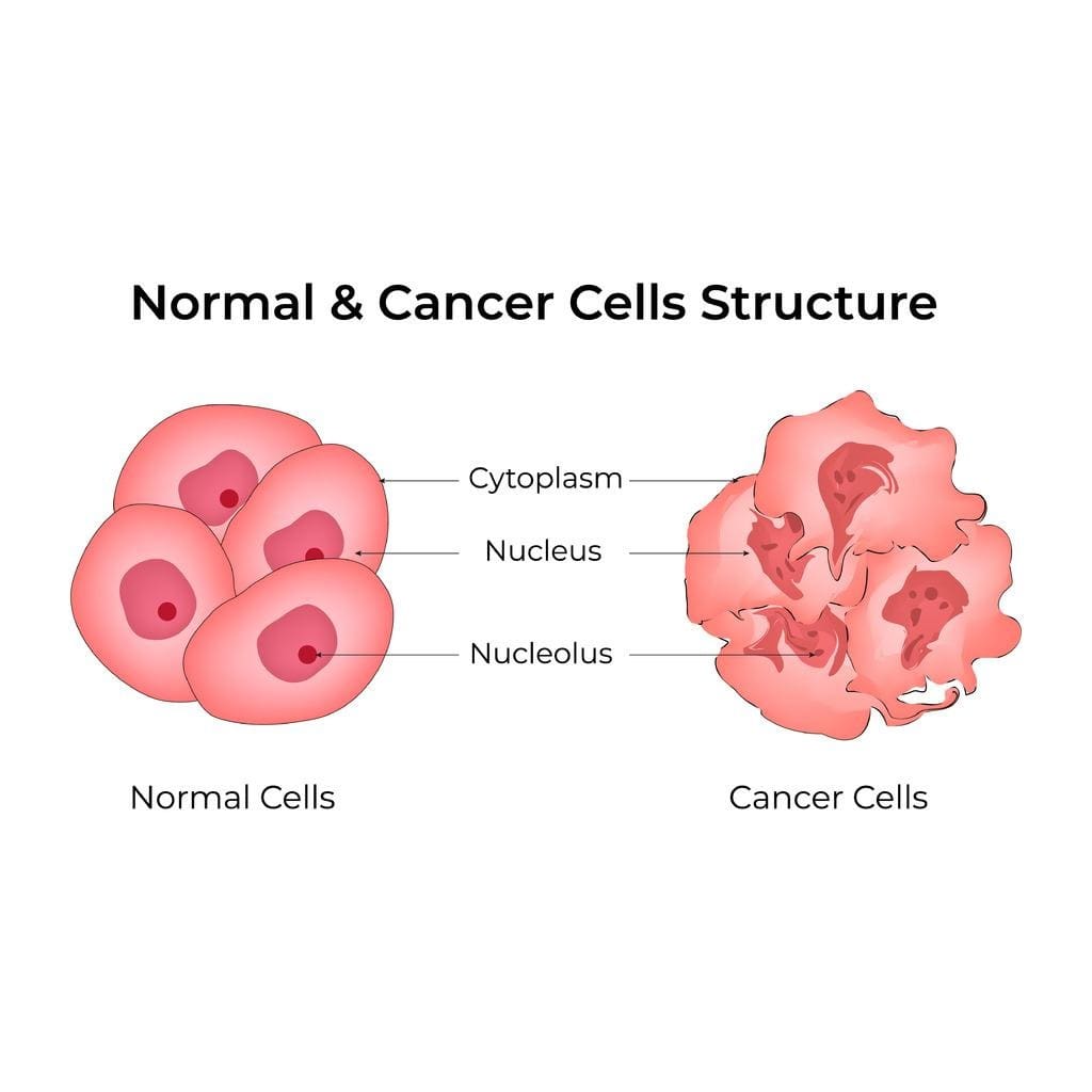
Cancer cells are irregular in shape and size. They often have big or many nuclei. Their structure is not like that of healthy cells.
Key characteristics of cancer cells include:
Normal cells are uniform in shape and size. They have one nucleus and a neat structure. They divide at a steady rate.
Comparing cancer cells to normal cells shows big differences. These differences are in shape, size, and how they work.
| Cell Characteristics | Normal Cells | Cancer Cells |
| Cell Shape and Size | Uniform | Pleomorphic |
| Nuclear Characteristics | Single nucleus | Enlarged or multiple nuclei |
| Cellular Structure | Organized | Disorganized |
| Nuclear-to-Cytoplasmic Ratio | Normal | Increased |
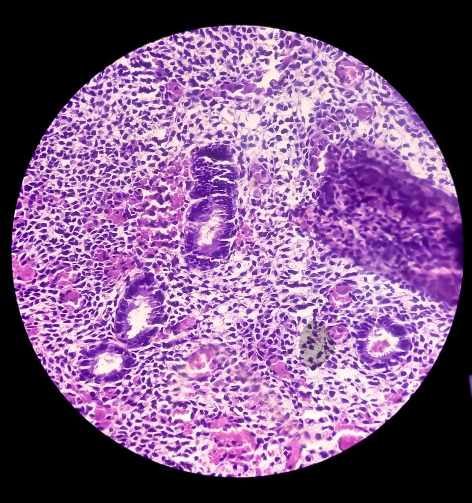
Looking at cells under a microscope can show if they are cancerous. We see mitotic figures, which mean cells are dividing. We also look for cells invading nearby tissues.
“The microscopic examination of cells is a critical step in cancer diagnosis, allowing pathologists to identify the characteristic features of malignancy and determine the aggressiveness of the tumor.”
Knowing these visual differences helps us diagnose and grade cancer better. This information guides treatment and improves patient care.
Tumors can look different in the body, affecting how they look and act. At Liv Hospital, we focus on these differences to help diagnose and treat them well.
Tumors are mainly two types: solid and liquid. Solid tumors are firm tissues found in places like the breast, prostate, or lungs. Liquid tumors, or hematological malignancies, affect the blood, bone marrow, or lymphatic system.
| Tumor Type | Characteristics | Common Locations |
| Solid Tumors | Firm masses of tissue | Breast, prostate, lungs |
| Liquid Tumors | Involve blood, bone marrow, or lymphatic system | Blood, bone marrow, lymph nodes |
Tumors can look different in color, texture, and size. They might be white, gray, or red. Their texture can be soft or hard. Size can range from small to large, affecting the area around them.
Tumors can affect tissues in different ways. Malignant tumors can invade and disrupt tissues. Benign tumors can compress tissues, causing symptoms. Knowing how tumors interact with tissues is key to treatment.
“The way a tumor interacts with its surrounding tissue environment is a critical factor in determining its growth pattern and response to treatment.”
Liv Hospital Oncology Team
At Liv Hospital, we offer detailed care and treatment plans. Our team works with patients to find the best care for their needs.
Looking at cancer cells under a microscope is key to understanding them. We search for special features that show they are different from normal cells.
Many microscopy methods help find cancer. These include:
Getting samples ready is very important for accurate viewing. We use several ways to prepare cancer cell samples, such as:
Staining is a key step to show cancer cells’ features. Common stains include:
By using these microscopy, preparation, and staining methods, we can fully understand cancer cells. This knowledge is essential for accurate diagnosis and treatment planning.
Looking at nuclear abnormalities in cancer cells can tell us a lot about the cancer. We study these changes to understand their role in diagnosing and tracking cancer.
Cancer cells often have nuclei that are different in size and shape. This is a key difference from normal cells. These changes can mean that something is wrong with how the cells work.
Nuclear pleomorphism is when nuclei vary a lot in size and shape. This is a common sign of cancer. It helps doctors know if a tumor is cancerous.
How chromatin is spread out in the nucleus is also important. In cancer cells, this can look different. Sometimes, nuclei look darker because they have more DNA. Other times, chromatin clumps together in certain spots.
These changes show that cancer cells are unstable genetically. This is a big part of what makes cancer cells different.
Nucleoli, where ribosomes are made, can also change in cancer cells. If nucleoli get bigger or more numerous, it’s called nucleolar prominence. This is seen in many cancers.
These changes in nucleoli mean that cancer cells need to make more proteins. This helps them grow and multiply quickly.
| Nuclear Abnormality | Description | Significance in Cancer |
| Size and Shape Variations | Nuclei vary in size and shape, showing pleomorphism | Indicates malignancy and disruption of normal cell processes |
| Chromatin Distribution Patterns | Altered chromatin distribution, hyperchromasia, chromatin clumping | Reflects genetic instability characteristic of cancer cells |
| Nucleoli Changes | Increased size or number of nucleoli | Supports rapid growth and proliferation of cancer cells |
More aggressive tumors often have more severe nuclear abnormalities. Knowing about these changes is key for accurate diagnosis and treatment planning.
Cancer cells have different cytoplasm than normal cells. These changes help us understand how cancer cells work and affect the body. We’ll look at the main changes in cancer cells, like changes in cellular parts, how they metabolize, and their cell membranes.
Cancer cells have big changes in their cytoplasm. One key change is in the mitochondria, which are like the cell’s batteries. This can affect how the cell uses energy.
Another change is the presence of abnormal protein aggregates. These can show certain cancers and help doctors diagnose them.
| Cellular Component | Normal Cell Characteristics | Cancer Cell Characteristics |
| Mitochondria | Normal number and structure | Altered number and structure |
| Protein Aggregates | Absent or normal distribution | Presence of abnormal aggregates |
Cancer cells have different metabolic patterns than normal cells. These differences can be seen under a microscope with special stains. For example, cancer cells might take up more glucose, which can be stained to show.
The cell membrane of cancer cells also changes a lot. Changes in the cell membrane can affect how cells stick together and talk to each other. These changes help cancer cells spread.
Knowing about these changes is key for diagnosing and studying cancer. By studying these changes, scientists and doctors can learn more about cancer cells. This helps them find better treatments.
Understanding how cancer changes cell and tissue structure is vital. Cancer tissues often have big changes in their structure. These changes help doctors diagnose and understand cancer’s growth.
In healthy tissues, cells follow a specific pattern for proper function. But, cancer tissues don’t follow this pattern. The loss of normal tissue architecture is a hallmark of cancer, leading to impaired cellular function and contributing to the development of tumors. Research has shown that the disruption of normal tissue patterns can be an early indicator of carcinogenesis (NCBI).
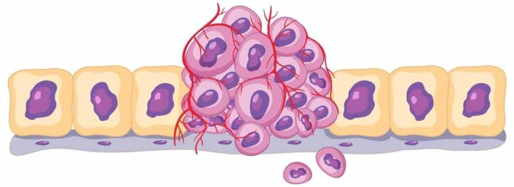
Cancer cells can invade surrounding tissues and lose their boundaries. This invasive property is a key factor in the malignancy of tumors. As cancer cells invade, they disrupt normal tissue architecture, leading to the loss of boundary between different tissue types. This loss of boundary is a critical aspect of cancer progression, allowing tumors to grow and spread more easily.
Another significant change in cancerous tissues is the formation of new blood vessels, a process known as angiogenesis. Angiogenesis provides tumors with the necessary nutrients and oxygen for growth, facilitating the progression of cancer. Also, vascular changes in tumors can lead to the development of abnormal blood vessels that are leaky and inefficient, further contributing to tumor growth and metastasis.
At Liv Hospital, we understand the importance of these changes for effective treatments. Our mission is to provide innovative healthcare solutions that align with the latest research in cancer diagnosis and visualization.
Looking at cancer cells and tissues is key in grading and staging cancer. This helps decide the best treatment. We use visual markers to see how severe and spread out the disease is.
Grading cancer means checking how tumors look to see how aggressive they are. Low-grade tumors look more like normal cells, showing a less aggressive cancer. On the other hand, high-grade tumors look very different, showing a more aggressive disease that needs quick and strong treatment.
Knowing if a tumor is low-grade or high-grade is very important. It helps us plan treatment and predict how well the patient will do. We look at how the tumor cells and tissues look to decide its grade and plan the best treatment.
The differentiation status of a tumor shows how much it looks like normal cells and tissues. Tumors that look a lot like normal tissues are well-differentiated. Those that don’t look much like normal tissues are poorly differentiated and are more aggressive.
Knowing the differentiation status is key for predicting how well a patient will do. Generally, well-differentiated tumors have a better outlook than poorly differentiated ones. This helps us make treatment plans that fit each patient’s needs, improving their chances of recovery.
Tumor heterogeneity means a tumor has different cell types, each with its own genetic and physical traits. Under the microscope, this shows up as different cell shapes, staining patterns, and other visual signs.
Understanding tumor heterogeneity is important because it can affect how well a tumor responds to treatment and how it grows. Tumors with a lot of heterogeneity might be harder to treat, so we need to be more careful and flexible with treatment plans.
By studying these visual markers, we can learn more about how cancer behaves. This helps us create effective treatment plans. Using visual checks along with other tests helps us give patients care that’s just right for them.
Cancer cells look different in each type of cancer, which affects how we treat them. We’ll look at the common visual patterns in various cancers. This includes carcinomas, sarcomas, hematological malignancies, and brain and nervous system tumors.
Carcinomas, the most common cancer, start from epithelial cells. They often have irregular cell arrangements and different sizes. Under a microscope, carcinomas show unique nuclear abnormalities, like big nuclei and uneven chromatin.
Sarcomas, from connective tissue, have different looks than carcinomas. They have a disorganized structure and may have odd blood vessels. Sarcomas can be classified by where they start, like in bones or fat tissue.
Hematological malignancies affect the blood and lymphatic system. They show unique visual characteristics. These cancers, like leukemias and lymphomas, have abnormal cell shapes and disrupted blood cell production. Looking at them under a microscope is key for diagnosis.
Brain and nervous system tumors have their own visual patterns. They can start from different cells, like glial cells or neurons. Their visual features, like cell arrangement and nucleus, help in diagnosis and grading.
| Cancer Type | Common Visual Patterns | Diagnostic Features |
| Carcinomas | Irregular cell arrangements, variable cell size | Nuclear abnormalities, irregular chromatin |
| Sarcomas | Disorganized tissue structure, abnormal blood vessels | Tissue-specific features (e.g., bone or fat) |
| Hematological Malignancies | Abnormal cell morphology, disrupted blood cell production | Abnormal white blood cell count, specific markers |
| Brain and Nervous System Tumors | Variable cellular arrangement, nuclear features | Cell type-specific characteristics (e.g., glial or neuronal) |
Knowing these visual patterns is key for accurate diagnosis and treatment planning. By recognizing the unique features of each cancer type, doctors can offer more tailored care.
We are committed to top-notch healthcare at Liv Hospital. We know that getting the right diagnosis is key to good treatment. That’s why we use the latest medical tech to give our patients the best care.
Liv Hospital uses the latest imaging tech, like high-resolution MRI and CT scans. These cutting-edge imaging technologies help our doctors spot problems more clearly.
We also use integrated pathology and molecular testing. This mix of old and new tech gives us a full picture of what’s going on inside a patient. It helps us create treatment plans that really work.
| Diagnostic Approach | Description | Benefits |
| Cutting-Edge Imaging | High-resolution MRI and CT scans | Precise diagnosis of internal conditions |
| Integrated Pathology | Combining traditional pathology with molecular testing | Comprehensive understanding of patient conditions |
| Personalized Diagnostics | Tailored diagnostic protocols based on individual patient needs | Effective treatment plans with minimal delay |
At Liv Hospital, we know every patient is different. So, we create personalized diagnostic protocols just for them. This way, they get the right tests for their needs.
Visual analysis is key in planning treatments. Our doctors look at images and data to find the best treatment. It might be surgery, medicine, or both.
We stay up-to-date with the latest medical methods and care standards. At Liv Hospital, we aim to give top-notch healthcare and support to patients from around the world.
Knowing how cancer cells look different from normal cells is key for spotting cancer early and treating it well. Cancer tumors grow in ways that normal cells don’t, and this is seen under a microscope. The size, shape, and feel of tumors help doctors figure out what to do next.
At Liv Hospital, we use the latest in imaging and lab tests to help diagnose cancer. We aim to give top-notch care and support to patients from around the world. By understanding cancer cells, we can tailor treatments that work best for each patient.
Looking closely at cancer cells is very important. It helps us understand tumors better, make treatment plans, and improve how well patients do. As we learn more about cancer, we’re committed to finding new ways to help our patients.
Cancer cells are often irregular in shape and size. They may have more than one nucleus or unusual patterns of chromatin.
Cancer cells have different shapes and sizes. They disrupt the normal structure of tissues. Their cytoplasm and cell membranes also show changes.
Signs of cancer include unusual nuclei and cytoplasm. These changes help identify cancer cells. They also show how cells are organized differently.
Tumors can look different in color, texture, and size. Solid tumors are visible as masses. Liquid tumors, like leukemia, might not be seen.
Tumors can grow into surrounding tissues. This can cause them to lose their boundaries.
Tumor heterogeneity means there are many different cell types in a tumor. This affects how cancer is graded and treated. It can mean the tumor is more aggressive or resistant.
Each type of cancer, like carcinomas or sarcomas, looks different under a microscope. Knowing these patterns helps doctors make accurate diagnoses.
Liv Hospital uses the latest imaging and testing. They combine pathology and molecular testing. This helps them give accurate diagnoses and plan treatments.
Looking at cancer cells and tissues helps doctors plan treatments. It tells them about the tumor’s behavior and how it might respond to treatment.
Knowing the differences is key for accurate diagnosis and treatment. It helps doctors identify cancer, determine the type, and plan targeted treatments.
References:
• National Center for Biotechnology Information. (2016). Detection and classification of cancer from microscopic biopsy images. https://www.ncbi.nlm.nih.gov/pmc/articles/PMC4782618/
• Wikipedia. (n.d.). Cancer cell. https://en.wikipedia.org/wiki/Cancer_cell
• National Cancer Institute. (n.d.). Tumor grade. https://www.cancer.gov/about-cancer/diagnosis-staging/diagnosis/tumor-grade
• UR Medicine. (n.d.). Grading and staging of cancer. https://www.urmc.rochester.edu/encyclopedia/content.aspx?contenttypeid=85&contentid=p00554
• Cancer Research UK. (n.d.). Types of cancer. https://www.cancerresearchuk.org/about-cancer/what-is-cancer/how-cancer-starts/types-of-cancer
Subscribe to our e-newsletter to stay informed about the latest innovations in the world of health and exclusive offers!