
Meta Description: Discover what bone marrow looks like under a microscope, its structure, and why it plays a vital role in blood cell production and overall health.
We look into the detailed structure of bone marrow and its key role in our bodies. Bone marrow is a complex tissue. It’s vital for making blood cells and boosting our immune system.
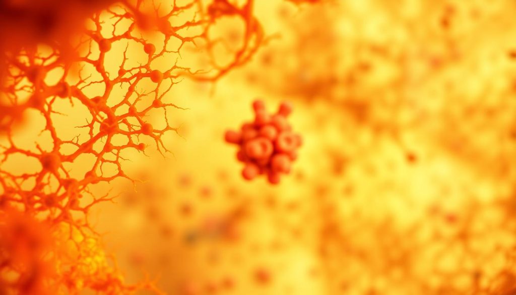
When we look at bone marrow under a microscope, we see a complex mix of cells. This includes hematopoietic cells, adipocytes, and stromal components. The makeup of bone marrow can change with age or illness. It’s divided into red and yellow marrow.
At Liv Hospital, we use top-notch diagnostic tools to study bone marrow. Knowing how bone marrow looks under a microscope is key to giving the best healthcare.

The bone marrow is a dynamic tissue that is essential for the continuous production of blood cells throughout our lives. It is responsible for generating the cells that make up our blood, including red blood cells, white blood cells, and platelets. These cells are vital for our immune function.
Bone marrow is where all blood cells are made. This process, called hematopoiesis, starts with hematopoietic stem cells. These cells turn into different types, each with its own role. Red blood cells carry oxygen, white blood cells fight off infections, and platelets help blood to clot.
“The bone marrow is the body’s blood factory, producing the cells necessary for oxygen delivery, immune defense, and hemostasis,” highlighting its critical role in maintaining our overall health.
The bone marrow is like a factory that never stops making blood cells. It makes sure the body has the right number and types of cells. This ability to adjust to the body’s needs, like during an infection, shows how dynamic it is.
Bone marrow is found in the cavities of bones, like in the pelvis and vertebrae. It has a network of blood vessels and different cell types. This setup helps blood cells to be made and released into the body efficiently.
Looking at bone marrow under a microscope is like going on a journey through a complex world. We see how bone marrow works and its role in health and sickness.
The bone marrow is made of a complex mix that helps blood cells grow. This mix has a network of fibers and cells that are key for making blood cells.
Looking closely, we see islands of hematopoietic cells in a jelly-like substance. These islands have cells at all stages, from new to mature. The jelly supports these cells as they grow and mature.
When we look even closer, we see important cells in the marrow. Megakaryocytes, the big cells that make platelets, stand out. We also see fat cells, or adipocytes, which help the marrow work right. Spotting these cells helps us understand the marrow and find problems.
By studying bone marrow under a microscope, we learn a lot about its structure and role. This knowledge is key for diagnosing and treating blood disorders. It shows how important bone marrow exams are in medicine.
Bone marrow is divided into red and yellow types. Red marrow makes blood cells, while yellow stores fat. Knowing the differences is key to understanding bone marrow’s role in health.
Red bone marrow is full of blood vessels. It’s actively involved in hematopoiesis, or making blood cells. It’s mainly in the pelvis, vertebrae, sternum, and the top parts of the femur and humerus.
“The presence of red marrow in certain bones is critical for maintaining the body’s blood cell count,” as noted in medical literature. Red marrow also helps the immune system by making lymphocytes and other immune cells.
Yellow bone marrow is mostly fat cells and has fewer blood vessels. It’s in the long bones and is more common in adults. It acts as a reserve for energy storage and can turn back to red marrow when needed.
As we age, bone marrow changes. In young ones, it’s mostly red, showing its blood-making role. With age, more of it turns yellow, like in long bones. This change is normal but can be affected by health and environment.
Understanding these changes helps doctors interpret bone marrow tests. It helps them spot problems related to marrow function.
Under the microscope, bone marrow shows a mix of different cells. Each cell type helps with blood cell production and the immune system. This mix is key for keeping blood cells healthy and the body well.
Hematopoietic stem cells (HSCs) are at the heart of bone marrow’s blood-making work. They can grow and change into many blood cell types. HSCs turn into progenitor cells, which focus on specific blood cell types. These cells are essential for making blood cells all our lives.
Megakaryocytes are big, many-nucleated cells that make platelets. Platelets are key for stopping bleeding. Under the microscope, megakaryocytes are easy to spot because of their size and complex nuclei. As they grow, they release platelets into our blood, helping to stop bleeding.
The bone marrow has two main blood cell types: erythroid and myeloid. Erythroid cells turn into red blood cells, which carry oxygen. Myeloid cells become white blood cells, like granulocytes and monocytes, which fight off infections. Keeping these cell types in balance is important for making the right amount of blood cells.
Besides blood cells, bone marrow has fat cells and stromal components. Fat cells store energy and affect the blood-making area. Stromal cells, like fibroblasts and endothelial cells, help hold everything together and support blood cell growth. All these cells work together to keep bone marrow healthy and functioning well.
The cellularity of bone marrow gives us important health insights. It changes with age and in response to diseases. Doctors look at bone marrow cellularity to see the balance between blood-making cells and fat cells.
Bone marrow cellularity changes a lot with age. Kids have a lot of blood-making cells, about 70% to 80%. As we get older, this number goes down, to about 30% by old age. This change is a natural part of aging.
| Age Group | Average Cellularity |
|---|---|
| Children | 70-80% |
| Adults | 50-60% |
| Elders | 30-40% |
Changes in cellularity can mean different things. Hypercellular marrow means too many cells, like in leukemia. Hypocellular marrow means too few cells, which can be a sign of bone marrow failure.
Changes in bone marrow cellularity are important for health. They can show early signs of disease. Knowing why cellularity changes helps doctors diagnose and treat bone marrow problems.
“Assessment of bone marrow cellularity is a fundamental aspect of diagnosing hematological disorders. It provides critical information about the functional status of the marrow and helps in identifying various pathological conditions.”
We know how important bone marrow cellularity is in medicine. It’s a key part of checking the bone marrow. By understanding normal and abnormal changes, doctors can better help patients with bone marrow issues.
The microscopic study of bone marrow uses advanced methods for diagnosis. These methods help us understand the bone marrow’s complex cells and spot diseases.
Bone marrow aspiration takes a sample with a needle. Then, it’s checked under a microscope for cell details and any oddities. The process looks at cell types, finds unusual cells, and checks marrow density.
A bone marrow biopsy takes a small bone and marrow piece for study. It’s needed for a detailed marrow look. It’s useful when aspiration fails or for marrow fibrosis checks.
Staining methods make bone marrow cells clearer under a microscope. Wright-Giemsa and hematoxylin and eosin (H&E) stains are common. They help spot different cells and show cell details.
Modern imaging, like digital microscopy, has changed bone marrow study. It lets us save and study images, helping in teaching and research. It also finds small issues missed by old methods.
Using these techniques, doctors can deeply understand bone marrow problems. This leads to better diagnoses and treatment plans.
Changes in bone marrow morphology can show signs of many diseases. The bone marrow is a dynamic organ that can change due to diseases. Knowing these changes helps doctors diagnose and treat bone marrow diseases.
Leukemia and lymphoma change the bone marrow’s look. In leukemia, the marrow gets too full of bad cells, stopping normal blood making. For example, in AML, the marrow has lots of myeloblasts.
Looking closely, you can see Auer rods in myeloblasts, which means AML.
Lymphoma in the bone marrow can look different, from a few cells to the marrow being full of cancer cells. Finding lymphoma cells in the marrow is key for knowing how serious the disease is.
Bone marrow failure syndromes, like aplastic anemia, mean the marrow can’t make enough blood cells. The marrow looks too empty, with fewer cells and more fat. Doctors check the marrow biopsy to see this.
Infectious diseases can also change the bone marrow. For example, tuberculosis can cause inflammation, and HIV can lead to changes or infections. Special tests can find certain germs.
Some cancers, like breast, prostate, and lung, can spread to the bone marrow. Finding these cancer cells in the marrow is important for knowing how serious the disease is. Looking closely, you can see cancer cells that don’t belong. Tests can tell where these cells come from.
Bone marrow microscopy is very important in medical practice. It helps us find and treat many blood-related diseases.
We use bone marrow tests in many cases. This includes finding blood cancers, checking for low blood counts, and staging cancers. For example, if someone feels very tired, loses weight without trying, or gets sick a lot, we might do a test.
Learning about leukemia symptoms helps us know when to test.
The results of bone marrow tests match what the patient is feeling. For example, seeing abnormal cells means the patient might have leukemia or lymphoma. We look at these results with the patient’s symptoms and other tests to make a diagnosis.
Some changes in the bone marrow tell us about diseases. For example, more blast cells might mean leukemia, and fibrosis could point to other blood disorders. We check the cells, how many there are, and other details to find these problems.
Even though bone marrow tests are helpful, they’re not perfect. The quality of the sample, technical problems, and how we interpret the results can affect the accuracy. We keep learning and improving to get the best results for our patients.
| Challenge | Description | Mitigation Strategy |
|---|---|---|
| Sample Quality Issues | Inadequate or contaminated samples | Strict adherence to sampling protocols |
| Technical Issues | Problems during staining or processing | Regular maintenance of equipment and training of staff |
| Interpretation Variability | Subjective interpretation of microscopic findings | Continuous education and peer review |
By facing these challenges and getting better, we can make bone marrow microscopy even more useful for our patients.
Today, bone marrow analysis uses old methods and new ones. This mix has made diagnosing diseases better in hematology.
Looking at bone marrow under a microscope is key. But now, we also use new molecular methods. These methods give us deeper insights into the cells.
For example, PCR and FISH help find genetic problems. These problems are important for diagnosing diseases like leukemia or lymphoma.
“The integration of molecular techniques with traditional microscopy has revolutionized the field of bone marrow analysis, enabling more accurate diagnoses and targeted therapies.”
Flow cytometry is great for studying cells in a fluid. It helps identify and count different cell types in bone marrow.
Immunohistochemistry uses antibodies to find proteins in tissues. It’s key for finding cancer cells by their markers.
| Technique | Application | Benefits |
|---|---|---|
| Flow Cytometry | Analyzing cell populations | Quantifies cell types based on surface markers |
| Immunohistochemistry | Detecting specific proteins | Identifies cancer cells and diagnoses malignancies |
Genetic and cytogenetic studies are vital. They help find the causes of bone marrow problems. Techniques like karyotyping and microarray analysis spot chromosomal issues.
These studies help diagnose and predict outcomes. They also guide treatment plans.
New research uses next-generation sequencing (NGS) to find mutations. This helps tailor treatments to each patient’s needs.
As research grows, so will our methods for studying bone marrow. This will help us better diagnose and treat diseases.
Our team at Liv Hospital is all about giving accurate and quick bone marrow tests. We use a team effort to care for our patients. We know how key a correct diagnosis is for treating bone marrow issues. Our goal is to offer top-notch healthcare services.
At Liv Hospital, we have a detailed test plan that brings together experts from different fields. This team effort makes sure every patient gets a complete check-up. We use the newest bone marrow test and analysis methods.
Our lab is packed with the latest tech for precise bone marrow tests and analysis. We keep our methods fresh with the latest in the field. This means our patients get the best in diagnostic care.
Key features of our laboratory include:
At Liv Hospital, we put patients first in our bone marrow diagnostics. We’re all about being open and honest in our tests. We make sure patients are in the loop and help them make choices.
We’re always pushing for better in diagnosing and treating bone marrow issues. We join global research and follow the latest guidelines. This way, we aim to give our patients the best results.
By using the latest tech, team work, and focusing on patients, Liv Hospital leads in bone marrow diagnostics.
Bone marrow microscopy is key in diagnosing many blood disorders. It helps doctors understand diseases like leukemia and lymphoma. By looking at bone marrow under a microscope, they get important clues.
We use bone marrow analysis to decide on treatments and track how diseases progress. This detailed look helps doctors spot changes that show what’s wrong. It’s a big help in diagnosing conditions.
New methods in microscopy and staining have made diagnosing better. Mixing microscopy with newer tools like flow cytometry and genetic analysis will keep improving care. This means more accurate diagnoses and better treatment for patients.
Bone marrow microscopy is essential in blood disease diagnosis. It combines old-school microscopy with new tech. This way, we can give top-notch care to those with complex blood conditions.
Bone marrow is a special tissue in our bones. It makes blood cells and helps our immune system. Looking at it under a microscope can tell us about our health.
Under a microscope, bone marrow shows a mix of cells and tissue. You see islands of blood-making cells and fat cells in a jelly-like background.
Red bone marrow makes blood cells. Yellow bone marrow stores fat. The mix of red and yellow marrow changes with age and can be affected by diseases.
The number of cells in bone marrow can tell us about our health. It shows normal changes with age and can point to diseases.
Bone marrow aspiration takes a sample of bone marrow fluid for tests. It helps diagnose and track blood disorders, cancers, and other bone marrow issues.
New methods mix microscopy with molecular tests, flow cytometry, and immunohistochemistry. This makes diagnoses more accurate and helps understand bone marrow disorders better.
Doctors do bone marrow tests to diagnose or monitor conditions like leukemia, lymphoma, and bone marrow failure. They also check for cancer spread.
Liv Hospital uses a team approach and the latest lab tech. They focus on patient care and ethics to offer detailed bone marrow tests and management.
Bone marrow histology gives insights into its microscopic structure. It helps diagnose and manage blood disorders.
Changes in bone marrow structure can show diseases like leukemia or lymphoma. They are key for correct diagnosis and treatment plans.
Basic Medical Key: Bone Marrow Pathology
Kenhub: Histology of Bone Marrow
SAGE Journals (International Journal of Hematology and Oncology): Article on Bone Marrow Cytomorphology
Bone marrow is a special tissue in our bones. It makes blood cells and helps our immune system. Looking at it under a microscope can tell us about our health.
Under a microscope, bone marrow shows a mix of cells and tissue. You see islands of blood-making cells and fat cells in a jelly-like background.
Red bone marrow makes blood cells. Yellow bone marrow stores fat. The mix of red and yellow marrow changes with age and can be affected by diseases.
The number of cells in bone marrow can tell us about our health. It shows normal changes with age and can point to diseases.
Bone marrow aspiration takes a sample of bone marrow fluid for tests. It helps diagnose and track blood disorders, cancers, and other bone marrow issues.
New methods mix microscopy with molecular tests, flow cytometry, and immunohistochemistry. This makes diagnoses more accurate and helps understand bone marrow disorders better.
Doctors do bone marrow tests to diagnose or monitor conditions like leukemia, lymphoma, and bone marrow failure. They also check for cancer spread.
Liv Hospital uses a team approach and the latest lab tech. They focus on patient care and ethics to offer detailed bone marrow tests and management.
Bone marrow histology gives insights into its microscopic structure. It helps diagnose and manage blood disorders.
Changes in bone marrow structure can show diseases like leukemia or lymphoma. They are key for correct diagnosis and treatment plans.