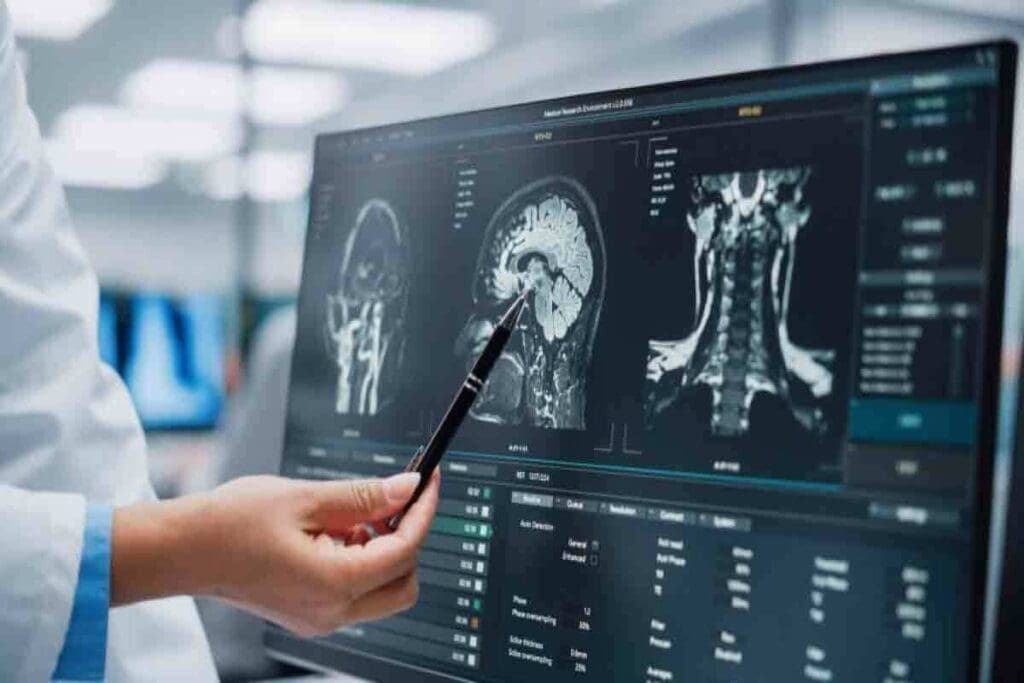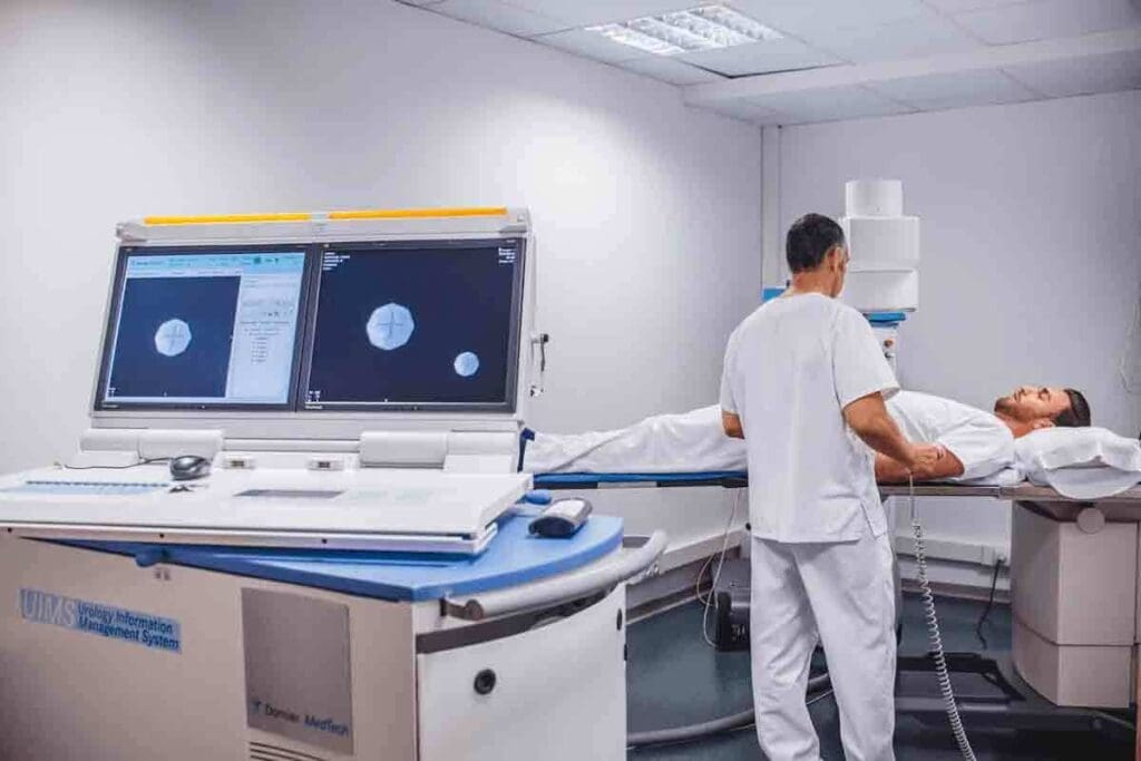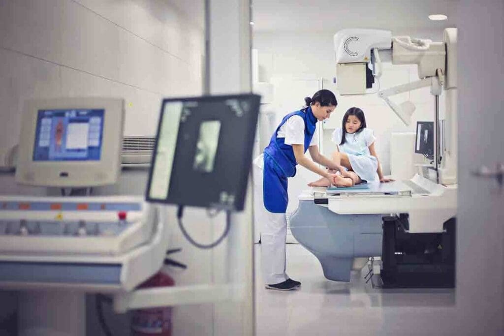
At Liv Hospital, we often get asked, “what is a renogram?” A renogram is an advanced nuclear medicine test used to check how well your kidneys are functioning. It’s a safe and non-invasive procedure that provides valuable insights into kidney health.
We use advanced tests like the renogram and MAG3 scan to assess kidney function. During a renal scan, a small amount of radioactive material is injected into your vein to help visualize how well your kidneys filter and drain urine.
This test is especially important for people with kidney diseases or those who have undergone a kidney transplant. By using specialized medicines and imaging techniques, we can accurately monitor kidney performance and provide the best possible care.

A renogram is a test that checks how well our kidneys work. It uses a tiny bit of radioactive material, called a tracer, injected into our blood. A special camera then takes pictures as the tracer moves through our kidneys.
These images help doctors see how our kidneys are doing. They can spot any problems early on.
A renogram shows how our kidneys function over time. It tracks the tracer’s journey through our kidneys. This gives doctors important information about how well our kidneys filter waste and fluids.
This test is safe and gives doctors the information they need to diagnose kidney issues. It’s great because it shows both how our kidneys work and their structure.
Renography has changed a lot over the years. New tech and tracers have made it better. It started with the first radioactive tracers in nuclear medicine.
Now, we have better tracers and imaging. Renography is a key tool in kidney disease management. It’s vital for doctors to make the right choices for their patients.

Nuclear kidney imaging uses radiolabeled pharmaceuticals to find kidney problems. These medicines have a special tag that shows up in the body.
This method, like the MAG3 scan, works by tracking how the kidneys handle certain medicines. It shows how well the kidneys work and if there are any blockages.
Radiolabeled medicines are key in checking the kidneys. They help doctors see how well the kidneys are working by detecting special rays. Technetium-99m MAG3 is a common one used for this.
Using these medicines has changed how we check the kidneys. It lets doctors do it without surgery and very accurately.
Radioactive tracers are special substances that give off rays. In kidney scans, these tracers are attached to medicines. The rays are then caught by a camera, making pictures of the kidneys.
The success of kidney scans depends on the tracers and the camera technology. They must work well together.
Diagnostic tools in nephrology have grown with new renogram procedures. These tests help doctors check how well the kidneys work and find many kidney problems.
A standard renogram is a key tool for checking kidney function. It uses a radioactive tracer injected into the blood. This shows how the kidneys work and can spot any issues.
A diuretic renogram, or Lasix renal scan, checks for urinary tract blockages. It uses a diuretic, like Lasix (furosemide), to make more urine. Doctors look at how the kidneys react to the diuretic to find blockages.
“The Lasix renogram is key for finding urinary tract blockages. It’s a safe way to check kidney function,” says a top nephrology expert.
A captopril renogram is used mainly to find renovascular hypertension. This is when kidney arteries get narrow or blocked. Captopril, an ACE inhibitor, is given to check kidney blood flow.
Each renogram procedure gives unique insights into kidney function. They can be adjusted for each patient’s needs. Knowing about these procedures helps doctors choose the best test for their patients.
The MAG3 scan is a top choice for checking how well the kidneys work. It uses advanced imaging to give detailed views of kidney function. This is key when a precise check is needed.
Technetium-99m MAG3 is a special medicine used in kidney scans. It’s better at getting out of the blood than other tracers. This helps doctors see the kidneys more clearly.
This clear view is great for spotting kidney problems. It’s very helpful when doctors think there might be an issue with the kidneys.
Using Technetium-99m MAG3 for kidney scans has many benefits. Some of these are:
To show why Technetium-99m MAG3 is so good, let’s compare it with other medicines used in kidney scans:
| Radiopharmaceutical | Extraction Efficiency | Primary Use |
| Technetium-99m MAG3 | High | Kidney function assessment |
| Technetium-99m DTPA | Moderate | Glomerular filtration rate measurement |
| Technetium-99m DMSA | High | Static renal imaging |
The table shows Technetium-99m MAG3 is the top for kidney scans. Its high extraction rate makes it the best choice for many doctors.
In the world of renal imaging, terms like renogram and renalgram can be confusing. They are often used in the same way to talk about nuclear medicine kidney scans. But knowing what they mean is key to clear talk between doctors and patients.
The words renogram and renalgram mean the same thing. They both talk about a test that checks how well the kidneys work using nuclear medicine. The “reno-” part means kidneys, and “-gram” means a picture or record. So, both words mean a picture or record of the kidneys.
This test uses a tiny bit of radioactive stuff that the kidneys take in. Then, a special camera picks up the radiation, making pictures of how the kidneys are doing.
You might also hear about the MAG3 scan and the diuretic renogram. A MAG3 scan uses Technetium-99m MAG3 to check kidney function. A diuretic renogram uses a diuretic to see how the kidneys work under stress.
Knowing these terms helps make the diagnostic process clearer. It also helps patients and doctors talk better. Remember, while renogram and renalgram are the same, a specific scan like MAG3 can give more detailed info about kidney function.
The renogram procedure is a simple test to check your kidney health. It helps find any problems early. We’re here to help you know what to expect.
To make the test go well, you need to prepare. Drink lots of water to help get clear images of your kidneys. Wear comfy clothes without metal parts to avoid any issues.
Preparation Checklist:
A small amount of a radioactive tracer is injected into your arm. This tracer goes to your kidneys. A gamma camera takes pictures as it moves.
The steps are:
| Procedure Step | Description | Duration |
| 1. Tracer Injection | A radioactive tracer is injected into a vein | Instantaneous |
| 2. Initial Imaging | Gamma camera captures initial kidney function images | 20-30 minutes |
| 3. Diuretic Administration (if applicable) | A diuretic is given to stimulate kidney function | Varies |
| 4. Continued Imaging | Further images are taken to assess kidney function over time | 30 minutes to 1 hour |
After the test, you can go back to your usual activities. The tracer leaves your body in a few hours. Drinking lots of water helps get it out.
Feeling a slight pinch when the tracer is injected is normal. But the test itself is usually painless. Your doctor will talk about the results with you. They might suggest more tests or treatment.
Most people don’t have any side effects after the test. But if you’re worried or notice anything strange, call your doctor.
Renogram machines use gamma camera technology to turn radiotracer photons into images. This tech is key for checking kidney health and spotting problems. We’ll look at how gamma cameras work and how images are analyzed in renogram tests.
Gamma cameras are vital in nuclear medicine, like in renograms. They catch the gamma rays from radiopharmaceuticals in the kidneys. This info helps make detailed images of kidney function and shape.
Key parts of gamma camera systems include:
The National Center for Biotechnology Information says gamma scintillation cameras are key. They turn radiotracer photons into images for diagnosing kidney issues.
After the gamma camera gets the data, special software works on the images. It fixes issues like scatter and quality problems. Then, the images are studied to check kidney function and more.
The analysis makes time-activity curves. These curves show how the kidneys take in and get rid of the radiotracer. They’re key for spotting blockages, checking kidney function, and seeing if treatments work.
| Parameter | Description | Clinical Significance |
| Differential Renal Function | The percentage of total renal function each kidney has | Helps see how each kidney is doing |
| Tracer Clearance Rate | How fast does the radiotracer leave the kidneys | Tells us about kidney efficiency |
| Time-Activity Curves | Graphs of radiotracer uptake and excretion over time | Crucial for finding blockages and other kidney issues |
Experts say gamma camera tech in renography has changed nuclear medicine. It lets for exact diagnoses and treatment plans for kidney problems. This shows how vital advanced imaging is in healthcare today.
Understanding renogram results is key to knowing how well our kidneys work. We’ll explain how to read these results. This includes looking at time-activity curves, spotting normal versus abnormal findings, and figuring out how each kidney functions.
Time-activity curves show how a radioactive tracer moves through our kidneys. These curves help us see how well our kidneys work and how well they drain. The shape and details of these curves tell us a lot about our kidney health.
We study these curves to see how fast the tracer is taken in, peaks, and then goes out. If the curve doesn’t follow this pattern, it might mean there’s a blockage or delay in drainage. For example, a slow increase in activity without a peak could suggest a blockage.
Telling normal from abnormal renogram results is very important for making the right diagnosis. Normal results show a quick uptake, a peak, and then a drop as the tracer is flushed out. Any change from this pattern could mean something’s not right.
Abnormal results might show a slow uptake, the tracer staying too long, or if one kidney works differently from the other. These signs can point to problems like blockages, infections, or less-than-ideal kidney function.
Figuring out how each kidney works is a big part of reading renogram results. This helps us see how well each kidney is doing. It’s really helpful when one kidney is sick or damaged.
By comparing how each kidney functions, doctors can tell if there’s a big difference. This info is key for deciding on treatments, like surgery or medicine.
We use the renogram data to find out how much each kidney does, usually as a percentage. This number helps us track changes in kidney function and see if treatments are working.
Nuclear kidney scans have many uses, from finding blockages to checking how well kidneys work. They are key in kidney care, helping doctors understand and treat kidney issues.
These scans are great at spotting kidney blockages. They use special tracers to see how urine flows and find any problems. This helps doctors diagnose issues like ureteropelvic junction obstruction (UPJO).
Key benefits of nuclear kidney scans in detecting obstructions include:
Scans also help check how well the kidneys are working. They look at how tracers are taken up and removed. This helps doctors manage kidney disease and see if treatments are working.
| Kidney Function Parameter | Normal Value | Abnormal Value |
| Differential Kidney Function | 45-55% | <40% or >60% |
| Time to Peak Activity | 3-5 minutes | >5 minutes |
Scans are also used to find specific kidney problems. They can spot pyelonephritis, renovascular hypertension, and other diseases. This info helps doctors create better treatment plans.
“Nuclear medicine techniques, including renal scans, provide unique functional information that complements anatomical imaging modalities.” – A Nuclear Medicine Specialist
Using nuclear kidney scans helps doctors better manage kidney diseases. This leads to better patient care and outcomes.
Renograms are very useful in special groups like kids and the elderly. They help us understand how their kidneys work and look. This is important because their bodies are different.
When we do pediatric nuclear kidney imaging, we have to think about a few things. Kids’ kidneys are growing and are smaller. So, we have to be careful with the medicine we use.
We also need to think about how much radiation they might get. This is because kids are more sensitive to it.
Renograms help us find problems in kids’ kidneys. This is important for their health and growth. We make sure the test is safe and works well for them.
In elderly patients, we use renograms to check their kidney function. This is important because age can affect their kidneys. We also look for problems like diabetes or high blood pressure.
When we look at the results, we have to think about their whole health. This helps us make better treatment plans for them.
Renal scans are safe, but they involve some risks. We’ll look at radiation, side effects, and precautions. This will help you know what to expect during a renal scan.
Renal scans use small amounts of radioactive tracers. This is to see how well your kidneys work. Even though there’s some radiation, the benefits are often greater than the risks.
We use Technetium-99m MAG3, which has a short half-life. This means less radiation for you. But it’s important to talk to your doctor about your personal risks.
| Radiopharmaceutical | Half-life | Effective Dose (mSv) |
| Technetium-99m MAG3 | 6 hours | 2.3 |
| Other tracers | Varies | Varies |
A Lasix renal scan uses furosemide (Lasix) to check kidney function. It’s usually safe, but some people might feel side effects.
Side effects can include more urine, dizziness, and headaches. Serious side effects are rare. But it’s good to know about them and talk to your doctor.
Renal scans are useful, but there are some things to watch out for. If you’re pregnant, it’s not safe because of radiation risks to the baby.
Also, if you’re breastfeeding, talk to your doctor first. They’ll help you decide if it’s safe.
Key Considerations:
Knowing about these safety points and precautions helps. This way, you can have a renal scan safely. It will give you important information about your health.
Renograms are key in checking how well the kidneys work and spotting problems in the urinary tract. We’ve learned how important they are in finding and treating kidney diseases. They give us a detailed look at kidney function and any blockages.
As we wrap up our talk on renograms, it’s clear they’re essential in patient care. Knowing how they work, their uses, and safety helps both patients and doctors. This knowledge is vital for making smart choices about kidney health.
To sum up, renograms give a full picture of kidney function. This helps doctors diagnose and treat kidney issues well. As medical tech keeps getting better, renograms will keep being a big help in kidney health checks.
A renogram is a test that checks how well your kidneys work. It also finds blockages in the urinary tract. It’s a non-invasive test.
Renogram and renalgram mean the same thing. They are tests that use nuclear medicine to check kidney function.
A MAG3 scan is a type of renogram. It uses technetium-99m MAG3 to see how the kidneys work and find blockages.
A renogram tracks a radioactive tracer in the kidneys. It shows how well the kidneys work and if there are blockages.
A diuretic renogram checks for urinary tract blockages. It sees how kidneys react to Lasix, a diuretic.
Side effects of a Lasix scan include more urine, dizziness, and headaches. These are usually mild and short-lived.
Yes, renograms are generally safe. But talk to your doctor about any worries about radiation.
To prepare for a renogram, arrive with a full bladder. Avoid certain meds and follow your doctor’s diet advice.
During a renogram, a tracer is injected into a vein. A gamma camera takes pictures as the tracer moves through the kidneys.
Results are analyzed through time-activity curves. They show kidney function and help find any problems.
Yes, renograms work for kids and the elderly, too. But special care is needed for their unique needs.
Nuclear kidney scans, like renograms, find blockages and check kidney function. They help diagnose and guide treatment for kidney issues.
Subscribe to our e-newsletter to stay informed about the latest innovations in the world of health and exclusive offers!