Heart disease is a major cause of death globally. Accurate diagnosis is key for effective treatment. Cardiac imaging is essential in this process. It helps doctors spot blockages and decide the best treatment. Patients often ask, “What is the best imaging for heart blockage? since several tests are available, but each serves a different role.
The nuclear stress test is a top choice for diagnosing heart blockages. It gives detailed images of the heart’s function under stress. This tool is great for finding coronary artery disease and checking the heart’s health.
At our institution, we know how important accurate diagnosis and personalized care are. We use advanced cardiac imaging techniques. This ensures our patients get the best treatment for heart blockages.
Key Takeaways
- Cardiac imaging is key for finding heart blockages.
- The nuclear stress test is a common tool.
- Diagnostic imaging helps check heart health.
- Knowing about imaging techniques is important for treatment.
- There are many cardiac imaging methods, each with its own benefits.
Understanding Heart Blockages and Their Impact on Cardiovascular Health
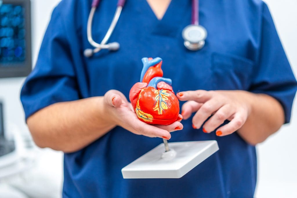
Heart blockages are a big threat to heart health. They happen when the arteries that bring blood to the heart get blocked. This is usually because of plaque buildup, known as coronary artery disease.
This blockage can reduce blood flow to the heart. It might cause chest pain, shortness of breath, or even a heart attack.
The Pathophysiology of Coronary Artery Disease
Coronary artery disease is when plaque builds up in the arteries. This makes them hard and narrow. It’s often caused by high blood pressure, high cholesterol, smoking, and diabetes.
As plaque grows, it can burst and cause a blood clot. This clot can block the artery and lead to a heart attack.
It’s key to understand how coronary artery disease works. This helps us find people at risk and take steps to prevent it.
Warning Signs and Symptoms of Heart Blockages
Knowing the signs of heart blockages is important. Symptoms include chest pain or discomfort, which can spread to the arms, back, neck, jaw, or stomach. You might also feel short of breath, tired, or dizzy.
Some people might not notice symptoms until it’s too late, like during a heart attack. It’s vital to seek help if you notice these signs.
Risk Factors That Necessitate Cardiac Imaging
Some factors increase the risk of heart blockages. These include a family history of heart disease, high blood pressure, high cholesterol, smoking, diabetes, and obesity. People with these risks should get regular heart checks.
Cardiac imaging is key for finding and managing heart blockages. We use tests like stress tests, CT scans, and MRI to see the heart and its blood vessels. This helps us diagnose and plan treatment.
The Importance of Early and Accurate Diagnosis
Getting a heart blockage diagnosed early and accurately is key. It helps doctors make the right treatment plans. Cardiac imaging is a big part of this process.
How Imaging Influences Treatment Decisions
Tests like nuclear stress tests and echocardiograms give detailed heart info. This info is vital for figuring out how bad heart blockages are. It helps doctors decide the best treatment.
Cardiac imaging affects how well patients do. Research shows early treatment of heart blockages can save lives. It lowers the chance of serious heart problems.
Mortality and Morbidity Statistics Related to Undiagnosed Blockages
Heart blockages that go unnoticed are a big risk. They can lead to serious heart issues and even death. Early detection through imaging can prevent these problems.
It’s important to know the dangers of not finding heart blockages early. Knowing these risks can help people get checked sooner.
When to Seek Cardiac Imaging Evaluation
Knowing when to get a cardiac imaging test is important. People with high blood pressure, diabetes, or heart disease in their family should get tested. Also, if you have chest pain or trouble breathing, see a doctor right away.
Talking to a doctor about getting a cardiac imaging test is a good idea. This way, you can get the care you need on time.
Overview of Non-Invasive Cardiac Imaging Techniques
Non-invasive cardiac imaging has changed how we diagnose heart blockages. It offers safer options than old methods. New medical tech has made these tests more accurate and comfortable for patients.
We’ll look at different non-invasive methods for diagnosing heart issues. We’ll talk about their uses, benefits, and what they can’t do.
Stress Tests: Exercise vs. Pharmacological
Stress tests are key in checking the heart’s health. They test how the heart works when stressed. There are two main types: exercise and pharmacological stress tests.
- Exercise Stress Tests: These tests make you move, like on a treadmill or bike. They help find heart problems and check your heart health.
- Pharmacological Stress Tests: If you can’t exercise, medicine is used to mimic exercise. This is good for people who can’t move much.
Imaging-Enhanced Stress Tests
Imaging-enhanced stress tests add more info by mixing stress tests with imaging. This gives a clearer picture of the heart.
| Imaging Modality | Description | Benefits |
| Nuclear Stress Test | Uses radioactive tracers to see how the heart works under stress. | Great at finding heart disease. |
| Stress Echocardiogram | Uses sound waves to see the heart’s shape and function during stress. | Shows the heart in action and checks valves. |
CT and MRI-Based Cardiac Imaging
CT and MRI scans give detailed views of the heart. They show its structure and how it works.
- Coronary CT Angiography: Uses CT scans to see the heart’s arteries. It finds blockages and checks plaque.
- Cardiac MRI: Uses MRI to look at the heart’s shape, function, and blood flow. It gives insights into heart tissue.
These imaging methods have changed how we diagnose and treat heart blockages. They help doctors make better plans for treatment.
The Nuclear Stress Test: Gold Standard for Detecting Heart Blockages
In cardiology, the nuclear stress test is key for spotting coronary artery disease. It’s a vital tool for checking heart health and finding blockages that could cause big problems.
Scientific Principles Behind Nuclear Cardiology
Nuclear cardiology uses tiny amounts of radioactive tracers to see the heart’s blood flow. These tracers build up in the heart muscle based on blood flow. This lets doctors check how well the heart works under stress and at rest.
Key aspects of nuclear cardiology include:
- The use of radioactive tracers to visualize heart function
- Assessment of myocardial perfusion under stress and at rest
- Identification of areas with reduced tracer uptake, indicative of blockages or scar tissue
Radioactive Tracers and How They Identify Blockages
Radioactive tracers are vital in nuclear stress tests. Injected into the blood, they light up the heart muscle. Areas with good blood flow show more tracer, while blockages show less. This helps doctors see where the heart might be blocked.
Technetium-99m and Thallium-201 are common tracers. Each has its own strengths. The right tracer depends on the patient and what the doctor needs to know.
Sensitivity and Specificity in Detecting Coronary Artery Disease
The nuclear stress test is known for its accuracy in finding coronary artery disease. Sensitivity means it correctly spots those with the disease. Specificity means it correctly spots those without the disease. Research shows it’s very good at both, making it a top choice for doctors.
How well the test works depends on a few things. These include who gets tested, the quality of the equipment, and the doctor’s skill. When done right, it helps doctors make better choices for their patients.
Nuclear Stress Test Procedure: A Patient’s Guide
Knowing what to expect during a nuclear stress test can make you feel more at ease. We’ll walk you through everything, from getting ready before the test to taking care of yourself after.
Pre-Test Preparations and Restrictions
Before your nuclear stress test, there are a few things you need to do. These steps help make sure the test is accurate and safe for you. Here’s what we recommend:
- Avoid eating or drinking anything except water for a few hours before the test.
- Tell us about any medications you’re taking. Some might need to be adjusted or stopped before the test.
- Wear comfortable clothing and shoes that are good for exercise.
The Day of Your Test: What to Expect
On the day of your test, here’s what will happen:
- A small intravenous (IV) line will be placed in your arm to administer the radioactive tracer.
- You will then undergo a stress test, typically involving walking on a treadmill or using a stationary bike.
- After the stress test, you’ll be injected with a radioactive tracer, which helps us visualize your heart’s function.
- Imaging will be performed both after the stress test and at rest, usually on the same day or the following day.
Post-Test Care and Follow-Up
After the test, you can usually go back to your normal activities unless your doctor tells you differently. The radioactive tracer will leave your body through urine or feces over the next few days.
Here’s what you can expect after the test:
| Activity | Instructions |
| Resuming Normal Activities | Usually immediate, unless specified differently |
| Hydration | Drink plenty of water to help flush out the radioactive tracer |
| Follow-Up | Schedule a follow-up appointment to discuss your test results and further treatment |
Understanding the nuclear stress test procedure can help reduce your anxiety and make the experience smoother. If you have any questions or concerns, don’t hesitate to contact your healthcare provider.
Echocardiography and Stress Echocardiograms
Echocardiography, or heart ultrasound, is key for checking the heart’s health. It’s a safe way to see how the heart works and blood flows. This helps doctors spot heart problems early.
Visualizing Heart Function with Ultrasound
Ultrasound uses sound waves to show the heart’s inside. It lets us look at the heart’s parts and how well they work. Real-time imaging shows the heart’s movement and blood flow. This is important for finding heart issues.
Exercise and Pharmacological Stress Echo Protocols
Stress echocardiography tests how the heart handles stress. There are two main ways to do this:
- Exercise Stress Echo: Patients exercise on a treadmill or bike to raise their heart rate.
- Pharmacological Stress Echo: Medicine is used to make the heart work hard, good for those who can’t exercise much.
Both methods help find heart areas that might not get enough blood when stressed. This could mean there’s a blockage.
Advantages in Specific Populations
Stress echocardiography is great for some patients. It’s good for those who can’t have nuclear tests because of health issues. Here are some benefits:
| Patient Group | Advantages of Stress Echocardiography |
| Patients with kidney issues | Avoids harmful radioactive tracers and contrast agents. |
| Patients who cannot exercise | Pharmacological stress echo is a good alternative. |
| Patients with certain medical implants | It’s safe without worries about MRI or other imaging. |
Knowing how echocardiography and stress echocardiograms work helps us diagnose and treat heart issues better. This leads to better health outcomes for patients.
Advanced Imaging Options for Coronary Blockages
Several advanced imaging options are available for diagnosing coronary artery disease. Each has its own benefits and uses. These techniques give detailed views of the heart’s structure and function. This helps doctors diagnose blockages more accurately.
Coronary CT Angiography: Visualizing the Arteries
Coronary CT angiography is a non-invasive test that uses X-rays to show the coronary arteries. It helps us see the arteries and spot any problems. This method is great for catching coronary artery disease early, which can prevent serious issues.
Key Features of Coronary CT Angiography:
- Non-invasive procedure
- High-resolution images of coronary arteries
- Early detection of coronary artery disease
Cardiac MRI: Tissue Characterization and Perfusion
Cardiac MRI gives detailed images of the heart’s structure and function. It’s excellent for checking tissue health and blood flow. This helps us see if the heart is working right and find any damaged areas.
Benefits of Cardiac MRI:
- Detailed assessment of cardiac structure and function
- Evaluation of tissue characterization and perfusion
- No radiation exposure
PET Cardiac Imaging: Metabolic Assessment
PET cardiac imaging looks at the heart’s metabolic activity. It’s great for checking if heart tissue is alive and finding ischemic areas.
Advantages of PET Cardiac Imaging:
- Assessment of myocardial viability
- Identification of areas of ischemia
- Guidance for revascularization decisions
Invasive Coronary Angiography: The Definitive Test
Invasive coronary angiography is the top choice for diagnosing coronary artery disease. It directly looks at the coronary arteries with contrast agents. This gives a clear view of the heart’s anatomy.
| Imaging Modality | Key Features | Benefits |
| Coronary CT Angiography | Non-invasive, high-resolution images | Early detection of CAD |
| Cardiac MRI | Tissue characterization, perfusion assessment | No radiation, detailed cardiac assessment |
| PET Cardiac Imaging | Metabolic assessment, viability evaluation | Guidance for revascularization decisions |
| Invasive Coronary Angiography | Direct visualization of coronary arteries | Gold standard for CAD diagnosis |
Safety Considerations and Risks of Cardiac Imaging
Cardiac imaging is key for accurate diagnoses. But, it’s important to know the safety risks. These tests are vital for diagnosing heart issues but come with risks that must be managed.
Radiation Exposure from Nuclear Tests: Putting Risk in Perspective
Nuclear stress tests expose you to small amounts of radiation. The benefits usually outweigh the risks. We use the least amount of radiation needed for clear images.
Radiation Risk Management: We follow strict rules to lower radiation exposure. This ensures the benefits of the test are greater than the risks.
Contrast Agent Reactions and Kidney Concerns
Contrast agents in some tests can cause reactions or kidney problems. People with kidney issues or allergies are at higher risk. We check patients carefully before using these agents.
Pre-Test Assessment: We review patients’ medical history before tests with contrast agents. This helps us spot risks and take precautions.
Exercise-Related Risks During Stress Testing
Exercise stress tests are mostly safe but can lead to heart problems. We watch patients closely during these tests. This way, we can act fast if needed.
Monitoring During Stress Tests: Our team keeps a close eye on patients’ vital signs and heart activity. This ensures quick action if there’s a problem.
Alternatives for High-Risk Patients
For high-risk patients or those who can’t have certain tests, we have other options. We choose the safest and most fitting test for each patient.
Knowing the risks of cardiac imaging helps us use these tools wisely. This way, we can give our patients the best care possible.
Understanding Your Test Results and Next Steps
Getting your test results is a big moment for your heart health. It’s a time to figure out what’s next for your care. We know it can be tough to understand cardiac imaging results. So, we’re here to help you through it.
Normal vs. Abnormal Findings Across Different Imaging Modalities
Different tests give different views of your heart health. For example, a nuclear stress test shows how your heart works under stress. A coronary CT angiography shows your coronary arteries in detail. Knowing if your results are normal or abnormal is key.
Normal findings mean your heart is working well, with no big blockages. Abnormal findings might show heart function issues or blockages.
Grading the Severity of Blockages
If you have blockages, knowing how severe they are is important. Blockages are graded from mild to severe. Mild blockages might not need quick action but will be watched. Severe blockages often need more serious treatments like angioplasty or surgery.
Knowing the severity helps your healthcare team plan your treatment.
Follow-Up Testing and Treatment Pathways
Your healthcare provider might suggest more tests or changes to your treatment plan. This could include more imaging, lifestyle changes, medication, or procedures. We work with you to create a treatment pathway that meets your needs and health goals.
When a Second Opinion Is Warranted
Getting a second opinion can be helpful. If you’re not sure about your diagnosis or treatment, it’s okay to ask for another look. A second opinion is great for complex cases or when considering big treatments.
We’re here to support you every step of the way. By understanding your test results and what comes next, you can take charge of your heart health.
Conclusion: Selecting the Optimal Imaging Approach for Heart Blockage Detection
Choosing the right imaging technique is key for accurate diagnosis and treatment planning. We’ve looked at different imaging options for finding heart blockages. These include nuclear stress tests, echocardiography, and advanced methods like coronary CT angiography and cardiac MRI.
Understanding the strengths and weaknesses of these imaging tools helps healthcare providers make better choices. The best imaging approach for finding heart blockages depends on the patient’s health history, risk factors, and the blockage’s details.
Using the best imaging modality helps patients get accurate diagnoses and targeted treatments. This improves their heart health outcomes. As cardiac care advances, staying updated on the latest imaging options is vital for detecting heart blockages.
FAQ
What is a nuclear stress test, and how does it detect heart blockages?
A nuclear stress test uses a tiny amount of radioactive material. It shows how the heart’s blood flow changes under stress. This stress can be from exercise or medicine. It spots heart blockages by finding areas where blood flow is low.
What are the differences between a nuclear stress test and a stress echocardiogram?
A nuclear stress test uses radioactive tracers to see the heart. On the other hand, a stress echocardiogram uses ultrasound. Both check how the heart works under stress but in different ways.
How accurate is a nuclear stress test in detecting coronary artery disease?
Nuclear stress tests are very good at finding coronary artery disease. They give detailed info on the heart’s blood flow and function under stress. This helps spot blockages and decide on treatment.
What are the risks associated with nuclear stress tests?
Nuclear stress tests have some risks, like radiation from the tracers. But, the benefits often outweigh these risks, mainly for those at high risk of heart disease.
Can a nuclear stress test show blocked arteries?
Yes, it can. The test shows where blood flow to the heart muscle is low, meaning arteries are blocked or narrowed. It helps diagnose heart disease and plan treatment.
What are the alternatives to nuclear stress tests for diagnosing heart blockages?
Alternatives include stress echocardiograms, coronary CT angiography, cardiac MRI, and PET cardiac imaging. Each has its own benefits and is chosen based on the patient’s condition and history.
How do I prepare for a nuclear stress test?
Preparation includes eating certain foods, avoiding some medicines, and wearing comfy clothes. Your doctor will give you detailed instructions for a smooth test.
What should I expect during and after a nuclear stress test?
During the test, you’ll do stress testing and then imaging. After, you’ll get care instructions and follow-up advice. Your doctor will talk about the results and what treatment you might need.
How are test results interpreted, and what do they mean for treatment?
Results are compared to images taken at rest and under stress. If there are abnormal findings, it might mean blockages or low blood flow. This guides treatment, like lifestyle changes, medicine, or more procedures.
When is a second opinion warranted after cardiac imaging?
Getting a second opinion is wise if you’re unsure about your diagnosis or treatment. It’s also a good idea if the results are unclear or if you’re at high risk of heart disease.


