Every year, 1.5 million Positron Emission Tomography (PET) scans are done in the United States. They are used for cancer detection and monitoring. A PET scan is a medical imaging test that helps doctors understand and manage different health issues.
The main goal of a PET scan is to show how active the body’s tissues and organs are. It uses a tiny bit of radioactive material. This helps doctors find areas that are not working right, which could mean cancer, neurological problems, or heart disease.

PET scans are key in healthcare. They help diagnose and manage many medical conditions. Understanding their functionality is crucial.
PET means Positron Emission Tomography. It’s a way to see how the body works using special tracers. Doctors use it to check for diseases at a small scale.
PET scans use a radioactive tracer that’s injected into the body. This tracer goes to areas that are very active, like cancer cells. When it decays, it sends out positrons.
These positrons meet electrons and make gamma rays. The PET scanner picks up these rays. It makes detailed pictures of how the body works.
What PET scans show is very important. They help find and track diseases like cancer and heart problems. Understanding their functionality is crucial.e make better choices about health.
PET scan technology has come a long way. It started with early development and has grown a lot. Now, it’s used in many ways to help doctors diagnose diseases.
The idea of PET scans began in the 1950s. Scientists were looking into using special isotopes for medical images. The initial development was boosted by nuclear medicine and new radiopharmaceuticals.
In the 1970s, the first PET scanners were made. They were simple but started a new chapter in imaging. Today, we have much better technology.
PET imaging has made big leaps forward. Scanners, algorithms, and drugs have all improved. Now, we have PET-CT and PET-MRI, which give even more information.
New drugs have also opened up more uses for PET scans. This lets doctors see how the body works and find diseases better.
| Advancement | Description | Impact |
| Improved Scanner Design | Enhanced sensitivity and resolution | Better image quality and diagnostic accuracy |
| Advanced Image Reconstruction Algorithms | More accurate and detailed images | Improved diagnostic confidence |
| New Radiopharmaceuticals | Expanded range of applications | Increased versatility in diagnosing various conditions |
PET scan technology is always getting better. Scientists are working hard to make images clearer, scans faster, and use PET scans for more things.
PET scan technology uses radioactive tracers to show how the body works. These tracers are injected into the body and gather in areas that are very active. Then, the PET scan detects the radiation from these tracers.
Radioactive tracers are special substances with a bit of radioactive material. The most used one is Fluorodeoxyglucose (FDG), a radioactive sugar. Cancer cells use more sugar than normal cells, making FDG great for finding cancer.
Other tracers help check blood flow or find specific receptors in the body. The right tracer depends on what disease or condition is being looked at.
After the tracer is given, it goes to the area it’s meant for. The PET scanner picks up the radiation from the tracer. It uses this to make images of the body’s activity.
The scanner catches gamma rays from the tracer’s breakdown. It uses these rays to build detailed pictures.
The scanner’s data is turned into images that show the body’s activity. These images help find many conditions, like cancer or brain problems.
PET scans give doctors a deep look into how the body works. They help doctors see how serious a disease is, check if treatments are working, and decide the best care for patients.
PET scans help find changes in cells that show early signs of disease. This is key for spotting and treating many health issues, like cancer.
They’re great for finding cancer early. Early detection can really help with treatment.
PET scans are a type of functional imaging. They show how cells and tissues work, not just what they look like. This is different from structural imaging like CT and MRI, which show the body’s layout.
Using both types of imaging gives a full picture of diseases. Structural imaging shows the body’s shape, while PET scans reveal metabolic activity. This combo helps doctors understand diseases better.
Knowing the difference between these imaging types helps doctors pick the best tools for patients. This way, they use each method’s strengths to help patients the most.
PET scans are key in the fight against cancer. They help find cancer, see how far it has spread, and check if treatments are working. This makes them essential in oncology.
PET scans spot cancer by looking at how cells use sugar. Cancer cells use more sugar than normal cells. A special sugar, called FDG, is given to the body. It goes to cancer cells, showing up on the PET scan.
Knowing the cancer stage is key for the right treatment. PET scans help by showing how far the cancer has spread. This helps doctors plan the best treatment, like surgery or chemo.
PET scans help plan better treatments, leading to better patient results.
PET scans also track how well treatments are working. They compare scans before, during, and after treatment. This helps doctors see if the treatment is working and make changes if needed.
Key benefits of using PET scans for monitoring treatment response include:
PET scans are vital in changing treatment plans as cancer care evolves.
PET imaging is key in neurology, showing how the brain works. It helps understand brain conditions and plan treatments.
PET scans check brain function and metabolism. They use radioactive tracers to find brain areas that don’t work right. Understanding brain metabolism is key for diagnosing and treating neurological issues.
When using PET scans, a radioactive tracer is given to the brain. It shows how brain cells work, giving clues about brain health.
PET imaging is vital for spotting Alzheimer’s and other dementias. It looks at brain activity to find patterns linked to these diseases. Early detection of Alzheimer’s is important for starting the right care.
PET scans also help diagnose other dementias like frontotemporal dementia and Lewy body dementia. They show different metabolic patterns, helping doctors tell these conditions apart.
PET scans are used in epilepsy evaluation and surgery planning. They find where seizures start, helping plan surgery. Finding seizure origins is key for surgery success.
In epilepsy, PET scans show abnormal brain activity. This info is vital for surgeons planning to remove the seizure area. It could lead to better results for patients with hard-to-treat epilepsy.
PET scans are key in cardiology, showing how the heart works and blood flows. They help doctors see the heart’s activity, which is vital for planning treatments.
PET scans mainly check the heart’s function and blood flow. They use special tracers to see how active the heart is. This helps spot problems like blocked arteries or damaged heart areas.
This detailed view helps doctors choose the best treatment. It could be medicine, surgery, or other options.
After a heart attack, PET scans are key in finding healthy heart tissue. They show which parts of the heart can recover. This info guides treatments like bypass surgery or angioplasty.
Using PET scans helps doctors create personalized treatment plans. This can lead to better results and fewer heart problems later on.
In summary, PET scans are essential in cardiology. They give insights into the heart’s function, blood flow, and tissue health. This helps doctors make accurate diagnoses and effective treatments, improving patient care.
PET-CT scans are a powerful tool that combines PET and CT images. This technology helps doctors diagnose and manage diseases, like cancer, better.
PET-CT scans merge PET’s metabolic activity with CT’s detailed images. This is done through special software that aligns the images. It gives a clearer picture of the disease.
Key aspects of PET-CT fusion include:
PET and CT imaging together have many advantages. They improve how well doctors can diagnose and plan treatments. This is because they get both the metabolic activity and detailed images of the body.
| Benefit | Description |
| Improved Diagnostic Accuracy | Combining functional PET data with anatomical CT images enhances the ability to detect diseases accurately. |
| Enhanced Treatment Planning | PET-CT scans provide detailed information necessary for planning effective treatment strategies. |
| Better Disease Staging | The fusion of PET and CT images allows for more accurate staging of diseases, particularlly cancer. |
Recent studies show PET and CT together have made healthcare better. They help doctors make more accurate diagnoses and treatments. As one study says, “PET-CT combines the functional information from PET with the anatomical information from CT, providing a more complete understanding of disease.” This shows how much PET-CT has changed healthcare.
Getting ready for a PET scan means paying attention to several important details. This ensures the scan goes smoothly and gives accurate results. Patients need to follow specific steps to make sure the scan works well.
One key part of preparing for a PET scan is following dietary rules. Patients usually need to fast for a while before the scan. The exact time can vary based on the type of scan and doctor’s orders. It’s also important to avoid sugary foods and drinks, as they can impact the scan’s results.
Patients also need to talk to their doctor about any medicines they’re taking. Some medicines might need to be changed or stopped before the scan. This is to prevent any issues with the radioactive tracer used in the scan.
On the day of the scan, wear comfy, loose clothes without metal parts. Avoid jewelry or metal objects that could mess with the scan.
Bring all needed documents, like insurance cards and ID. Also, list your current medicines and any past health issues. It’s a good idea to have a friend or family member there for support.
Diabetic patients need special care when getting ready for a PET scan. They should talk to their doctor about managing their blood sugar before and after the scan. The doctor might need to adjust their medicine or insulin to make sure the scan is safe and effective.
| Preparation Step | Instructions | Additional Notes |
| Dietary Restrictions | Fast for the recommended period | Avoid sugary foods and drinks |
| Medication Considerations | Inform your healthcare provider about current medications | Adjust or discontinue medications as advised |
| Clothing and Accessories | Wear loose, metal-free clothing | Avoid jewelry and other metal objects |
| Diabetic Patients | Consult with your healthcare provider about managing blood sugar levels | Adjust medication or insulin regimen as necessary |
By following these guidelines, patients can help make their PET scan a success. This ensures accurate results. Always check with a healthcare provider for personalized advice that fits your needs.
Getting a PET scan is straightforward when you know what to expect. The process includes preparation, scanning, and recovery. Each step is important for a successful PET scan.
The first step is getting a radioactive tracer through an IV line in your arm. The tracer used varies by scan type, but FDG (fluorodeoxyglucose) is common for cancer scans.
After the tracer is given, you wait for 30 minutes to an hour. This is called the uptake period. It’s important to stay calm and not move during this time. This helps the tracer spread evenly in your body.
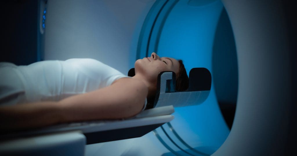
When the uptake period ends, you’ll go to the PET scanner. It’s a large, doughnut-shaped machine. You’ll lie on a table that slides into the scanner. It’s important to stay very quiet and not move during the scan.
The PET scan is painless. You won’t feel anything during the scan.
After the scan, the IV line will be removed. You won’t need to rest after a PET scan. You can go back to your usual activities right away, unless your doctor says not to.
The images from your scan will be looked at by a specialist. Your doctor will then talk to you about the results. This can take a few hours to a few days, depending on how busy the facility is.
In summary, knowing what to expect during a PET scan makes the process easier. If you have any questions or concerns, always talk to your healthcare provider.
It’s important to know about the side effects and safety of PET scans. These scans are useful for medical diagnosis but involve radioactive tracers. This can cause problems for some people.
PET scans use radioactive tracers. These tracers emit positrons that create gamma rays. The scanner detects these rays. Even though the radiation is small, it’s key to know the risks, mainly for those needing many scans.
Some people might feel side effects or discomfort during or after a PET scan. These can include:
These side effects are usually mild and short-lived. But, it’s important to tell your doctor if you have any unusual or severe reactions.
Some people should not get PET scans or need special care. These include:
Talking to your healthcare provider about your medical history and concerns is vital before a PET scan.
PET scans are among several imaging tools used in diagnosis. Each has its own benefits. Knowing the differences helps choose the best tool for a patient’s needs.
PET and CT scans have different main uses. CT scans use X-rays to show body structures. PET scans look at how tissues work by using a radioactive tracer.
Key differences include:
MRI is another key tool that shows internal details without harmful radiation. The choice between PET and MRI depends on what you’re looking for.
Considerations for choosing between PET and MRI include:
Imaging techniques work together to fully understand a patient’s health. For example, PET-CT or PET-MRI combines functional and anatomical views. This improves diagnosis and helps plan treatments.
Using many imaging methods shows the value of a customized approach. The right choice depends on the patient’s situation and what each method can do best.
PET scan technology is on the verge of a new era. This is thanks to better imaging and artificial intelligence. These advancements will make PET scans even more useful in diagnosing and treating diseases.
New PET scan technology is improving how well it can spot diseases early. This means doctors can treat problems sooner and more effectively. High-resolution PET scans are key in this, allowing for detailed images of small areas.
New materials and designs in PET scanners have made them more sensitive and clear. For example, digital PET detectors have better timing, leading to clearer images and less noise.
| Feature | Traditional PET | Advanced PET |
| Resolution | 5-6 mm | 2-3 mm |
| Sensitivity | Moderate | High |
| Scan Time | 20-30 minutes | 10-15 minutes |
The use of artificial intelligence (AI) and machine learning (ML) with PET scans is changing medical imaging. AI helps make images clearer and reduces noise. ML can spot patterns in images that humans might miss, helping find diseases sooner.
AI in PET imaging could also lead to more personalized treatments. By analyzing lots of data, AI can predict how well a patient will respond to different treatments. This could mean treatments that are more tailored to each patient.
As PET scan technology keeps getting better, we can look forward to even more accurate diagnoses and better care for patients. The future of PET scans is about giving detailed, functional information about the body. This will help doctors make better treatment decisions and improve patient outcomes.
PET scans have changed medical imaging. They help doctors diagnose and treat better. PET-CT scans combine images of the body’s structure and function.
As medical imaging grows, so will PET scans’ role. They will help find and treat diseases better. Their importance in healthcare is clear, and they will keep being a big part of it.
PET stands for Positron Emission Tomography. It’s a way to see how the body works by using special tracers.
PET scans help find changes in the body’s cells. They are key in diagnosing and managing diseases, like cancer.
A PET scan uses a special tracer that goes into the body. It shows where the body is most active. The scanner then makes detailed images of this activity.
The most common tracer is Fluorodeoxyglucose (FDG). It shows where the body uses a lot of sugar. Other tracers are used for different needs, like checking the heart or finding certain cancers.
A PET scan usually takes 30-60 minutes. But getting ready and waiting for the tracer can take hours.
PET-CT fusion combines PET’s function with CT’s structure. This gives a clearer view of the body’s activity and its layout.
Before a PET scan, you need to fast for hours. This means no food or sugary drinks. It helps the tracer work better.
Side effects are usually mild. You might feel a bit uncomfortable from the injection or nervous during the scan. But the benefits usually outweigh the risks.
PET scans show how active the body is. CT and MRI scans show the body’s structure. PET scans are great for finding cancer and checking how treatments work. CT and MRI are better for looking at body parts.
PET scans are getting better. They will have higher resolution and work with new tech like AI. This will make them even more accurate.
Yes, diabetic patients can have a PET scan. They need to follow special rules to manage their sugar levels. This ensures the best results.
You can usually go back to normal activities right after a PET scan. Just drink lots of water to get rid of the tracer.
Subscribe to our e-newsletter to stay informed about the latest innovations in the world of health and exclusive offers!