Choosing the right imaging technique is key when diagnosing and treating brain, head, and cancer issues. We have two main tools: CT and MRI scans. Each has its own benefits, depending on the situation.
Many patients wonder which is best, CT or MRI scan, for their condition. MRI scans are great for soft tissue contrast, perfect for spotting brain tumors and detailed brain checks. CT scans, though, are quicker and better for emergencies like trauma, bleeding, or stroke.
The right choice between CT and MRI scans depends on the situation and the patient’s health. Knowing what each scan does best helps doctors make better decisions and ensures accurate diagnosis.

It’s key to know how CT and MRI scans work for accurate diagnoses. These technologies have changed how we see inside the body. They give us new ways to understand health.
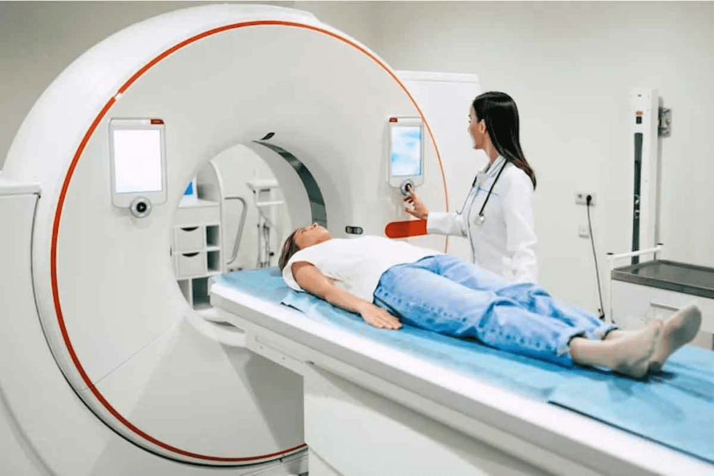
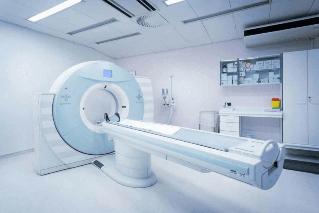
CT scans use X-rays to make detailed images of the body. A CT scanner, shaped like a doughnut, moves around the patient. It measures how much X-ray energy different body parts absorb.
Then, computers turn these measurements into images. These images show bones, soft tissues, and blood vessels clearly.
MRI scans use a magnetic field and radio waves to create images. The patient lies in a strong magnetic field. This field aligns the body’s hydrogen atoms.
Radio waves disturb these atoms, sending signals to the MRI machine. These signals help make detailed images, mainly of soft tissues.
CT and MRI scans work in different ways. CT scans use X-rays, while MRI scans use magnetic fields. This difference affects what each scan can show and how they’re used.
| Characteristics | CT Scan | MRI Scan |
| Imaging Principle | X-ray energy absorption | Magnetic field and radio waves |
| Best for Imaging | Bones, lung tissue, and acute bleeding | Soft tissues, including organs and tumors |
| Scan Time | Typically faster, often just a few minutes | Can be longer, sometimes up to an hour |
| Radiation Exposure | Yes, uses X-rays | No ionizing radiation |
Knowing these differences helps doctors choose the right scan for each patient. This improves care and outcomes.
Diagnostic imaging is key in today’s medicine. Knowing the differences between CT and MRI scans is vital for accurate diagnoses. The right choice depends on the tissue type and suspected condition.
MRI scans are top for soft tissue contrast. They’re best for the brain, spinal cord, and other soft tissues. MRI’s high contrast resolution helps spot tumors, inflammation, and other issues in these areas. CT scans are good for many things but have lower soft tissue contrast.
For brain tumors, MRI’s detailed soft tissue images are key for diagnosis and treatment. Research shows MRI is better than CT for finding small or hard-to-see brain lesions.
While MRI is great for soft tissues, CT scans are better for bones and hard structures. CT scans show bones in high detail, making them useful for trauma or osteoporosis. They help diagnose fractures, check bone density, and plan surgeries.
Both CT and MRI scans are good at finding abnormalities, but their sensitivity varies. MRI is great at spotting small changes in soft tissues. This is important for early diagnosis and treatment of conditions like multiple sclerosis or cancer.
In summary, choosing between CT and MRI depends on the specific clinical question. Consider each modality’s strengths and limitations in image quality and resolution.
Diagnostic imaging’s speed and accessibility are key in patient care. The choice between CT and MRI scans depends on several factors. These factors can greatly affect diagnosis and treatment.
CT scans are generally faster than MRI scans. They usually take just a few minutes. This makes CT scans great for emergency situations where time is critical.
MRI scans, on the other hand, can take from 15 to 90 minutes. This longer time can be tough for patients who are claustrophobic or have trouble staying calm for long. But new MRI technology is making scans faster and more comfortable.
Where you can get a CT or MRI scan is also important. CT scanners are more commonly found in hospitals and diagnostic centers. They are used a lot in emergencies and are cheaper than MRI machines.
MRI scanners are less common but can be found in bigger hospitals and specialized centers. The availability of these machines can vary a lot. Urban areas usually have more access to both CT and MRI scanners than rural areas.
Choosing between CT and MRI scans also depends on patient comfort. CT scans are quicker and less likely to cause claustrophobia. But, they involve radiation.
MRI scans don’t use radiation but can be harder for patients. This is because of the scanner’s enclosed space and the loud noises during the scan. To make MRI scans more comfortable, many places offer sedation for anxious patients. Open MRI machines and wide-bore scanners are also becoming more common, providing a better experience for patients.

Choosing between CT and MRI scans depends on the clinical situation. We look at when each is best, helping doctors make the right choice.
In emergencies like trauma or bleeding, CT scans are often first. They are fast and widely available, key in urgent cases where quick diagnosis is vital.
For example, in head trauma, CT scans quickly spot fractures, bleeding, or injuries needing fast care. This makes CT scans essential in emergency rooms.
For long-term conditions like neurological disorders or soft tissue diseases, MRI is usually better. MRI shows soft tissues better, helping with detailed checks of conditions like multiple sclerosis or tumors.
MRI’s clear images of soft tissues are key for diagnosing and tracking chronic conditions. This helps in treatment planning and checking how well treatments work.
Choosing between CT and MRI for follow-ups depends on the first diagnosis and what’s needed next. If soft tissue was involved initially, MRI might be best for follow-ups. But if bony structures or areas where CT is better are involved, CT scans could be more fitting.
| Clinical Scenario | Preferred Imaging Modality | Key Advantages |
| Emergency Situations (Trauma, Acute Bleeding) | CT Scan | Speed, Availability, Quick Detection of Injuries |
| Chronic Conditions (Neurological Disorders, Soft Tissue Diseases) | MRI Scan | Superior Soft Tissue Contrast, Detailed Evaluation |
| Follow-up Examinations | Depends on Initial Diagnosis | Specific to the Condition Being Monitored |
Knowing the strengths of CT and MRI and using them right can improve patient care and results.
CT and MRI scans are key tools for brain imaging. They help us find and understand brain issues like tumors and strokes. These tools are essential for diagnosing many brain-related problems.
MRI scans are better at finding brain tumors and small problems. MRI gives us detailed images of soft tissues, which is key to finding brain tumors. This helps us know the tumor’s size, type, and where it is, which guides treatment.
CT scans are good in emergencies, but don’t show soft tissues as well as MRI. Yet, they’re useful for spotting big tumors or those that change the brain’s structure a lot.
| Imaging Modality | Tumor Detection Capability | Soft Tissue Detail |
| CT Scan | Good for larger tumors or those causing structural changes | Limited |
| MRI Scan | Excellent for detecting small tumors and subtle abnormalities | High |
When a stroke is suspected, CT scans are often first because they’re fast and easy to get. They help us see if a patient has a hemorrhagic stroke, which needs quick treatment.
MRI scans, like diffusion-weighted imaging (DWI), are great for finding ischemic strokes. They show how much brain tissue is affected by the stroke.
MRI scans are top-notch for seeing neurological disorders like multiple sclerosis and Alzheimer’s. They give us detailed images that help us diagnose and track these conditions.
While CT scans can be used sometimes, MRI’s better soft tissue contrast makes it the best choice for these disorders.
Knowing what CT and MRI scans can do helps us choose the right tool for each case. This leads to better care for our patients.
Head imaging is key to accurate patient care. Knowing when to use CT or MRI scans is vital. The choice depends on the clinical scenario and needed information.
For trauma and acute head injuries, CT scans are preferred. They are fast and good at showing bones. CT scans offer quick and accurate head injury assessments, which is critical in emergencies.
A study compared CT and MRI in head trauma. CT scans were better at finding acute hemorrhage and fractures. Here’s a table showing CT’s advantages in trauma cases.
| Imaging Modality | Advantages in Trauma |
| CT Scan | Quick, effective for bone imaging, good for detecting acute hemorrhage |
| MRI | Better for soft tissue evaluation, useful for detecting subtle injuries |
For sinus and facial structure evaluation, CT scans are preferred. They offer detailed bone images. CT scans help diagnose sinusitis, fractures, and facial abnormalities.
MRI is useful when soft tissue evaluation is needed. This is true for tumors or infections in sinuses and facial structures.
MRI is preferred for inner ear and temporal bone assessment. It has better soft tissue contrast. MRI shows detailed inner ear structures and cranial nerves, key for diagnosing conditions like acoustic neuromas.
In summary, choosing between CT and MRI for head imaging depends on the application and clinical question. Understanding each modality’s strengths helps healthcare providers make better decisions for patient care.
Accurate detection and staging are key to managing cancer. CT and MRI scans have different roles in this process. The choice between them depends on the cancer type, location, and stage.
Both CT and MRI scans are strong for initial cancer diagnosis. CT scans are often first because they’re fast and widely available. They’re great for finding tumors in the lungs, liver, and other organs.
MRI scans, with their better soft tissue contrast, are best for tumors in complex areas like the brain, spine, and pelvis. They provide detailed images without using radiation, which is a big plus for cancer diagnosis.
MRI scans are top-notch for tumor characterization. They can tell the difference between various tissue types. This helps figure out if a tumor is benign or malignant and guides treatment plans.
Tumor characterization looks at the tumor’s size, location, and how it relates to nearby structures. MRI’s high-resolution images are key for this, even in delicate areas like the brain.
Checking for metastasis is vital for cancer staging and predicting outcomes. MRI is best for finding metastases in the brain and head. It does this because of its excellent soft tissue contrast and ability to spot lesions.
| Imaging Modality | Cancer Detection Strengths | Limitations |
| CT Scan | Fast, widely available, good for lung and liver tumors | Less effective for soft tissue tumors, involves radiation |
| MRI Scan | Excellent soft tissue contrast, no radiation, ideal for brain and spine tumors | More expensive, less available than CT, longer scan times |
Knowing the strengths and weaknesses of CT and MRI scans helps doctors choose the best imaging for each patient. This ensures the most accurate cancer detection and staging.
It’s important to know the differences between CT and MRI contrast agents. These agents help make diagnostic images clearer. This helps doctors diagnose and treat conditions better.
CT scans use iodine-based contrast agents to show structures or lesions in the body. These agents contain iodine, which absorbs x-rays. This makes areas with the agent appear brighter on CT images.
Iodine-based contrast is great for imaging blood vessels, organs, and tumors. But, it’s important to check for iodine allergies or kidney disease. These can increase the risk of bad reactions.
MRI scans often use gadolinium-based contrast agents. Gadolinium changes the magnetic properties of hydrogen nuclei. This makes MRI images clearer. Gadolinium agents are good for seeing tumors, inflammation, and neurological conditions.
Gadolinium-based agents are usually safe. But, they can be risky for those with severe kidney problems. This is because they can cause a rare condition called nephrogenic systemic fibrosis (NSF).
The safety of contrast agents in CT and MRI scans is key. Both iodine-based and gadolinium-based agents can have side effects. These range from mild allergic reactions to serious problems. Doctors weigh the risks and benefits for each patient.
New contrast agent technologies are making them safer and more effective. Research is ongoing to find better agents with fewer risks.
In summary, choosing between CT and MRI contrast agents depends on many factors. These include the diagnostic needs, patient health, and the risks of the agents. Understanding these agents helps doctors make the best choices for patient care.
When choosing between a CT or MRI scan, we must think about radiation exposure and safety. It’s important to make sure patients are safe while getting the images needed for diagnosis.
CT scans use ionizing radiation, which can increase cancer risk, mainly in children. The amount of radiation varies based on the scan type and body part.
Key considerations for CT scan radiation risks include:
MRI scans don’t use ionizing radiation, making them safer for repeated scans. But, MRI scans have their own safety issues, like claustrophobia and metal implant risks.
Important MRI safety considerations include:
For patients needing repeated scans, we must balance the risks and benefits of each option. MRI is safer for repeated scans because it doesn’t use ionizing radiation. But, the choice between CT and MRI depends on the specific case and needed diagnostic information.
Factors to consider when choosing between CT and MRI for repeated imaging include:
Understanding the cost of CT and MRI scans is key when choosing imaging options. The cost of medical imaging affects both patients and healthcare providers.
The price of CT and MRI scans changes based on the facility and location. Generally, CT scans are less expensive than MRI scans. But, prices can change based on the scan’s complexity and technology used.
CT scans usually cost between $250 and $500 for a basic scan. MRI scans can cost from $800 to $2,500 or more. These costs are estimates and can vary widely.
Insurance coverage for CT and MRI scans varies. Most plans cover both scans when they’re medically necessary. It’s essential for patients to check their insurance policies to know what’s covered and what they’ll have to pay out-of-pocket.
A medical billing expert notes, “Understanding insurance coverage can significantly reduce unexpected medical bills.” This shows how important it is to know about insurance for medical procedures.
“The cost of medical imaging is a critical factor in healthcare decisions. Patients should be aware of the costs associated with different imaging modalities.”
When comparing CT and MRI scans, several factors are important. MRI scans are more expensive but might give more detailed information in some cases. This could mean fewer additional tests or treatments.
A cost-effectiveness analysis looks at the initial scan cost and long-term benefits. For example, an MRI might avoid the need for exploratory surgeries or long treatments.
In conclusion, while cost matters, it’s not the only thing to consider. Patients and healthcare providers must weigh the costs against the diagnostic value of CT and MRI scans. This helps make informed decisions.
Choosing the right imaging modality is key. It’s not just about the condition being checked. It also depends on the patient’s health and needs.
CT scans use ionizing radiation, which is a worry for some. Pregnant women should avoid CT scans unless it’s really needed. Also, those who have had many CT scans should watch out for too much radiation.
MRI scans don’t use ionizing radiation but have their own limits. Patients with metal implants, like pacemakers, can’t have MRI scans because of the strong magnetic fields.
Some patients need special care when choosing between CT and MRI scans. For example, pediatric patients might do better with MRI to avoid radiation. But, in emergencies, CT scans are often quicker and more useful.
The right choice between CT and MRI depends on the patient’s health and needs. It’s about their medical history, current health, and any risks from each scan.
Choosing between CT and MRI scans is important. CT scans are fast, lasting just a few minutes. They’re often used in emergencies like head traumas and fractures. MRI scans, on the other hand, give clearer images of soft tissues. They’re great for finding tumors, strokes, and infections.
The right choice depends on the situation and what the patient needs. MRI scans are safer for those with metal implants. CT scans are quicker and less confining, making them better for those with claustrophobia.
Understanding these differences helps you make a smart choice. This way, you get the most accurate diagnosis. We guide patients to ensure they get the best care.
MRI is better for brain imaging. It shows soft tissues more clearly. This helps doctors see brain structures and problems better.
MRI is better for soft tissue tumors. CT scans are used for initial cancer checks and to see if cancer has spread.
CT scans use X-rays and involve radiation. MRI uses magnetic fields and radio waves. MRI is safer for repeated scans.
CT scans are faster. They are better for emergencies when time is important.
MRI might not be safe with metal implants. CT scans are safer. Always check with a doctor first.
MRI scans cost more than CT scans. Prices vary by location, facility, and insurance.
CT scans are first for head injuries. They’re fast and show bones well.
MRI scans are hard for those with claustrophobia. CT scans might be easier. Talk to a doctor about your fears.
CT and MRI contrast agents have different safety levels. Always tell your doctor about any allergies before imaging.
Choosing depends on your condition and what the doctor needs to see. Talk to a doctor to find the best scan for you.
Subscribe to our e-newsletter to stay informed about the latest innovations in the world of health and exclusive offers!