Choosing between X-ray, CT, and MRI scans can be tough. At LivHospital, we focus on advanced, patient-centered care. We make sure every imaging option is tailored for the best medical outcomes.
Diagnostic imaging is key in modern healthcare. It helps us diagnose and treat many medical conditions. X-rays, CT scans, and MRIs each have their own benefits and uses. For example, knowing the differences helps patients and healthcare providers make better choices.
We will look at the main differences between X-ray, CT, and MRI scans. We’ll talk about their uses and what each scan shows. This will give valuable insights to those seeking healthcare. Compare X ray vs CT vs MRI! Learn 7 essential differences in imaging technology, best uses, and what each scan can reveal.
Key Takeaways
- X-rays are great for finding fractures and checking bone alignment.
- MRIs are best for looking at soft tissues like nerves, discs, and muscles.
- CT scans are excellent for detailed bone checks and are often used before surgery.
- CT scans and MRIs may use contrast dye to improve image quality.
- MRIs are not good for people with certain implanted devices.
Understanding Medical Imaging: The Basics
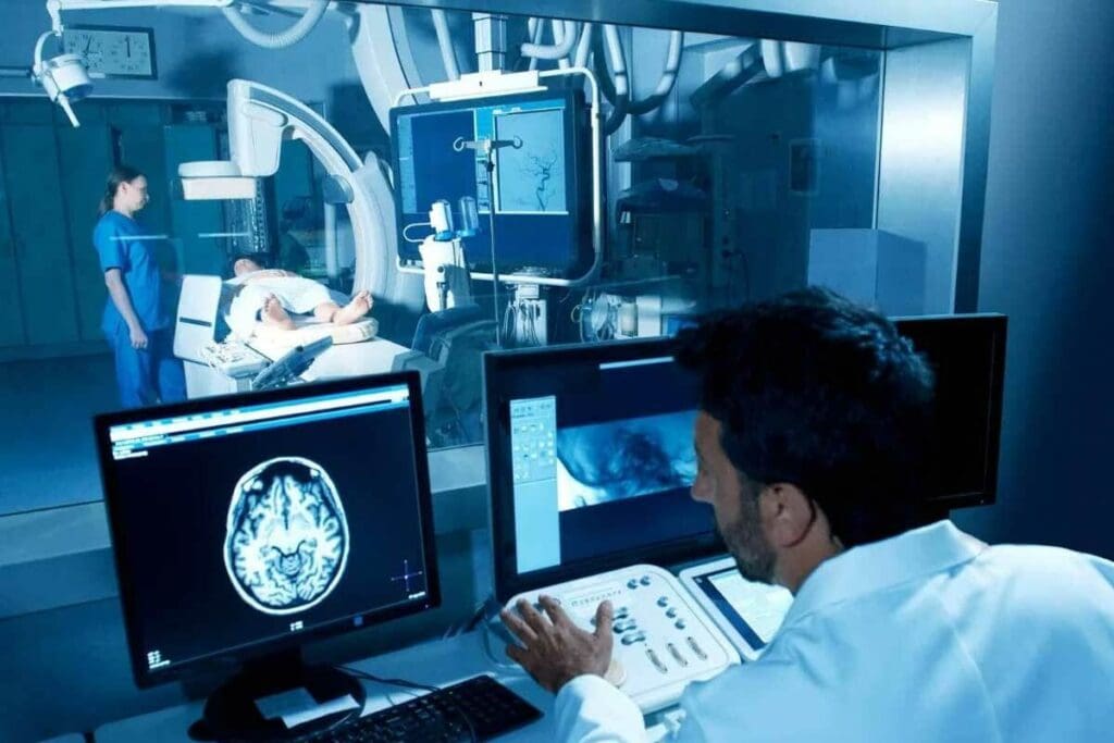
Medical imaging is key in today’s healthcare. It lets doctors see inside the body. This helps them find and treat many health issues, like broken bones and tumors.
The Evolution of Medical Imaging Technology
Medical imaging has grown a lot over time. Wilhelm Conrad Röntgen found X-rays in 1895. Later, MRI and CT scans were developed. These changes have made diagnosing diseases more accurate and care better.
CT scans came out in the 1970s. They let doctors see body parts in slices. MRI came next, showing soft tissues without harmful radiation. Each tech has its own uses and benefits.
Why Different Imaging Methods Are Necessary
Each imaging method is needed for different reasons. X-rays are great for bones and fractures. MRI is best for soft tissues like tendons and organs. CT scans are good for both bones and soft tissues.
| Imaging Modality | Primary Use | Key Benefits |
| X-ray | Bone fractures, lung conditions | Quick, widely available, low cost |
| CT Scan | Complex injuries, internal organs | Detailed cross-sectional images, fast |
| MRI | Soft tissue injuries, neurological conditions | High detail without radiation, versatile |
How Imaging Helps Guide Medical Decisions
Medical imaging is vital for making medical choices. It helps doctors find the right treatment. For example, it shows how bad an injury is and if treatment is working.
In short, knowing about medical imaging is important. It helps us see how it helps in healthcare. By understanding the different types and their uses, we can better follow our health journey and the choices our doctors make.
How X-Rays Work: Technology and What They Reveal
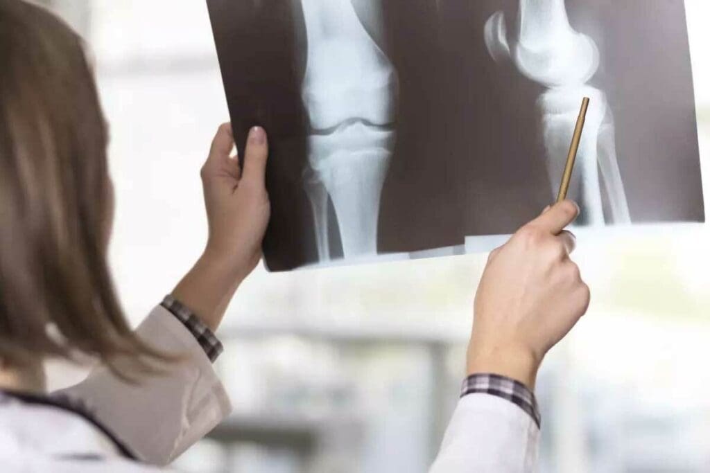
X-rays changed how doctors see inside the body without surgery. They use X-rays to show bones and other internal parts.
The Science Behind X-Ray Imaging
X-rays work because different things block X-rays in different ways. Bones block a lot, showing up white on the image. Softer parts show up gray or black. This helps doctors see bones and some problems.
What X-Rays Can Show: Bones, Fractures, and Calcifications
X-rays are great for checking bones. They can spot fractures, bone spurs, and even stones in the kidneys. This makes X-rays key in fixing bones and in emergency care.
“X-rays are invaluable for diagnosing bone fractures and certain other conditions affecting the skeletal system.”
Limitations: What X-Rays Cannot Detect
But X-rays can’t see everything. They can’t show soft tissues like tendons and ligaments well. So, they can’t always find sprains or tears in these tissues.
| Condition | X-Ray Visibility |
| Bone Fractures | High |
| Ligament Sprains | Low |
| Tendon Tears | Low |
| Calcifications | High |
Knowing what X-rays can and can’t do is important. They’re a big help in diagnosing, but other tests like MRI and CT scans are needed too. These tests help see soft tissue injuries better.
CT Scan Technology: Three-Dimensional X-Ray Imaging
CT scan technology has changed how we diagnose injuries and conditions. It gives doctors a clear view of the body’s inside. Unlike X-rays, CT scans show more of what’s inside us.
Creating 3D Images
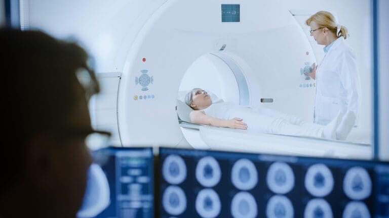
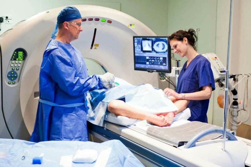
CT scans make detailed 3D images by combining X-rays from different angles. A CT scanner moves around the patient, taking X-ray measurements. These measurements are then turned into detailed images by computers.
These images show both bones and soft tissues clearly. This helps doctors diagnose complex conditions better. For example, CT scans can spot internal injuries that X-rays miss.
Revealing Complex Injuries and Internal Organs
CT scans are great for showing complex injuries and internal organs. The 3D images they create give a full view of the affected area. This helps doctors understand the injury or condition better.
- Detailed visualization of complex fractures
- Assessment of internal organ damage
- Identification of conditions such as tumors or cysts
For more info on X-rays, CT scans, and MRI, check out this resource.
When CT Scans Are Recommended Over X-Rays
Doctors often choose CT scans over X-rays for a closer look. This is true for severe injuries or suspected internal damage that X-rays can’t see.
The choice between CT and X-ray depends on the patient’s situation and what’s needed for diagnosis. For example, in emergencies, a CT scan can quickly show internal injuries.
MRI Technology: A Different Approach
CT scans use X-rays, but MRI uses magnets and radio waves instead. This makes MRI great for soft tissue conditions. It’s a safe choice because it doesn’t use radiation.
MRI’s high-resolution images of soft tissue are valuable. But whether to use CT or MRI depends on the specific case and what’s needed for diagnosis.
X Ray vs CT vs MRI: 7 Key Differences Compared
In medical imaging, X-ray, CT, and MRI are key tools. They differ in technology, use, and what they can show. Knowing these differences helps doctors and patients choose the right tests.
Difference 1: Imaging Technology and Radiation Exposure
X-rays, CT scans, and MRI scans work in different ways. X-rays use radiation to show bones and dense tissues. CT scans use X-rays too, but make detailed images by combining many views.
MRI uses a magnetic field and radio waves to create images without radiation. This makes MRI safer for some, like pregnant women and kids.
Difference 2: Image Detail and Dimensional Capabilities
X-rays show two-dimensional images, mainly bones and teeth. CT scans give more detailed images and can show three-dimensional views. This is great for complex injuries and organs.
MRI shows detailed images of bones and soft tissues without radiation. It’s best for brain, spine, and joint issues. MRI’s ability to show images in different planes is key for surgery planning.
Difference 3: Soft Tissue vs. Bone Visualization
MRI is top for soft tissue images, showing different soft tissues well. X-rays and CT scans are better for bones. X-rays check for bone fractures, while CT scans show complex bone injuries.
Difference 4: Scan Duration and Patient Experience
Scan time and patient experience vary. X-rays are quick, taking just a few minutes. CT scans are also fast, usually done in minutes. MRI scans, though, can take up to an hour.
MRI scans can be tough for those with claustrophobia. Open MRI machines help. CT scans and X-rays are generally easier, but CT scans might use contrast agents that can cause allergies.
Can X-Rays Show Tendon or Ligament Damage?
Tendon and ligament damage can be hard to spot without the right tools. X-rays are often used first, but they’re not great at showing soft tissue injuries.
Soft Tissue Injuries and X-Ray Limitations
X-rays are mainly for looking at bones and finding fractures. They don’t work well for soft tissue problems like tendon or ligament damage. This is because X-rays mostly show dense things like bones, not soft tissues.
There are a few reasons why X-rays aren’t good for this:
- Soft tissues like tendons and ligaments don’t block X-rays much, so they’re hard to see.
- X-rays can’t show the fine details needed to spot tears or strains in soft tissues.
- You might see signs like swelling or changes in joint space, but these aren’t clear signs of damage.
CT Scans for Tendon and Ligament Assessment
CT scans give more detailed pictures than X-rays and can make 3D models of injuries. But, they’re not perfect for looking at soft tissues.
CT scans can:
- Show bone fragments and how they relate to soft tissues better.
- Spot some signs of soft tissue injuries, like swelling or bleeding.
Even with these benefits, CT scans aren’t the best for finding tendon or ligament tears because they don’t show soft tissues well.
MRI: The Gold Standard for Soft Tissue Imaging
MRI is the top choice for seeing soft tissue injuries, like tendon and ligament damage. It shows soft tissues very clearly, making it great for finding tears and strains.
MRI has many benefits:
- It gives high-quality images of soft tissues, helping to check tendon and ligament health.
- It can find many soft tissue problems, from small strains to big tears.
- It doesn’t use radiation, making it safer for patients who need many scans.
In short, while X-rays have their uses, they’re not good for finding tendon or ligament damage. CT scans help a bit but are not perfect. MRI is the best for soft tissue injuries, giving the clear images needed for diagnosis and treatment.
Alternatives to MRI: Options When MRI Isn’t Possible
When MRI isn’t an option, finding other ways to diagnose is key. This is true for people with metal implants, claustrophobia, or other issues that make MRI scans hard. Other imaging methods can give the needed info.
Ultrasound Imaging for Soft Tissue Evaluation
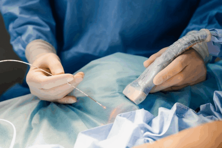
Ultrasound is a great choice for soft tissue injuries or conditions. It uses sound waves to see inside the body. It’s good for checking tendons, ligaments, gallbladder issues, and some blood vessel problems.
Ultrasound is safe and doesn’t hurt. It’s fast and doesn’t use radiation. This makes it perfect for those who can’t have MRI or CT scans.
When CT Might Substitute for MRI
CT scans can replace MRI in emergencies or for detailed organ or bone injury images. They use X-rays to show body parts in slices.
CT scans have radiation but are quicker and might be better for those afraid of tight spaces. Yet, they’re not as good as MRI for soft tissue.
Considerations for Patients with Contraindications to MRI
For those who can’t have MRI, like those with metal implants or pacemakers, picking the right imaging is important. Ultrasound and CT scans are good options, but it depends on the condition.
We look at the patient’s health, their condition, and what info is needed. This way, we make sure they get the best care for their situation.
International Standards in Medical Imaging
Medical imaging is key in today’s healthcare. International standards make it consistent, reliable, and safe for everyone. They ensure that imaging practices are top-notch worldwide.
Academic Protocols and Care Pathways in Imaging
Academic protocols and care pathways guide doctors in making accurate diagnoses. Standardized protocols reduce variability in imaging. This leads to better patient outcomes. For example, LivHospital follows strict academic protocols for high-quality imaging.
Here are the main points of academic protocols in medical imaging:
- Evidence-based guidelines for imaging procedures
- Continuous education and training for healthcare professionals
- Quality control measures to ensure accuracy and reliability
Patient Safety and Experience in Medical Imaging
Patient safety is a top priority in medical imaging. International standards aim to reduce radiation exposure and ensure patient comfort. Patient-centered care focuses on reducing anxiety and improving the experience.
Improving patient safety and experience includes:
- Using the lowest effective dose of radiation
- Implementing patient-friendly imaging protocols
- Ensuring clear communication between healthcare providers and patients
Innovative Solutions in Modern Imaging Centers
Modern imaging centers lead in adopting new solutions. Advances like AI-assisted imaging improve diagnostic accuracy and patient care. These technologies enhance image analysis and streamline workflows.
- Advanced imaging modalities like 3D and 4D imaging
- Integration of artificial intelligence in image analysis
- Personalized imaging protocols based on patient needs
Conclusion: Choosing the Right Imaging Test
Choosing the right imaging test is key when diagnosing medical conditions. We’ve looked at X-ray, CT, and MRI scans, each with its own strengths. X-rays are great for checking bones, CT scans for internal organs, and MRI for soft tissues like ligaments and discs.
Knowing what each scan can do helps doctors and patients pick the best one. The choice depends on the condition, how detailed the scan needs to be, and the patient’s situation. Talking to healthcare experts helps patients make smart choices about their care.
In short, knowing the differences between X-ray, CT, and MRI scans helps patients make better choices. This knowledge lets them navigate the diagnostic process confidently. It leads to better health outcomes.
FAQ
Can X-rays detect tendon or ligament damage?
X-rays are not good for finding soft tissue injuries like tendon or ligament damage. They work better for seeing bones, fractures, and calcifications.
What is the difference between an X-ray and a CT scan?
X-rays use one beam of radiation for a 2D image. CT scans use many X-ray beams for detailed 3D images. CT scans give more info, great for complex injuries.
How does MRI differ from X-ray and CT scans?
MRI uses magnets and radio waves, not radiation. It’s perfect for soft tissues like tendons, ligaments, and organs. It shows detailed images without radiation.
Are there alternatives to MRI for soft tissue evaluation?
Yes, ultrasound imaging is good for some soft tissue checks. CT scans can also work in some cases. But MRI is the best for many soft tissue injuries.
Can CT scans show tendon or ligament damage?
CT scans can show more than X-rays, but they’re not the best for soft tissue injuries. MRI is usually better for diagnosing tendon or ligament damage.
What are the key differences between X-ray, CT, and MRI scans?
The main differences are in technology, radiation, image detail, and patient experience. X-rays use radiation for 2D bone images. CT scans use radiation for 3D images. MRI uses magnets and radio waves for soft tissue details.
Is there an alternative to MRI for patients with contraindications?
Yes, for those who can’t have MRI, ultrasound or CT scans might be options. It depends on the medical condition being checked.
Do X-rays show ligaments or tendons?
X-rays can’t show ligaments or tendons well because they’re soft tissues. They’re better for bones and finding fractures or calcifications.
What imaging modality is best for visualizing torn tendons or ligaments?
MRI is the top choice for seeing torn tendons and ligaments. It gives detailed images of soft tissues.
How do international standards impact medical imaging practices?
International standards help ensure medical imaging follows strict safety and quality rules. They guide places like LivHospital to provide top-notch care.
Reference
- American Academy of Orthopaedic Surgeons. (2022, December 31). X-rays, CT Scans, and MRI Scans. OrthoInfo. https://orthoinfo.aaos.org/en/treatment/x-rays-ct-scans-and-mris/


