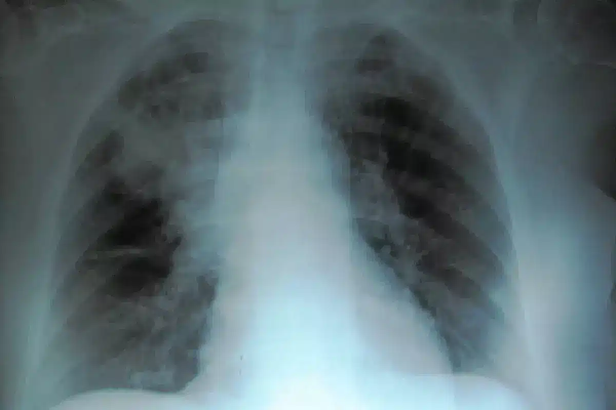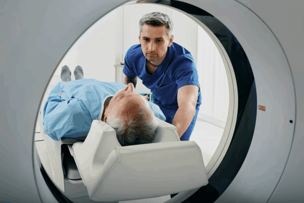
A gallbladder CT scan is a key tool for checking gallbladder health. It gives detailed images that doctors use to find and treat diseases.
The gallbladder is a small, pear-shaped organ. It stores and releases bile from the liver, helping with digestion. A CT scan for gallbladder is great for detailed checks, like when there’s a chance of acute cholecystitis or cancer.
Knowing about gallbladder imaging helps patients see why it’s so important. It leads to better diagnosis and results.
Key Takeaways
- CT scans provide detailed cross-sectional images of the gallbladder.
- A gallbladder CT scan is useful for diagnosing various gallbladder conditions.
- It helps healthcare professionals manage gallbladder diseases.
- A CT scan is specially useful in cases of suspected acute cholecystitis or cancer.
- Understanding key facts about gallbladder imaging can improve patient outcomes.
What Is a Gallbladder CT Scan and When Is It Used?
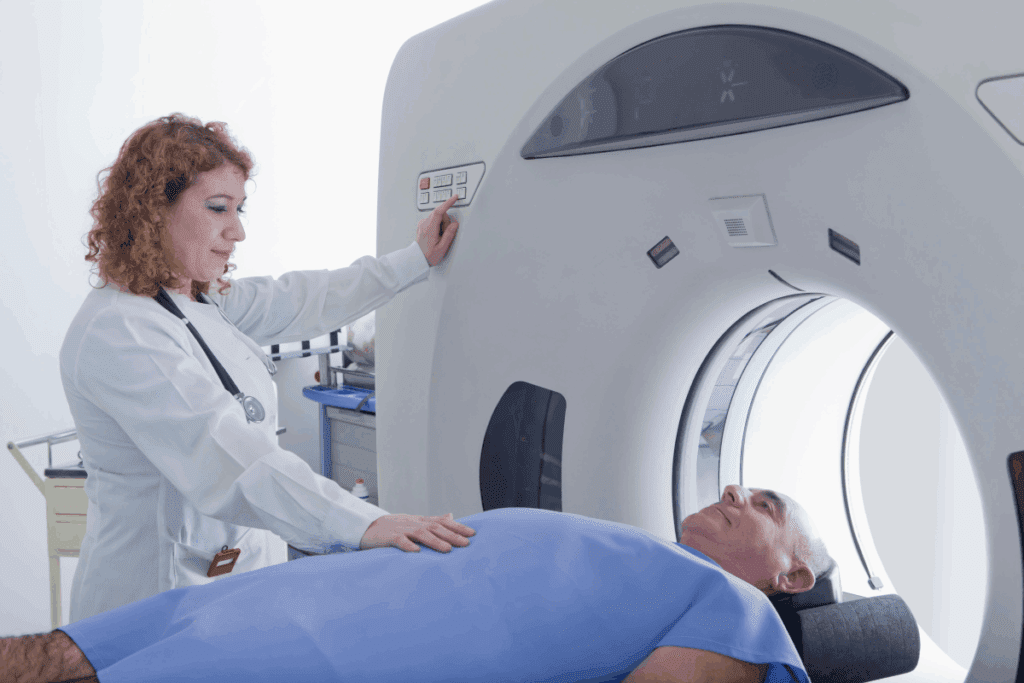
A gallbladder CT scan is a detailed test to check the gallbladder and nearby areas. It gives clear images that doctors use to find and treat gallbladder problems.
Definition and Basic Principles
A gallbladder CT scan is a non-invasive test that uses X-rays and computers to see the gallbladder. It works by moving an X-ray source and detector around the body. This captures data to make detailed images.
Contrast agents are used to make some parts clearer on the ct of gallbladder. They can be taken by mouth or through an IV, depending on the scan’s needs.
Cross-Sectional Imaging Technology
CT scans give cross-sectional images of the gallbladder. This lets doctors see its structure and any problems in detail. It’s great for finding issues that other tests can’t see.
Common Clinical Indications
A gallbladder CT scan is often used to find and check gallbladder issues. This includes acute cholecystitis, gallstones, and tumors. It helps spot inflammation, blockages, or other problems causing symptoms.
| Clinical Indication | Description | Role of CT Scan |
| Acute Cholecystitis | Inflammation of the gallbladder | Identify signs of inflammation, such as wall thickening and pericholecystic fluid |
| Gallstones | Presence of stones in the gallbladder | Detect radiopaque stones and assess for possible obstruction |
| Gallbladder Tumors | Mass lesions within the gallbladder | Evaluate the extent of the tumor and check for metastasis |
Using gallbladder ct with contrast makes the scan even better. It’s very helpful when you need to see blood vessels or tumors clearly.
The Anatomy of the Gallbladder on CT Imaging
Knowing the gallbladder’s anatomy is key to reading CT scans right. The gallbladder’s look on CT scans, along with nearby structures, helps doctors spot gallbladder diseases.
Normal Gallbladder Appearance
A normal gallbladder CT scan shows the gallbladder as a pear-shaped thing. It has a thin wall and a smooth edge. The wall should be under 3 mm thick when the gallbladder is empty.
On a CT scan, a normal gallbladder looks like a clear, defined space. No stones, masses, or abnormal thickening should be present in a normal gallbladder.
Surrounding Structures and Relationships
The gallbladder sits under the liver, linked to the liver, bile ducts, and blood vessels. On a CT scan gallbladder imaging, these structures are seen. This gives a full view of the gallbladder’s area.
The gallbladder’s spot next to the liver is key. It’s in a fossa on the liver’s lower right part.
Anatomical Variants to Be Aware Of
There are gallbladder variations like a duplicated or misplaced gallbladder seen on gallbladder anatomy CT scans. Knowing these variations is important for correct diagnosis. This helps avoid mistaking normal variations for diseases.
Other variations include gallbladder septa, diverticula, or an odd-shaped gallbladder.
Healthcare pros need to know the normal and varied gallbladder anatomy on CT scans. This knowledge helps them read CT scans well and make good medical choices.
Preparation and Procedure for a Gallbladder CT Scan
Getting ready for a gallbladder CT scan is important. It helps get clear images for diagnosing gallbladder diseases.
Pre-Scan Instructions
Before the scan, you’ll get instructions. These tell you what foods or medicines to avoid. It’s key to follow these steps for the best scan results.
- Remove any jewelry or metal objects that could interfere with the CT scan.
- Wear loose, comfortable clothing without metal fasteners.
- Inform your doctor about any allergies or sensitivities, specially to contrast agents.
Fasting Requirements
Fasting is needed before the scan. This fasting period is usually 4 to 8 hours. You should not eat or drink anything during this time. It helps get clear images of your gallbladder.
What to Expect During the Procedure
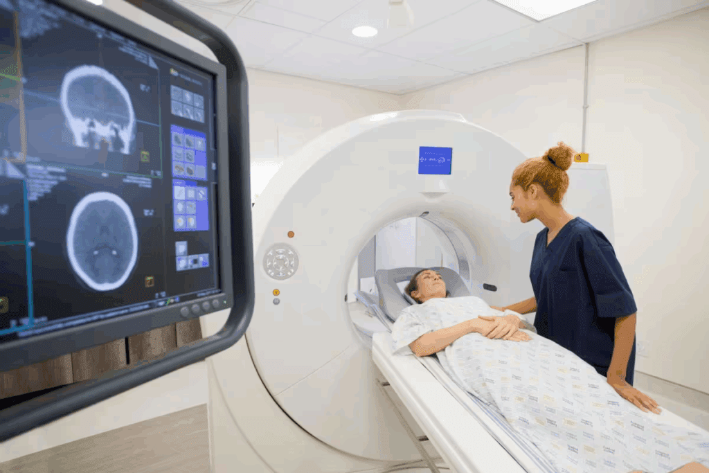
During the scan, you’ll lie on a CT scanner table. The scan takes just a few minutes. X-rays are used to create detailed images of your gallbladder. Contrast agents may be given orally or through an IV to make certain parts more visible.
You’ll need to stay very quiet during the scan. This is to avoid blurry images. The scan is usually painless, but some might feel a bit uncomfortable. This could be from the contrast agent or staying very quiet for a while.
“The key to a successful gallbladder CT scan lies in proper preparation and understanding the procedure,” says Dr. John Smith, a radiologist with over a decade of experience.
By following the pre-scan instructions and knowing what to expect, you can help make sure your scan is accurate and helpful.
Gallbladder CT Scan With Contrast: Enhanced Diagnostic Capabilities
Using contrast in gallbladder CT scans changes how we find problems. These scans with contrast media give a clearer and more precise diagnosis than scans without contrast.
Types of Contrast Media Used
Contrast agents in gallbladder CT scans are usually iodine-based or barium sulfate. Iodine-based contrasts are preferred because they are safe and show vascular structures and some pathologies well.
The right contrast media depends on the patient’s allergies, kidney health, and what the doctor needs to see.
| Type of Contrast | Characteristics | Common Uses |
| Iodine-based | High-density contrast, good vascular enhancement | Vascular studies, tumor detection |
| Barium Sulfate | Used for gastrointestinal tract imaging | Gastrointestinal studies |
Benefits of Contrast Enhancement
Contrast in gallbladder CT scans makes diagnosis more accurate. It helps see inflammation, tumors, and blood vessels better.
- Improved detection of gallbladder pathology
- Enhanced visualization of surrounding structures
- Better differentiation between various types of lesions
A study in the Journal of Radiology says, “Contrast-enhanced CT scans are key in diagnosing abdominal problems, including gallbladder disease.”
“Contrast-enhanced CT is invaluable for assessing the extent of gallbladder disease and planning appropriate treatment strategies.”
— Radiology Expert
Potential Risks and Contraindications
While contrast-enhanced CT scans have many benefits, there are risks and things to avoid. These include allergic reactions and kidney problems from the contrast.
To lower these risks, doctors check for allergies and kidney health before the scan.
Precautions for Contrast Use:
- Assess patient for contrast allergies
- Evaluate kidney function
- Hydrate patient before and after scan
Knowing the good and bad of contrast-enhanced gallbladder CT scans helps doctors decide when to use them.
Interpreting a Normal Gallbladder CT Scan
Understanding what a normal gallbladder looks like on a CT scan is key. It helps doctors spot problems. A normal scan is a baseline for finding issues.
Normal Wall Thickness and Density
A normal gallbladder wall is thin and even, less than 3 mm thick. It should look smooth and have the same density everywhere. There should be no thickening or irregularities.
Expected Gallbladder Size and Shape
The size of a normal gallbladder can vary. It’s usually pear-shaped and sits on the liver’s right lobe. It shouldn’t be too big, and its inside should look the same all over.
Normal Surrounding Tissues
The areas around the gallbladder, like the liver and bile ducts, should look normal on a CT scan. There should be no swelling, fluid, or other issues nearby.
| Characteristics | Normal Findings |
| Gallbladder Wall Thickness | Less than 3 mm |
| Gallbladder Shape | Pear-shaped |
| Surrounding Tissues | No signs of inflammation or abnormalities |
By knowing these normal signs, doctors can spot problems on a gallbladder CT scan. This helps them make better care plans for patients.
Abnormal Gallbladder CT Scan Findings and Their Clinical Significance
Abnormal gallbladder CT scans can show many important findings. These can include changes in the gallbladder wall, size, or shape. They can also show fluid or masses around it.
Wall Thickening and Inflammation
Wall thickening is a common finding on gallbladder CT scans. It often means inflammation or disease. Wall thickening greater than 3 mm is generally considered abnormal and may suggest conditions such as acute cholecystitis, chronic cholecystitis, or other inflammatory diseases.
Inflammation can be further assessed by looking for signs such as pericholecystic fat stranding or the presence of pericholecystic fluid. These findings help determine the severity of the inflammation and guide treatment.
Gallbladder Distention
Gallbladder distention is another abnormal finding on CT scans. It is characterized by an enlarged gallbladder. This can be caused by bile duct obstruction or other conditions that impede bile flow.
A distended gallbladder is a sign of an underlying issue that needs medical attention. The CT scan can identify the cause and guide further management.
Pericholecystic Fluid and Stranding
Pericholecystic fluid and stranding around the gallbladder indicate inflammation or infection. Pericholecystic fluid appears as a collection of fluid around the gallbladder, while stranding refers to increased density in the surrounding fat, suggesting inflammation.
| Finding | Description | Clinical Significance |
| Wall Thickening | Thickening of the gallbladder wall beyond 3 mm | Suggests inflammation or other pathology |
| Gallbladder Distention | Enlargement of the gallbladder | May indicate bile duct obstruction or other conditions |
| Pericholecystic Fluid and Stranding | Presence of fluid and increased density in surrounding fat | Indicates inflammation or infection |
Mass Lesions and Tumors
Mass lesions and tumors on gallbladder CT scans can be benign or malignant. The CT scan can identify the presence of a mass and provide information about its size, location, and characteristics.
Further evaluation with additional imaging modalities or biopsy may be necessary to determine the nature of the mass lesion. Understanding the characteristics of the mass is key for planning treatment and management.
Detecting Gallstones on CT: Capabilities and Limitations
CT scans are great for many health issues, but they have limits when it comes to gallstones. How well a CT scan can spot gallstones depends on a few things. These include the size and what the stones are made of.
Sensitivity of CT Scans for Cholelithiasis
CT scans can find gallstones, but they’re not always perfect. Big stones or those with lots of calcium are easier to see. The sensitivity of cholelithiasis CT scans is better for these kinds of stones.
The type of gallstone also matters. Stones mostly made of cholesterol are harder to spot than those with calcium.
Factors Affecting Gallstone Visibility
Several things can change how well CT scans can see gallstones. These include:
- Size: Bigger stones are easier to spot.
- Composition: Stones with calcium are more visible.
- Location: Some spots in the body are harder to see stones in.
Radiopaque vs. Radiolucent Stones
Gallstones can be either radiopaque or radiolucent. Radiopaque stones have calcium and show up on CT scans. On the other hand, radiolucent stones are mostly cholesterol and might not be seen.
It’s important to know what CT scans can and can’t do for gallstones. This helps doctors make the right plans for treatment. By understanding what affects how well gallstones show up, doctors can make better choices.
GB CT for Acute Cholecystitis: Diagnostic Criteria
Accurate diagnosis of acute cholecystitis is key, and gallbladder CT scans are a top tool. This serious condition needs quick medical care to avoid serious issues.
Primary Signs of Acute Inflammation
CT scans help spot primary signs of inflammation in acute cholecystitis. These include:
- Gallbladder distention
- Wall thickening
- Pericholecystic fluid
Gallbladder distention is a major sign, often with wall thickening from inflammation. Pericholecystic fluid shows inflammation spreading beyond the gallbladder.
Secondary Signs and Complications
CT scans also show secondary signs and complications. These include:
- Inflammation of surrounding tissues
- Gangrene
- Perforation
These signs suggest a more serious disease, possibly needing urgent surgery.
Diagnostic Accuracy Rates
CT scans are very accurate for diagnosing acute cholecystitis. A study compared different diagnostic methods. It found CT scans were as good as, or better than, ultrasound in spotting this condition.
| Diagnostic Modality | Sensitivity (%) | Specificity (%) |
| CT Scan | 92 | 95 |
| Ultrasound | 90 | 93 |
The table shows CT scans are very accurate in diagnosing acute cholecystitis. This makes them a trusted tool for doctors.
“The use of CT scans in diagnosing acute cholecystitis represents a significant advancement in the management of this condition, enabling timely and appropriate treatment.”
CT of the Gallbladder for Cancer Detection and Staging
CT scans are key in finding gallbladder cancer and figuring out its stage. They show detailed images of the gallbladder and nearby tissues. This makes them very useful for spotting tumors and seeing how big they are.
Imaging Features of Gallbladder Malignancy
Gallbladder cancer can show up on CT scans in different ways. It might look like mass lesions, wall thickening, or a mix of both. These signs are important for spotting cancer on CT scans.
Advanced CT techniques help make these signs clearer. This makes it easier to find cancer early. Using contrast media also helps show the tumor and how it relates to nearby areas.
Staging Capabilities
Knowing the stage of gallbladder cancer is key for choosing the right treatment. CT scans help by showing how far the cancer has spread, if it’s in lymph nodes, and if it has gone to other parts of the body. This info helps doctors decide if surgery is needed or if other treatments are better.
Integration with Other Diagnostic Methods
Diagnosing and staging gallbladder cancer often involves using CT scans along with other tests like ultrasound, MRI, and PET scans. This way, doctors get a full picture of the disease. This helps them make better treatment plans and improve patient care.
By using different imaging methods together, doctors can get a clearer view of gallbladder cancer. This leads to better care for patients.
Comparing Gallbladder CT Scans to Other Imaging Modalities
There are many ways to check for gallbladder disease, each with its own benefits and drawbacks. The right test depends on the patient’s situation, what doctors think might be wrong, and what’s available.
CT vs. Ultrasound
Ultrasound is often the first choice because it’s easy, available everywhere, and doesn’t use harmful radiation. But, CT scans give clearer pictures and can spot some problems better, like cancer or serious inflammation. A study showed CT scans are better at seeing thickened gallbladder walls and fluid around it.
A top radiologist said, “CT scans have changed how we handle tough gallbladder cases.” This shows why CT scans are key when ultrasound isn’t clear or when more info is needed.
CT vs. MRI
MRI gives detailed images without radiation, which is good for some patients. But, MRI costs more and isn’t as common as CT. MRI is great for looking at complex issues or the biliary system.
Both CT and MRI have their own strengths for gallbladder disease. CT is better for finding stones and some complications. MRI is better at showing soft tissues.
CT vs. HIDA Scan
HIDA scans check how well the gallbladder works. They’re good for acute cholecystitis and checking how well the gallbladder empties. But, HIDA scans don’t give as much detail as CT scans.
When it’s hard to tell what’s wrong, doctors might use more than one test. For example, someone with suspected acute cholecystitis might get both ultrasound and a HIDA scan.
Choosing the Right Imaging Test
Choosing the right test for gallbladder disease depends on many things. A radiologist helps pick the best test for each patient.
As radiology gets better, new tools and techniques will help us diagnose and treat gallbladder disease even better.
Advances in Gallbladder CT Scan Technology and Techniques
Recent CT scan advancements have greatly improved gallbladder disease diagnosis and treatment. These updates make CT scans more accurate and efficient. This leads to better health outcomes for patients.
Low-Dose CT Protocols
Low-dose CT protocols have been introduced to lower radiation exposure for patients. This is key for those needing repeated scans. These protocols use better scanner settings and algorithms to achieve this.
Low-dose CT scans are safer, which is great for pregnant women and those needing frequent scans. Studies have shown they keep diagnostic accuracy high while cutting radiation doses.
Dual-Energy CT Applications
Dual-energy CT technology is another big step forward. It scans at two energy levels to give more detailed tissue information.
This tech is great for identifying gallstones and other gallbladder issues. It can spot different stone types and tissue changes, boosting diagnostic confidence.
AI and Computer-Aided Diagnosis
AI and computer-aided diagnosis (CAD) systems are changing how we read CT scans. They use algorithms to spot issues and measure them.
AI-CAD helps radiologists by pointing out important areas. It lowers the chance of missing diagnoses and speeds up reading times. It’s very helpful in complex cases or when experts are scarce.
These technologies will keep improving gallbladder CT scans. With low-dose CT protocols, dual-energy CT applications, and AI-driven CAD systems, doctors can give more precise diagnoses. This leads to better treatment plans for gallbladder disease patients.
Conclusion: The Future of CT in Gallbladder Disease Management
The future of CT in managing gallbladder disease looks bright. New technology and techniques are making diagnoses more accurate and care better. We can look forward to clearer images and less radiation.
AI and computer-aided diagnosis will become more important in reading CT scans. This will help doctors make better choices for their patients. By keeping up with these advancements, doctors can give the best care to those with gallbladder disease.
New CT tech, like low-dose scans and dual-energy CT, will make CT scans even more useful. These improvements will keep CT scans a key tool in treating gallbladder disease.
FAQ
What is a gallbladder CT scan?
A gallbladder CT scan is a test that uses X-rays and computer tech. It makes detailed images of the gallbladder.
When is a gallbladder CT scan used?
Doctors use it to check for gallbladder problems. This includes symptoms like pain, gallstones, or tumors.
What is the difference between a CT scan with and without contrast?
A CT scan with contrast uses special agents to show more details. A non-contrast scan doesn’t use these agents.
How do I prepare for a gallbladder CT scan?
You’ll need to fast for a few hours before. This helps the gallbladder be ready for the scan. You might also get instructions on what to avoid eating or taking.
Can a CT scan detect gallstones?
Yes, it can find gallstones. But, it depends on the stone’s size and type. Bigger or calcium-rich stones are easier to spot.
What are the signs of acute cholecystitis on a CT scan?
Signs include a swollen gallbladder, thick walls, and fluid around it. Other signs might be inflammation, gangrene, or a hole in the gallbladder.
How is gallbladder cancer detected and staged using CT scans?
CT scans can find gallbladder cancer and tell how far it has spread. This helps doctors plan treatment.
What are the advantages and disadvantages of CT scans compared to other imaging modalities?
CT scans give clear images and can spot some issues. But, they use radiation and contrast. Other tests like ultrasound or MRI have their own benefits and drawbacks.
What are the latest advances in gallbladder CT scan technology and techniques?
New tech includes lower-dose scans, dual-energy CT, and AI help. These advancements improve how accurate and helpful CT scans are.
Can a CT scan be used to diagnose other gallbladder conditions beside gallstones and cancer?
Yes, it can diagnose other issues like inflammation, tumors, or unusual shapes. It helps doctors plan the best treatment.
References
- Chemmanur, A. T., & Anand, B. S. (2025, May 15). Biliary disease workup: Laboratory studies, imaging studies, and staging. Medscape. https://emedicine.medscape.com/article/171386-workup
- Alessa, M. Y., Aljohani, S., Alhashem, F., & Alshammari, T. (2025). The association of liver enzymes with acute cholecystitis: A retrospective study. Journal of Clinical Gastroenterology and Hepatology, ( ?), ?-?. https://www.ncbi.nlm.nih.gov/pmc/articles/PMC12001050/
- Yuen, W. Y. R., Piteša, R., McHugh, T., Poole, G., & Singh, P. P. (2023). Liver function tests as predictors of choledocholithiasis: A scoping review. Annals of Hepato-Biliary Surgery, 3, ( ?). Retrieved from https://asj.amegroups.org/article/view/75800/html


