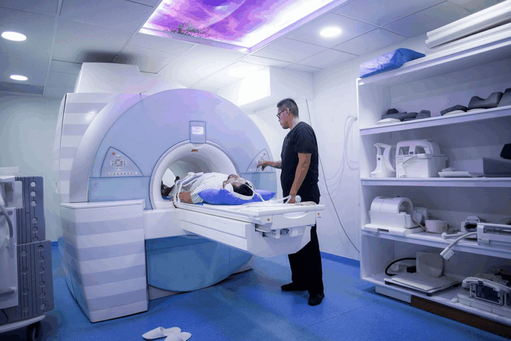
Finding the right hernia diagnostic imaging is key for good treatment. But, many wonder if CT scans or MRI is better. At Liv Hospital, we focus on top-notch care, making sure it’s safe, complete, and good for the patient.
Doctors often talk about the best ways to find hernias. We’ll look at what CT scans and MRI can do. This will help us figure out which is best for detecting hernias.
We’ll dive into the latest studies and data. Our goal is to give a clear view of how these tests work for finding hernias.
Key Takeaways
- CT scans give quick, detailed views for hernia detection.
- MRI shows soft tissue clearly, helping doctors diagnose.
- Choosing between CT scans and MRI depends on the patient’s needs.
- Good diagnostic care is vital for treating hernias well.
- Liv Hospital puts patient safety, thoroughness, and experience first in care.
Understanding Hernia Diagnosis and Imaging Needs
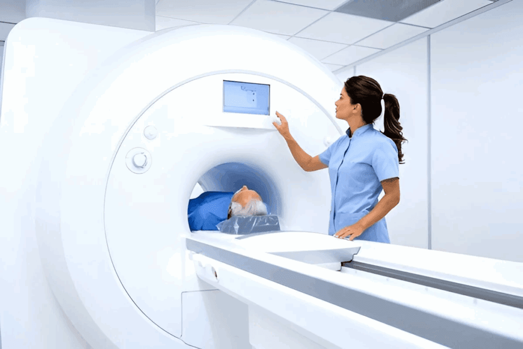
Getting a hernia diagnosis right is key, and tools like CT scans and MRI are leading the way. Hernias happen when an organ bulges through a weak spot in the muscle or fascia. While a physical check can spot many hernias, imaging is vital for unclear cases or to see how serious a hernia is.
What Is a Hernia and Why Imaging Matters
A hernia forms when a muscle or fascia weakens, letting an organ bulge out. Imaging is critical for finding hernias, like hidden or recurring ones. CT scans and MRI give detailed pictures that help doctors understand the hernia’s size, location, and how complex it is.
Most hernias can be found without imaging. But, imaging is key for:
- Hidden or recurring hernias
- Understanding how serious a hernia is
- Planning surgery
- Watching for complications after surgery
The Evolution of Hernia Diagnostic Techniques
Diagnosing hernias has changed a lot with new imaging tech. Before, doctors mostly used physical checks and simple X-rays. But now, CT scans and MRI give detailed views of hernias and the area around them.
Doctors say advanced imaging has made diagnosing and treating hernias better. A top surgeon noted, “Modern imaging has greatly improved how we find and fix hernias.”
“The precision offered by modern imaging techniques has significantly enhanced our ability to diagnose and treat hernias effectively.”
Research keeps improving how we diagnose and manage hernias. New imaging methods and tech are being explored, helping us understand and treat hernias better.
The Fundamentals of Hernia Types and Presentations
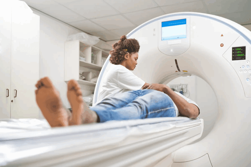
Hernias come in different forms and locations, making them a challenge to diagnose. We will look at the various types of hernias, their characteristics, and the challenges in diagnosing them.
Common Hernia Types and Their Characteristics
There are several common types of hernias, each with its own features. These include:
- Inguinal Hernias: These happen when tissue, like part of the intestine, bulges through a weak spot in the abdominal muscles in the groin area.
- Ventral Hernias: These occur in the abdominal wall, often at the site of a previous surgical incision.
- Hiatal Hernias: These happen when part of the stomach pushes up through the diaphragm into the chest cavity.
Each hernia type has its own presentation and diagnostic needs. For example, inguinal hernias are more common in men. Hiatal hernias are often linked with symptoms like gastroesophageal reflux disease (GERD).
Clinical Challenges in Hernia Diagnosis
Diagnosing hernias can be tricky because of their varied presentations and similar symptoms to other conditions. Doctors often use a combination of physical exams and patient history to diagnose. Sometimes, imaging is needed to confirm the diagnosis or plan treatment.
The choice to use imaging like CT scans or MRI depends on several factors. These include the suspected hernia type and the patient’s health. For instance, a CT scan is useful for complex or recurrent hernias, as it shows detailed images of the abdominal cavity.
When Imaging Becomes Necessary
Imaging is needed when the diagnosis is unclear or when the hernia’s extent needs to be assessed for surgery. Both CT scans and MRI can diagnose hernias, each with its own benefits. A CT scan is quick and often used in emergencies. MRI is better for soft tissue evaluation and doesn’t use radiation.
The choice between CT and MRI depends on the situation and the hernia’s characteristics. For example, if there’s a suspicion of a complicated or strangulated hernia, a CT scan might be preferred for its speed and detailed images.
In conclusion, knowing about hernia types and presentations is key for accurate diagnosis and treatment planning. By using clinical evaluation and the right imaging, healthcare professionals can offer the best care for patients with hernias.
CT Scan Technology: How It Works for Hernia Detection
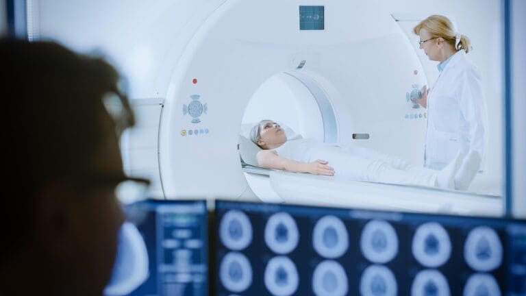
CT scan technology is key in finding hernias. It uses X-rays and computers to make detailed images. These scans help us see inside the body clearly, which is important for diagnosing hernias.
The Technical Principles of CT Scanning
CT scans use X-rays and computers to make detailed images. They rotate around the patient, taking pictures from different angles. Then, a computer puts these images together into a 3D picture.
This technology lets us see where a hernia is, how big it is, and what kind it is. Knowing this helps us figure out the best treatment.
Cross-Sectional Imaging Advantages
CT scans have many benefits for finding hernias. They let us see the hernia from different sides. This is very helpful when the hernia is hard to see or is hidden by other parts of the body.
For example, a CT scan can tell us if a hernia is stuck or strangulated. This is important for deciding if surgery is needed and what kind of surgery.
To learn more about diagnosing hernias, visit Diagnostic Testing for Hernias.
Contrast Enhancement in Hernia Visualization
Contrast enhancement makes CT scans even better for finding hernias. It uses a special dye to make certain areas stand out. This helps us see the hernia more clearly.
Contrast agents are very helpful when the hernia is small or hard to see. They also help us spot problems like bowel obstruction. This makes sure we can give the right diagnosis and treatment.
Can CT Detect Hernia? Effectiveness and Accuracy Rates
Computed Tomography (CT) scans are key in finding hernias. But how well do they work? We’ll look at how good CT scans are at spotting hernias. We’ll focus on their sensitivity, specificity, and what kinds of hernias they can find best.
Sensitivity and Specificity Statistics
Research shows CT scans are great at finding some hernias. For example, they can spot inguinal hernias with a 94% sensitivity and 96% specificity. This means they’re very good at finding these hernias and not many false positives.
CT scans are so good because they give detailed images of the belly and pelvis. This lets doctors see small hernias that other tests might miss.
Types of Hernias Best Visualized by CT
CT scans are best at showing certain hernias, like:
- Incisional hernias
- Inguinal hernias
- Ventral hernias
- Spigelian hernias
These hernias are easier to see on CT scans because of where they are and the nearby body parts. For example, incisional hernias through old scars are clear on CT scans because of the surgical clips and changed anatomy.
Limitations of CT in Hernia Detection
Even though CT scans are great at finding hernias, they have some downsides. Small hernias or tricky ones might get missed. Also, CT scans use radiation, which is a worry, mainly for young people or those needing many scans.
The accuracy of CT scans can also depend on the scan quality, the doctor’s skill, and any image problems. So, while CT scans are very useful, they should be part of a full check-up plan.
MRI Technology: Principles and Applications in Hernia Diagnosis
Magnetic Resonance Imaging (MRI) is key in finding hernias. It shows soft tissues clearly. This helps us see inside the body well, which is great for finding hernias.
How MRI Creates Soft Tissue Images
MRI uses a strong magnetic field and radio waves. It makes signals from hydrogen atoms in the body. Then, it turns these signals into detailed pictures of what’s inside us.
It’s really good at showing soft tissues. This makes it perfect for finding problems like hernias.
The steps are:
- Aligning hydrogen atoms in the body using a strong magnetic field.
- Disturbing these atoms with radio waves to generate signals.
- Capturing and processing these signals to create detailed images.
Specialized MRI Sequences for Hernia Evaluation
For hernia checks, we use special MRI sequences. These make the images clearer and more accurate. They help us see the hernia and the tissues around it better.
Some sequences are:
- T1-weighted images for detailed anatomy.
- T2-weighted images for assessing tissue edema and fluid collections.
- Cine MRI for evaluating dynamic movement and possible hernia occurrence.
Patient Experience During MRI for Hernia
Getting an MRI for a hernia is pretty simple. But, you might need to get ready first. You’ll have to take off any metal and wear a gown.
“The MRI scan is a painless procedure, but it does require remaining very quiet for a long time. Some people might feel scared because of the tight space. We try to make them comfortable and sometimes give them a little sleep medicine.”
During the scan, you might hear loud noises. We give you ear protection for this. The whole thing takes 15 to 60 minutes, depending on what we need to check.
MRI Effectiveness in Detecting Different Hernia Types
MRI is a top choice for finding different hernias. It’s great at showing soft tissues, which helps with complex hernias.
Superior Soft Tissue Contrast Benefits
MRI shines in hernia diagnosis because of its clear soft tissue images. This is key for accurate diagnosis. A leading radiologist says, “MRI offers unmatched detail, making it essential for doctors and surgeons.“
With MRI, we can tell apart various hernias like ventral, inguinal, and incisional. Knowing the type is important for the right treatment.
Recurrent and Occult Hernia Detection Rates
MRI is great at spotting recurrent and hidden hernias. It’s more accurate than other methods. A study shows MRI found recurrent hernias in 95% of cases, beating CT scans and ultrasound.
Recurrent hernias are hard to find because of scar tissue and changed anatomy. MRI’s detailed images help us understand these cases better.
Post-Surgical Mesh Visualization Capabilities
MRI also excels at showing mesh after surgery. We can see if the mesh is in the right place and working well. A medical journal notes, “
MRI is key for checking hernia repair success, spotting problems early
.”
In summary, MRI is great for finding hernias, showing soft tissues clearly, and checking mesh after surgery. It’s a top tool for hernia diagnosis.
Direct Comparison: CT Scan vs. MRI for Hernia Diagnosis
When it comes to finding hernias, CT scans and MRI have their own strengths and weaknesses. The choice between them depends on how fast you need the scan, how easy it is to get one, the quality of the images, and how much radiation it uses.
Speed and Accessibility Factors
CT scans are quicker and easier to get than MRI scans. This is key in emergencies when fast diagnosis is vital. CT scans can spot hernias fast, helping doctors act quickly. MRI scans, though, take longer and might not be available everywhere.
Because CT scans are faster, they’re often the first choice in emergency rooms. But, they do use radiation, which might worry some patients.
Image Quality and Diagnostic Confidence
MRI scans are known for their superior soft tissue contrast. This helps in finding complex or recurring hernias. MRI’s detailed images can make doctors more confident in their diagnoses, sometimes avoiding more tests.
CT scans can also find hernias well, but MRI’s quality without radiation is a big plus. It’s better for younger patients or those needing many scans.
Radiation Exposure Considerations
MRI scans don’t use radiation, making them safer for some patients. For those needing many scans or are worried about radiation, MRI for hernia diagnosis is a better choice. CT scans, though they use radiation, are often good enough and easier to find.
In the end, picking between CT scans and MRI for hernia diagnosis depends on how fast you need it, how good the images are, and what’s best for the patient, like avoiding radiation.
Clinical Context: Choosing the Right Imaging Test
Choosing the right imaging test for hernias is complex. It depends on the situation, the patient, and the surgeon’s team. We must think about how urgent it is, the patient’s health, and what the surgeon prefers.
Emergency vs. Elective Evaluation
In emergencies, like severe pain or bowel blockage, quick imaging is key. CT scans are often the first choice because they’re fast and show the abdomen well. They help spot serious problems like strangulation.
For less urgent cases, the choice between CT and MRI is more complex. The patient’s health, the type of hernia, and the need for soft tissue detail are important. These factors help decide the best test.
Patient-Specific Considerations
Each patient’s needs are unique when it comes to imaging. For example, people with metal implants or claustrophobia might not do well with MRI. Pregnant women or those worried about radiation might prefer MRI’s safety.
Each patient’s health, like BMI and other conditions, also matters. For example, CT scans might work better for obese patients because they can see through fat better.
Surgeon Preferences and Institutional Protocols
Surgeons and hospitals also have a say in imaging choices. Surgeons might pick one over the other based on their experience. Hospitals might have rules that guide which test to use for certain problems.
- Surgeons might choose CT for emergencies because it’s fast and detailed.
- Hospitals might use MRI for non-emergencies because it shows soft tissues better.
Value Assessment: Diagnostic Yield vs. Expense
Lastly, we must think about the cost and benefits of each test. MRI is better for soft tissues but is more expensive and less available than CT. We must weigh these factors for each case.
In summary, picking the right imaging test for hernias is complex. We must consider urgency, patient factors, surgeon preferences, and cost. By doing this, we can improve diagnosis and care for patients.
Alternative and Complementary Diagnostic Methods
Several methods are used to diagnose hernias, along with CT scans and MRI. These methods help give more clarity and are chosen based on the hernia’s type and the patient’s situation.
Ultrasound for Hernia Detection
Ultrasound is great for finding hernias, mainly in the groin area. It’s non-invasive and quick. Ultrasound for hernia is good when physical exams don’t show enough or when other tests are risky.
Ultrasound has many benefits:
- It shows images in real-time
- It doesn’t use radiation
- It’s cheaper than CT or MRI
- It can show how things move (like during coughing)
Physical Examination Techniques
A good physical exam is key in finding hernias. Doctors use touch and cough tests to check for hernias. Physical examination for hernia is often the first step. It can work well for simple cases.
| Examination Technique | Description | Clinical Utility |
| Palpation | Feeling with the hands to detect abnormalities | Identifies hernia presence, size, and tenderness |
| Cough Impulse | Assessing for a palpable bulge during coughing | Helps confirm hernia presence and assess reducibility |
Herniography and Other Specialized Tests
Herniography is less common but injects contrast to see hernias. It’s good for hard-to-find hernias. Other tests, like herniography, give detailed info for complex cases.
For tricky or recurring hernias, these tests help doctors make better treatment plans.
Future Developments in Hernia Imaging Technology
New imaging technologies are changing how we detect and diagnose hernias. The field is growing fast, with new tools and methods being developed. These advancements aim to make diagnosis more accurate and efficient.
Emerging Imaging Modalities
New ways to image the body are being explored for better hernia diagnosis. Contrast-Enhanced Ultrasound uses a special agent to make blood flow and tissue clearer. This helps spot and understand hernias better.
Diffusion-Weighted Imaging (DWI) is another new method. It’s an MRI technique that shows how water moves in tissues. DWI can help find hernias and check tissue health.
| Imaging Modality | Advantages | Potential Applications |
| Contrast-Enhanced Ultrasound | Improved visibility of blood flow and tissue perfusion | Detecting and characterizing hernias |
| Diffusion-Weighted Imaging (DWI) | Measures diffusion of water molecules in tissues | Detecting hernias and assessing tissue integrity |
Artificial Intelligence in Hernia Detection
Artificial Intelligence (AI) is transforming hernia imaging. AI can look through lots of images to find patterns and problems that humans might miss.
AI helps make diagnoses more consistent and accurate. For example, it can help spot hernias on scans, giving doctors a second opinion.
Personalized Imaging Protocols
The future of hernia imaging is in personalized care. Imaging plans will be made just for each patient, based on their health history and other factors. This approach aims to improve diagnostic results.
Personalized imaging can also reduce radiation exposure and costs. It makes imaging more comfortable for patients. For instance, those with a history of hernias might need more detailed scans.
- Customized imaging plans based on patient history and symptoms
- Reduced unnecessary radiation exposure
- Improved patient comfort and diagnostic outcomes
Conclusion: Making the Optimal Choice for Hernia Imaging
We’ve looked at how CT scans and MRI help find hernias. We’ve seen their good points and what they can’t do. The right choice between them depends on the situation, the patient, and the hernia’s details.
When picking the best imaging test, we think about a few things. We look at if it’s an emergency, what’s best for the patient, and what the doctor prefers. Both CT scans and MRI give important info, but the best one varies by case.
Choosing the best imaging for hernias means looking at what each offers. CT scans give quick, detailed pictures. MRI shows soft tissues well without using radiation. Knowing these benefits helps us pick the right test for diagnosing hernias.
Choosing the best imaging for hernias isn’t simple. It needs careful thought about the patient’s needs and the situation. Whether to use a CT scan or MRI, we must consider how well they work, how comfortable they are for the patient, and any risks.
FAQ
Does a CT scan show a hernia?
Yes, CT scans are very good at finding hernias, like inguinal hernias. They show detailed images of the body’s cross-sections.
Can a CT scan detect all types of hernias?
CT scans work well for many hernias. But, they might miss some, depending on where and how big the hernia is.
Will an MRI show a hernia?
Yes, MRI is great for finding hernias, even the tricky ones. It’s best when you need to see soft tissue details clearly.
What is the best imaging test for diagnosing a hernia?
Choosing between CT scans and MRI depends on several things. These include the situation, the patient, and what the doctor prefers.
Can MRI detect recurrent hernias?
Yes, MRI is excellent for spotting hernias that come back. It gives clear views of soft tissues.
How does CT scan technology work for hernia detection?
CT scans use X-rays to make detailed images of the body. This helps see hernias and the tissues around them.
What are the advantages of using MRI for hernia diagnosis?
MRI is top-notch for showing soft tissue details. This makes it great for finding hernias that CT scans might miss.
Can ultrasound detect hernias?
Yes, ultrasound is a good tool for finding hernias. It’s often used when CT or MRI isn’t the best choice.
What is the role of physical examination in hernia diagnosis?
Physical exams are key in the first steps of diagnosing hernias. They might be used with imaging tests to confirm the diagnosis.
Are there any emerging technologies in hernia imaging?
Yes, new tech like artificial intelligence and personalized imaging is coming. It’s expected to improve how we diagnose hernias.
Can CT scans or MRI be used for post-surgical mesh visualization?
Yes, both CT scans and MRI can show post-surgery mesh. MRI gives more detailed soft tissue info.
What factors influence the choice between CT scans and MRI for hernia diagnosis?
Several things affect the choice between CT scans and MRI. These include how fast they are, how easy they are to get, image quality, and radiation risks.
References
- Shogan, B. D., et al. (2024). The American Society of Colon and Rectal Surgeons Clinical Practice Guidelines for the treatment of colorectal diseases: Minimally invasive colorectal surgery. Diseases of the Colon & Rectum. https://pmc.ncbi.nlm.nih.gov/articles/PMC11640238/


